保脂下睑睑成形术:经结膜入路还是经皮入路?——系统综述
2026年8月19日39 min read
Plastic and Reconstructive Surgery Global Open;13(12):e7345
Plastic and Reconstructive Surgery Global Open;13(12):e7345
结论与实践要点
Bishara Atiyeh, Edwin Chrabieh, Kareem Makkawi, Paul Beaineh, Oussama Issa, Saif Emsieh
DOI: 10.1097/GOX.0000000000007345 | PMCID: PMC12708136 | License: CC BY(开放获取)
Lower lid blepharoplasty (LLB) has evolved significantly since its early description. The traditional subciliary incision is no longer the standard, and primary use of fat pad excision has been challenged by orbital fat preservation and repositioning. The current review aimed at determining whether effective pedicled orbital fat repositioning is better performed with a subciliary transcutaneous or a transconjunctival incision.
A comprehensive population, intervention, comparison, and outcome review of cohort clinical studies published from 2000 up to May 2025 in the English-language literature, adhering to Preferred Reporting Items for Systematic Reviews and Meta-Analyses guidelines, was conducted using the PubMed, Embase, and Web of Science databases.
Twenty-four studies met the inclusion criteria and were retrieved for analysis. LLB with fat excision or flap transposition can be safely performed with both approaches. However, the desired outcome seems to be equally achievable with both pedicle fat flap transposition and fat grafting.
LLB may be safely performed with both transcutaneous and transconjunctival access incisions. Orbital fat transposition is also possible with both approaches, although it is probably easier with the transcutaneous approach. However, fat transposition may not be the ideal technique for blending the lid-cheek junction or correcting tear trough deformity. Fat grafting may be more appropriate and easier to perform. Unfortunately, the LLB literature lacks rigorous reporting of outcomes and complications, greatly limiting the identification of the most appropriate approach. This deficiency needs to be seriously considered and corrected in future studies.
TakeawaysQuestion: Is transconjunctival (TC) or transcutaneous access more effective for performing orbital fat flap repositioning in lower lid blepharoplasty?Findings: This population, intervention, comparison, and outcome-based systematic review of 24 cohort studies found that both subciliary transcutaneous (STC) and TC approaches can safely achieve effective fat excision or repositioning, with no significant difference in outcomes. However, STC offers easier access for pedicled flap manipulation, whereas TC may be more suited for younger patients requiring no skin excision.Meaning: Both STC and TC approaches are safe and effective, but STC may offer better exposure for complex maneuvers, whereas fat grafting may be more reliable for correcting tear trough deformity.
The correction of periorbital signs of aging, including excess skin, lid laxity, and orbital fat “pseudo-herniation,” while minimizing risks, is the goal of lower lid blepharoplasty (LLB).[1]–[4] Traditionally, the technique involved subciliary transcutaneous (STC) incision with varied skin, muscle, or fat resection, with or without lateral canthus support.[5]–[7] Since it was first described, LLB has evolved significantly.[6],[8],[9] Fat removal alone is now believed to offer partial correction. Paradoxically, rather than rejuvenation, in the long term, it results in a hollow-eyed appearance, a form of iatrogenic aging.[5],[8],[10],[11] Depending on the particular demands, the STC incision with skin-only, composite skin muscle, or separate skin and muscle flaps has been described.[2] Complications of this approach are rounding of the lateral canthal angle, lid malposition, scleral show, ectropion secondary to excessive skin removal and vertical contracture forces, disruption of anterior and middle lamellar structures, and possible denervation of the orbicularis muscle.[8],[12]–[20] To eliminate visible scarring, but most importantly, to reduce complications by allowing direct and safe access to orbital fat pads,[2],[12],[17],[21] transconjunctival (TC) blepharoplasty with retroseptal fat removal was described. First reported in the 1920s,[4],[22] it was popularized by Zarem and Resnick[23] in the 1990s. It mainly addresses younger patients with true orbital fat excess.[2],[4],[10],[15],[17],[21],[24],[25] Additional noninvasive skin tightening or skin pinch excision is usually necessary for patients with more advanced signs of aging.[3],[4],[15],[18] Since the advantages of orbital fat preservation, first described by Loeb[26] and then championed by Hamra,[27] became evident, a paradigm shift has occurred. There was an evolution toward better periorbital contouring and a natural-appearing, smoother, and fuller lower eyelid, with a youthful transition at the lid-cheek junction, blending well with the full face.[5],[28] LLB continues to be varied. Distinct approaches with a range of challenges and benefits have been described. Currently, there is no general consensus on the ideal and most effective approach. The LLB access incision remains a major controversial topic with continued debate regarding fat management, lateral canthal manipulation, and other concomitant procedures.[5],[29]–[31] Whether TC access allows the same exposure for precision and ease of execution of fat preservation and transposition of traditional STC, and results in a similar aesthetic outcome, remains a subject of anecdotal reports. There remains also wide disagreement about the best technique with the lowest complication rates.[2],[15],[17],[32] The current review aimed at determining whether effective fat transposition (FT) is better performed through an STC or TC incision.
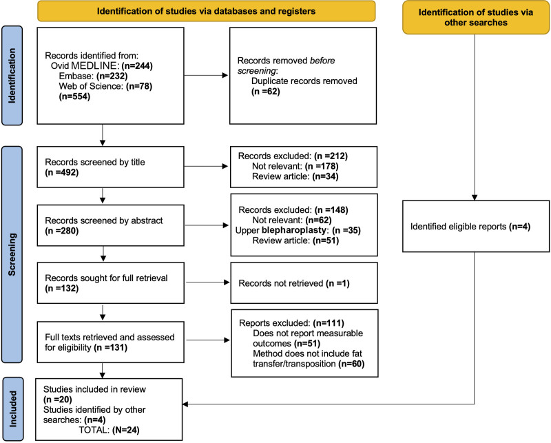
A comprehensive population, intervention, comparison, and outcome review of clinical studies published from 2000 up to May 2025 in the English-language literature was conducted using PubMed, Embase, and Web of Science databases, following the Preferred Reporting Items for Systematic Reviews and Meta-Analyses guidelines to identify studies describing LLB with fat preservation. Two investigators screened titles and abstracts and analyzed full texts of selected papers for eligibility. Disagreements were resolved by consensus or with the senior investigator, and data extraction from selected studies was conducted. Inclusion and exclusion criteria are detailed in Table 1, and the Preferred Reporting Items for Systematic Reviews and Meta-Analyses search algorithm is illustrated in Figure 1. No patient data were collected or used for the preparation of this study to warrant IRB approval.
Table 1.. Inclusion and Exclusion Criteria and Literature Search Methodology
| Criteria | Inclusion | Exclusion |
|---|---|---|
| Study design | • Peer-reviewed cohort studies | • Reviews, case reports, comments, and letters to the editor |
| Population | • Subjects undergoing LLB | • Subjects undergoing facial rejuvenation with LLB not a major component of the procedure |
| Outcome | • Reported improvement in TTD and/or blending of lid-cheek junction | • Description of the surgical intervention without explicit reporting of outcome |
| Methodological quality | • Clearly defined methodologies and measuring tools for determining improvement in TTD and/or blending of the lid-cheek junction | • Studies with poor methodological quality or insufficient reporting of outcome |
| Publication date | • Studies published since 2000 to capture the most recent evidence related to LLD techniques | • Studies published before this time frame |
| Language | • Studies published in English | • Non-English publications |
Fig. 1.. Preferred Reporting Items for Systematic Reviews and Meta-Analyses search algorithm.
Twenty-four studies involving a total of 3623 patients, mostly women, were retrieved for analysis. (See figure, Supplemental Digital Content 1, which displays demographic data and details of studies, https://links.lww.com/PRSGO/E532.) Gender distribution was not reported in 3 studies.[24],[33],[34] Ethnic background of patients was explicitly reported in only 2 studies[35],[36]; however, it could be deduced from patients’ illustrations in Asian studies. In other studies, illustrations were all of White patients (Table 2).
Table 2.. LLB Studies Retrieved for Analysis With Listing of Year of Publication, Country of First Author, Number and Ethnic Background of Patients, and Level of Evidence Provided
| Authors | Year of Publication | Country of First Author | No. Patients | Patients’ Ethnic Background | Level of Evidence |
|---|---|---|---|---|---|
| Huahui et al 37 | 2025 | China | 112 | Not explicitly mentioned. Photographs are for Asian patients | IV |
| Zhang et al 38 | 2025 | China | 37 | Not explicitly mentioned. Photographs are for Asian patients | IV |
| Lin et al 39 | 2025 | China | 17 | Not explicitly mentioned. Photographs are for Asian patients | IV |
| Chen et al 35 | 2025 | China | 97 | Chinese patients | IV |
| Gu et al 34 | 2024 | China | 22 | Not explicitly mentioned. Photographs are for Asian patients | IV |
| Xu et al 22 | 2024 | China | 104 | Not explicitly mentioned. Photographs are for Asian patients | II |
| Abdelhamid Elhendawy et al 40 | 2024 | Egypt | 15 | Not explicitly mentioned | IV |
| Karimi et al 32 | 2023 | United States | 142 | Not explicitly mentioned | IV |
| Bernardini et al 41 | 2023 | Italy | 30 | Not explicitly mentioned | IV |
| Liu et al 25 | 2022 | China | 913 | Not explicitly mentioned. Photographs are for Asian patients | IV |
| Wu et al 42 | 2022 | China | 51 | Not explicitly mentioned. Photographs are for Asian patients | IV |
| Jin et al 24 | 2021 | Philippines | 184 | Not explicitly mentioned. Photographs are for Asian patients | IV |
| Majidian Ba et al 14 | 2021 | United States | 41 | Not explicitly mentioned | IV |
| Cheng et al 43 | 2021 | China | 492 | Not explicitly mentioned. Photographs are for Asian patients | IV |
| Chen et al 44 | 2021 | China | 226 | Not explicitly mentioned. Photographs are for Asian patients | IV |
| Chang et al 45 | 2020 | South Korea | 545 | Not explicitly mentioned. Photographs are for Asian patients | IV |
| Wang et al 33 | 2020 | China | 36 | Not explicitly mentioned. Photographs are for Asian patients | IV |
| Gao et al 46 | 2019 | China | 14 | Not explicitly mentioned. Photographs are for Asian patients | IV |
| Ramesh et al 47 | 2019 | United States | 60 | Not explicitly mentioned | III |
| Youn et al 48 | 2014 | South Korea | 133 | Not explicitly mentioned. Photographs are for Asian patients | IV |
| Massry and Hartstein 28 | 2012 | United States | 54 | Not explicitly mentioned | IV |
| Liao and Wei 49 | 2011 | Taiwan | 212 | Not explicitly mentioned. Photographs are for Asian patients | IV |
| Momosawa et al 36 | 2008 | Japan | 20 | Japanese | IV |
| Mohadjer and Holds 50 | 2006 | United States | 66 | Not explicitly mentioned | IV |
Severity of deformity and outcome were documented in 10 studies based on various grading systems with different classification parameters (Table 3).[22],[25],[33],[35]–[38],[43]–[45],[48] The Barton scale is the most frequently mentioned. In 1 study, the depth of tear trough deformity (TTD) was measured with calipers.[49] In another, the depth of the nasojugal fold and pretarsal orbicularis crease were graded according to the Barton scale but not reported; instead, measurements of lower eyelid length, nasojugal fold depth, and pretarsal orbicularis depth were described.[47]
Table 3.. Summary of Studies in Which Measurement of Deformity Correction Was Reported
| Authors and Year | Surgical Technique | Grading System | Preoperative | Postoperative |
|---|---|---|---|---|
| Huahui et al, 2025 37 | TC preseptal; extension of fat flap; external transcutaneous fixation | Measurement of lower eyelid gray scale | eb-tt-z: 0.1812 ± 0.054 | eb-tt-z: 0.074 ± 0.049 |
| mc-tt-z: −0.432 ± 0.0616 | mc-tt-z: −0.531 ± 0.092 | |||
| mc-ftt-z: −0.581 ± 0.058 | mc-ftt-z: −0.629 ± 0.061 | |||
| pi-tt-z: 0.289 ± 0.044 | pi-tt-z: 0.227 ± 0.061 | |||
| Zhang et al, 2025 38 | TC preseptal; combined FT and injection of SVF-gel rich in vascular stromal components; external transcutaneous fixation | Measurement of lower eyelid gray scale | eb-tt-z: 0.1704 ± 0.0123 | eb-tt-z: 0.1000 ± 0.0322 |
| mc-tt-z: −0.5078 ± 0.0710 | mc-tt-z: −0.5368 ± 0.1230 | |||
| mc-ftt-z: −0.6147 ± 0.0537 | mc-ftt-z: −0.6122 ± 0.0597 | |||
| pi-tt-z: 0.2831 ± 0.0840 | pi-tt-z: 0.2391 ± 0.0516 | |||
| Chen et al, 2025 35 | STC preseptal skin-muscle flap; if necessary, excessive fat resection; external transcutaneous fixation | Barton | G I: 9 | G 0: 9 (100%) |
| G II: 47 | G 0: 46 (97.9%); G I: 1 (2.1%) | |||
| G III: 41 | G 0: 35 (85.4%); G I: 7 (7.2%) | |||
| Xu et al, 2024 22 | TC preseptal; external transcutaneous fixation combined with nasal alar base filling | Hirmand | G I: 28 | G 0:28 (100%) |
| G II: 69 | G 0: 60 (86.95%); G I: 9 (13.05%) | |||
| G III: 10 | G 0: 8 (80%); G I: 2 (20%) | |||
| Liu et al, 2022 25 | TC preseptal for G1; transcutaneous for other patients; fixation method not specified | Not specified | G I: 163 | Overall success rate: 97.81% |
| G II:259 | ||||
| G III: 313 | ||||
| G IV: 178 | ||||
| Cheng et al, 2021 43 | TC retroseptal; fabrication of fan-shaped thin fat flap for transposition; external transcutaneous fixation | TTD measurement with caliper | Mild TTD: 422 | TTD 0 mm: 400 (94.78%), TTD <5 mm: 22 (5.21%) |
| Moderate TTD: 492 | TTD 0 mm: 449 (91.26%), Mild TTD: 43 (8.74%) | |||
| Severe TTD: 70 | TTD 0 mm: 59 (84.29%), Mild TTD: 11 (15.71%) | |||
| Chen et al, 2021 44 | TC preseptal; external transcutaneous fixation | Hirmand | G I: 71 | G 0: 66 (93.0%); G I: 5 (7.0%) |
| G II: 146 | G 0: 124 (84.9%); G I: 22 (15.1%) | |||
| G III: 9 | G 0: 6 (66.7%); G I: 2 (22.2%); G II: 1 (0.5%) | |||
| Chang et al, 2020 45 | TC preseptal; excision of redundant skin and suspension of orbicularis muscle | Modified Barton | External fixation | |
| G 0: 35 (16.6%); G I: 121 (57.3%); G II: 50 (23.7%); G III: 5 (2.4%) | ||||
| Improvement: 191 (90.5%) | ||||
| No improvement 20 (9.5%) | ||||
| Excellent outcome: 70 (33.2%) | ||||
| Internal fixation | ||||
| G 0: 149 (44.6%); G I: 148 (44.3%); G II: 36 (10.8%); G III: 1 (0.3%) | ||||
| Improvement: 323 (96.7%) | ||||
| No improvement: 11 (3.3%) | ||||
| Excellent outcome: 196 (58.7%) | ||||
| Wang et al, 2020 33 | TC preseptal; internal fixation to soft tissues | Barton | Grade I: 22 | G 0: 19 (86.4%); G I: 3 (13.6%) |
| Grade II: 43 | G 0: 36 (83.7%); G I: 7 (16.3%) | |||
| Grade III: 14 | G 0: 5 (71.4%); G I: 1 (14.3%); G II: 1 (14.3%) | |||
| Youn et al, 2014 48 | TC retroseptal; internal periosteal fixation | Barton | FT | FT |
| G I: 17 | G 0: 12 (70.59%); G I: 5 (29.41%) | |||
| G II: 40 | G 0: 22 (55%); G I: 18 (45%) | |||
| G III: 14 | G 0: 3 (21.43%); G I: 9 (64.28%); G II: 2 (14.29%) | |||
| Septal rest | Septal reset | |||
| G I: 5 | G 0: 4 (80%); G I: 1 (20%) | |||
| G II: 28 | G 0: 22 (78.57%); G I: 6 (21.43%) | |||
| G III: 16 | G 0: 10 (62.5%); G I: 6 (37.5%) |
Reviewed reports are mostly cohort studies of various STC and TC LLB approaches. A wide range of techniques with various combinations of surgical steps are described. For TC, both retro- and preseptal dissections are reported. Subperiosteal, preperiosteal, sub–sub–orbicularis oculi fat (sub-SOOF), intra-SOOF, pre-SOOF, and subcutaneous FT are described, as well as internal and transcutaneous fat fixation and lipoaspirated or minced fat grafting[32],[38] (Figs. 2, 3). (See figure, Supplemental Digital Content 2, which displays details of various interventions performed, https://links.lww.com/PRSGO/E533.) Massry and Hartstein[28] reported both subperiosteal and preperiosteal FT without appreciable difference in outcome between the 2 techniques.
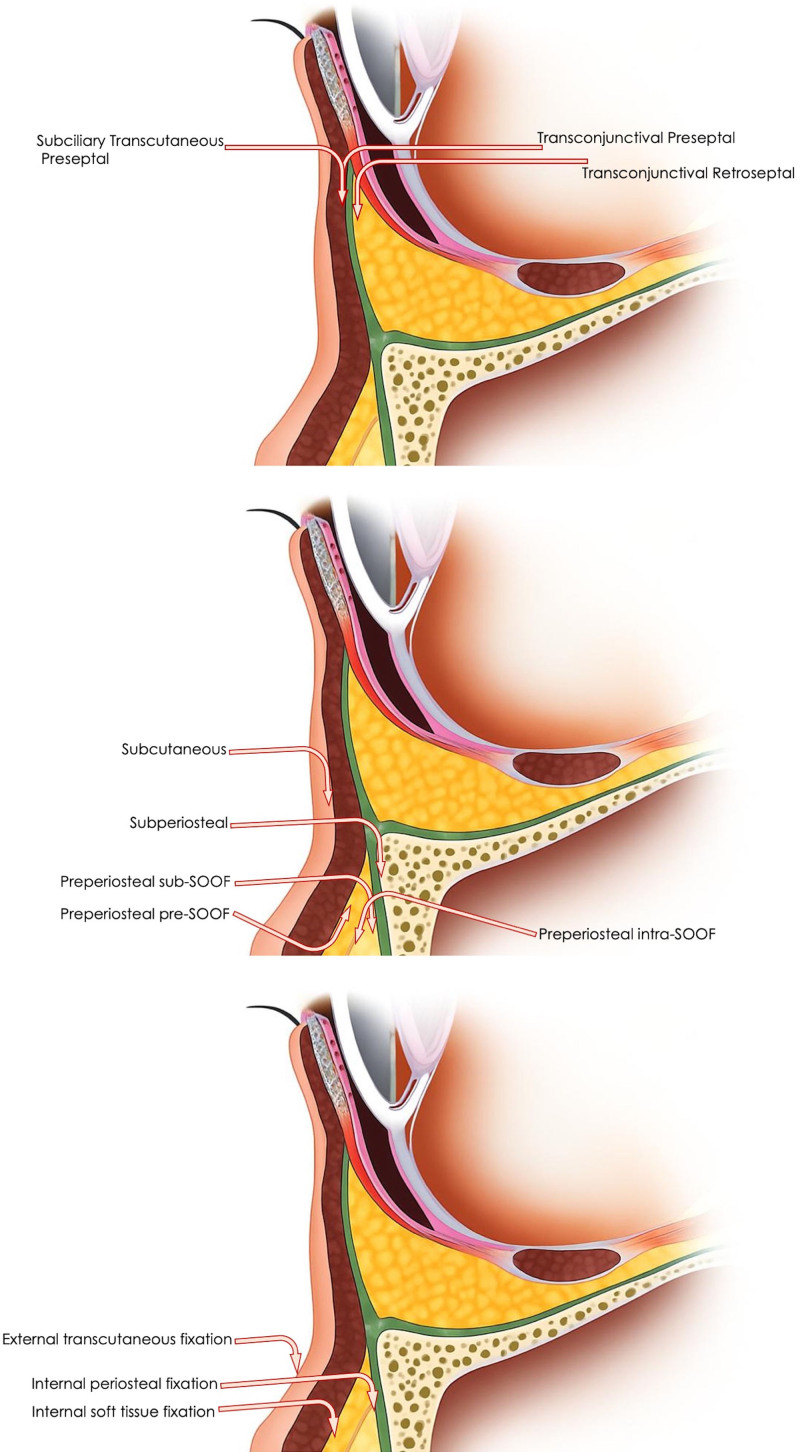
Fig. 2.. ChatGPT-generated figure demonstrating (A) STC and TC surgical access incisions and preseptal and retroseptal dissection planes, various pocket dissections for FT (B), and different fat fixation methods (C).
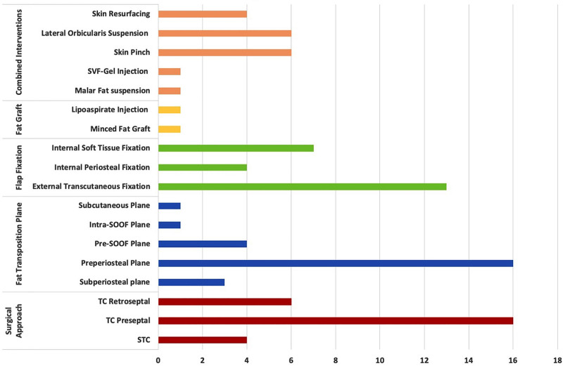
Fig. 3.. Sixteen different interventions performed in various combinations across the 24 studies retrieved for review. SVF-gel, stromal vascular fraction-gel.
Few studies are comparative. Two compared STC and TC with no significant difference in outcome.[25],[49] Ramesh et al[47] compared fat excision to FT. Mean nasojugal fold depth was effaced with both, but significantly more with transposition. Youn et al[48] compared TC subperiosteal FT to septal reset. In patients with grades II and III deformities, better outcomes were observed with septal reset. Transcutaneous and EZ-Tcon internal fixation of transposed fat were compared by Chang et al,[45] concluding that this original internal fixation could have advantages over conventional internal and external fixation techniques. FT and minced fat grafts were also compared by Karimi et al[32]; desired outcomes are equally achievable with both. Excellent correction of deformity is described by all studies. Patients’ satisfaction was measured for 179 STC (3 studies) and 553 TC (9 studies) patients. Despite sampling imbalance, the t test demonstrated that the 2 groups have comparable overall means with standard error and a high Pearson correlation (0.7316904). Overall, excellent and good patient satisfaction was 84.26% for TC and 74.10% for STC (analysis of variance P = 0.87602855) (Table 4). The difference is not significant; however, it must be interpreted with care due to incomplete reporting and the inherent subjectivity bias of several studies.[22],[32],[37],[38],[40]
Table 4.. Pooled Patients’ Reported Outcome Relative to Surgical Access Incision and FT
| Patients’ Reported Outcome | STC * (179 Patients), n (%) | TC † (553 Patients), n (%) | P |
|---|---|---|---|
| Highly satisfied | 69 (38.55) | 386 (69.80) | |
| Satisfied | 62 (34.64) | 80 (14.46) | |
| Moderately satisfied | 4 (0.72) | ||
| Mildly satisfied | 1 (0.56) | 8 (1.44) | |
| Highly satisfied and satisfied | 131 (73.19) | 466 (84.26) | 0.87602855 |
Reported complications were very few. No major complications were noted by most studies. The incidence was not significantly different between the 2 approaches (analysis of variance P = 0.06378577). Minimal swelling was most commonly observed in all patients.[24] Chen et al[44] reported that half of the patients developed transient granulomas of transposed fat. In 2 studies, all patients routinely complained about palpable firmness and developed a slight lumpy appearance early postoperatively.[43],[49] Complications are summarized in Table 5. They were not reported in 1 study.[37] (See graph, Supplemental Digital Content 3, which displays the statistical analysis, https://links.lww.com/PRSGO/E534.)
Table 5.. Pooled Reported Complications Relative to Surgical Access Incision and FT
| Complication | STC (182 Patients), n (%) | TC (2266 Patients), n (%) | STC and TC Combined (913 Patients), n (%) |
|---|---|---|---|
| Ectropion | 3 (2.6) | 1 (0.04) | |
| Temporary lower lid malposition | 12 (1.3) | ||
| Transitory blurring of vision | 1 (0.04) | ||
| Dry eye | 5 (0.2) | ||
| Diplopia | 1 (0.04) | ||
| Transient dry eye syndrome | 37 (4) | ||
| Conjunctival edema and chemosis | 8 (0.35) | ||
| Residual skin laxity | 1 (0.5) | ||
| Residual fat | 2 (1) | 1 (0.04) | |
| Undercorrection of eyelid bags and TTD | 2 (1) | 19 (0.8) | |
| Hematoma | 2 (1) | 7 (0.3) | |
| Delayed resolution of edema and ecchymosis, induration, and granuloma | 2 (1) | 68 (3) | |
| Poor-quality scars requiring secondary surgery | 18 (2) | ||
| Numbness | 7 (0.3) | ||
| Hyperpigmentation | 1 (0.04) | ||
| Mild oozing of blood on the first postoperative week | 5 (0.2) | ||
| Infection and suture abscess | 3 (0.1) | ||
| Total | 12 (6.6) | 127 (5.6) | 67 (7.3) |
| P : 0.06378577 |
Initially, LLB was focused on resecting skin, muscle, and “herniated” orbital fat.[51] How much fat to resect was subjective. Though early results were generally described to be satisfactory, overresection invariably resulted in a hollowed-out periorbital area with a more aged appearance and persistent TTD.[8],[13] Although there remains a role for fat debulking, fat excision alone may be adequate in only a handful of patients. Optimal rejuvenation for youthful and revitalized facial appearance in most patients requires restoration of the natural lower eyelid midface transition.[2],[5],[21],[29],[51] Addressing patient-specific lower eyelid, lid-cheek junction, and canthal changes in a graded approach, with judicious use of surgical maneuvers for correction of sharp demarcation along the orbital rim and malar depression with a smooth midface contour, is essential.[20],[25],[52],[53] This requires correction of the tear trough and palpebromalar grooves with no or minimal fat and skin excision, selective release of orbicularis retaining ligament, orbital fat and SOOF transposition, augmentation of deep malar fat compartment, and preservation of the orbicularis muscle, together with orbicularis and/or lateral canthal support and midface suspension.[2],[5],[10],[16],[20],[22],[41],[54]–[56] With the current gold standard trend of transposing previously discarded protruding fat,[2],[7],[12],[16],[32],[49],[52],[57] the TC technique, preferred when fat excision is the main focus,[10],[29],[58] has been adapted to FT.[8],[14],[24],[43] TC retroseptal extended subperiosteal dissection has been described to correct TTD with transposition of medial and central “herniated” infraorbital fat caudal to the orbital rim. Its theoretical major benefit is providing the most direct access to fat pads while preserving the orbital septum.[10],[29],[58] FT with this approach is challenging and associated with a steep learning curve.[12],[24],[59] Except for good infraorbital foramen visualization through a fairly avascular plane, it provides limited exposure of fat pads and other structures. It is also associated with difficulty in maneuvering instruments and positioning FT in the appropriate orientation, as well as securing midface soft tissues to stable landmarks.[2],[14],[55],[60] Though the maxillary periosteum near the levator alaeque nasi muscle origin is fairly loose, allowing easy FT, the subperiosteal space is invariably rigid. The required extended dissection causes prolonged periods of postoperative edema.[61],[62] The technique may not be adequate to achieve redefined blepharoplasty aesthetic goals because the tear trough and orbicularis retaining ligaments, which constitute the anatomical basis of aging lid-cheek junction deformity, are not released.[24],[55] TC incision certainly has some appeal even if it offers limited ability to tighten the anterior lamella.[8],[13]–[16],[63],[64] Though described as making fat excision easier, ironically, inadequate fat removal is often the most reported complication of this approach due to limited exposure, specifically of the lateral compartment, that may be particularly difficult to retrieve.[8],[11],[60],[65] Fat excision may be performed comparably with STC and TC incisions. In a comparison of the same patient, with STC fat removal from 1 side and TC from the other, Wong and Mendelson[10] reported comparable outcomes. It is important to note that with STC, the scar was not problematic for patients. Furthermore, whenever skin pinch excision and canthal suspension are indicated, the advantage of a nonvisible scar with TC blepharoplasty is lost. On the contrary, adding a skin incision to the TC incision may seem counterintuitive. With TC preseptal dissection, fat excision or transposition requires orbital septum transgression. This approach has a similar outcome and risk profile to retroseptal subperiosteal dissection,[1],[8],[28],[51] indicating that the impact of middle lamella scaring has been overemphasized. “Isolated” orbital septal scars are not likely related to eyelid retraction. No statistically significant difference in lid retraction has been observed comparing excision of the septum versus no manipulation. Lid malposition may still occur with TC blepharoplasty and can be just as much of an issue as with STC blepharoplasty.[6],[55],[64],[66] It is most likely due to multilamellar scarring secondary to transgressing the functional orbicularis muscle from the front or the lid retractors from behind.[55] Lower lid malposition that occurs following STC lamellar manipulation is mostly observed in patients with preoperative orbicularis weakness or increased lid laxity and is primarily associated with a negative vector orbital-malar relationship, which is documented as an important risk factor.[67],[68] It can be most effectively prevented by maintaining lateral canthal support, orbicularis muscle tone, and tarsoligamentous sling integrity, and by correcting eyelid laxity.[20],[56],[69] Proper lateral canthal tightening and lower lid muscle suspension procedures have become the mainstay of STC lower lid surgery in virtually all patients.[7],[17],[20],[56],[70] This has greatly reduced the incidence of previously reported eyelid malposition and canthal angle rounding.[2],[3],[8],[16],[71] Currently, a comparably low 0.4% incidence of symptomatic lid malposition is being reported with both TC and STC approaches.[72],[73] This further negates the claimed advantage of TC blepharoplasty. Muscle denervation is cited as one of the causes of STC blepharoplasty eyelid malposition.[20] The claim that wide orbicularis muscle dissection contributes to complications and results in partial denervation with unbalanced contraction has been proven to be ill-founded. Subciliary skin-muscle flap surgery does not cause any significant orbicularis muscle denervation.[55] A study about blepharoplasty with separation of skin and muscle did not demonstrate any significant postoperative changes in compound muscle action potential. Detailed anatomical description of muscle innervation and investigation with electroneurography has shown that the buccal and medial branches of the zygomatic facial nerve remain untouched by surgery and innervate the muscle efficiently.[59],[74],[75] Effective FT requires extensive orbital fat release and advancement without tension.[34] It also requires secure fixation, which is often difficult to perform.[42] Various flap preparation and fixation techniques either transcutaneously or in situ with internal sutures have been described.[33],[37],[42]–[45] Several modifications of TC blepharoplasty, including preseptal dissection plane, supraperiosteal, SOOF premaxillary and prezygomatic, intra-SOOF, and suborbicularis or subcutaneous planes for easier FT, have been reported, each with some advantages and inherent complications.[2],[10],[12],[14],[21],[24],[50],[61] Subcutaneous FT violates anterior lamellar structures and may potentially increase the risk of lower lid retraction.[73] It is unclear how this approach would be superior to what can be performed with STC blepharoplasty. Various fixation techniques have been described as well, reflecting difficulties encountered with FT regardless of dissection and transposition planes.[14],[33],[42],[45],[46] Though the preperiosteal suborbicularis plane is mostly favored,[16] extended TC approaches have not been widely used because of the invariable lid margin anterior restriction and limited visualization, as well as uncertainty with midcheek surgical anatomy.[10],[55] Moreover, they are all associated with more bruising, prolonged edema, fat granulomas, soft-tissue irregularities, and contour deformities.[12],[16],[43] The technique also has its limitations due to advancement directional discrepancy for medial and lateral hollows, and distance discrepancy for the central hollow. Fat trauma during flap preparation and possible upward retraction secondary to inadequate release and fixation are additional limitations.[32],[37] Whether the STC approach is better at overcoming these limitations cannot be ascertained due to the very limited studies retrieved for review. FT may not entirely eliminate the risk of orbital hollowing. At times, it may be inadequate due to restricted fat volume and pedicle length particularly for full correction of the central V deformity located too inferior to be effaced by a pedicle flap.[32],[38],[43] Furthermore, if orbital fat is not sufficiently released, it can lead to uneven distribution, postoperative asymmetry, or incomplete correction, creating new or worsened depressions and negatively affecting the aesthetic outcome.[38] Volume augmentation by autologous fat grafting may be an alternative option.[2],[9],[52] “Fat pearls” harvested from excess herniated fat pads have been used since the 1980s concomitantly with STC blepharoplasty. Grafting of resected fat is claimed to be safe and effective in some selected patients. It allows more flexibility of distribution in any desired pattern and location with precise volume enhancement, and is relatively easy to learn and perform. It provides not only a biologically superior source of fat graft but also obviates the need for an additional harvesting site.[10],[18],[30],[32],[73],[76]–[78] The effectiveness of FT or grafting of only excised orbital fat has been questioned. The average weight of orbital fat that can be dissected for transposition or for subsequent grafting may be far less than what is needed. Micro-autologous fat transplantation from aspirated abdominal or medial thigh fat has been described as a more suitable alternative.[5],[38],[78] Despite good outcomes, the survival of fat grafts, whether excised orbital fat or lipoaspirated from other areas, is not always predictable with highly variable take between 20% and 90%. Visible nodulation and fibrosis are potential complications not to be underestimated.[14],[37],[78] It has been reported that the combination of orbital FT and stromal vascular fraction-gel injection provides superior correction of TTD in cases of insufficient orbital fat.[38] Regardless, it is important to note that the outcome may not be adequate if it falls short of release of retaining ligaments.[32] Combined orbital FT and fat grafting may effectively overcome limitations of individual applications and maximize their advantages.[38] Patients have varying degrees of aging changes, and thus, 1 procedure cannot fit all.[5] Surgeons should be able to balance techniques to deliver a customized aesthetic result.[47] The decision regarding the particular technical approach depends on clinical findings with special consideration of the sagittal cornea-bony orbital relationship.[11] TC is not a substitute for STC blepharoplasty.[16] Although merits of both techniques are widely debated, regardless of arguments in favor of one or the other, lower blepharoplasty can be safely and efficiently performed with both.[3],[31],[49] STC blepharoplasty remains a time-honored technique.[20],[51] Despite claims that fat pads may be equally exposed with both TC and STC approaches,[11] STC allows better visualization with more space to perform the required multiple maneuvers. It maximizes cosmetic outcomes even if it may result in more aggressive remodeling with an external scar.[20],[37],[55],[71] It is the preferred method for an increasingly aging population in whom lower lid-tightening, shortening, or canthoplasty is necessary, or whenever orbicularis oculi hypertrophy necessitates muscle excision.[15] Furthermore, with the STC approach, simultaneous SOOF relocation and midface lift may be performed together with lateral canthopexy/canthoplasty and septal reset.[16],[17],[54],[57],[79] Compared with transtemporal midface lift with lower lid skin pinch, LLB with FT can yield similar successful improvement in lower lid and midface contour.[80] Combined with SOOF elevation, it allows greater ability for lid-cheek junction blending and results in gratifying midface rejuvenation.[29],[54]
In addition to inherent variability in the design of studies retrieved for analysis, major limitations of this review include the wide variability in clinical presentations and degrees of lower eyelid skin laxity, fat herniation, sagging of facial tissue, or TTD, in addition to variation in parameters used to measure and determine outcome, criteria for aesthetic improvement, and patients’ satisfaction.[36],[39] Moreover, the techniques in the reviewed studies included a variety of surgical manipulations, making the determination of outcome attributed to FT rather difficult, preventing accurate comparison and determination of the efficacy of various techniques. Most studies provided a low level of evidence and carried a high risk of bias due to confounding factors, lack of blinded outcome measurement, and selective reporting of results (Fig. 4).[81] Furthermore, 16 studies (66.66%) were about TC blepharoplasty involving relatively young Asian patients (mean age range: 27.7–50.6 y), likely reflecting the prevalence of specific lower lid anatomy and aesthetic preferences, limiting generalization to Western populations.
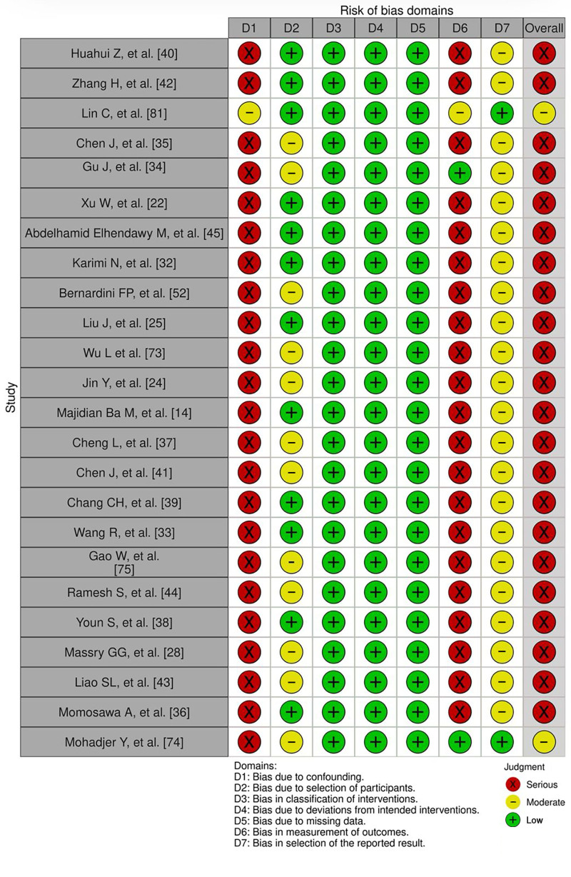
Fig. 4.. Assessment of the risk of bias of reviewed studies using the ROBINS-I tool for assessing risk of bias in nonrandomized studies of interventions. 81 ROBINS-I, risk of bias In non-randomized studies of interventions.
LLB may be safely performed with both STC and TC access incisions. Orbital fat excision or FT can be executed with both, yielding comparable outcomes. The TC approach is suitable for relatively young patients in whom skin excision or canthal support is not needed. The outcome of TC fat excision and/or transposition may be easily enhanced in these patients with fat grafting and noninvasive skin tightening. Whenever a cutaneous incision is required for any reason, STC is probably more suitable. In some patients, FT may not be adequate. Fat grafting would be more applicable. Despite the wide popularity of LLB, the body of LLB literature is unfortunately very scarce and lacks rigorous reporting of outcomes and complications, greatly limiting the identification of the optimal approach. This deficiency needs to be seriously addressed. Further studies are needed to clarify the advantages of either approach, as well as the best option for blending the lid-cheek junction, and to identify which patients may benefit most from these treatments.
The authors have no financial interest to declare in relation to the content of this article.
Published online 16 December 2025. Disclosure statements are at the end of this article, following the correspondence information. Related Digital Media are available in the full-text version of the article on www.PRSGlobalOpen.com.Bishara Atiyeh, Edwin Chrabieh, Kareem Makkawi, Paul Beaineh, Oussama Issa, and Saif Emsieh contributed equally to this work.
1.Raggio BS, Winters R. Lower Lid Transconjunctival Blepharoplasty. StatPearls Publishing; 2025. Updated July 24, 2023.2.Zoumalan CI, Roostaeian J. Simplifying blepharoplasty. Plast Reconstr Surg. 2016;137:196e–213e.3.Drolet BC, Sullivan PK. Evidence-based medicine: blepharoplasty. Plast Reconstr Surg. 2014;133:1195–1205. 4.Palmer FR, III, Rice DH, Churukian MM. Transconjunctival blepharoplasty. Complications and their avoidance: a retrospective analysis and review of the literature. Arch Otolaryngol Head Neck Surg. 1993;119:993–999. 5.Rohrich RJ, Mahedia M, Hidalgo D, et al. The evolving role of blending of the lid-cheek junction in lower blepharoplasty. Plast Reconstr Surg. 2018;142:377–382. 6.Core GB, Steele AW. Minimal access lateral approach in lower eyelid blepharoplasty for three-dimensional recontouring. Clin Plast Surg. 2025;52:73–81. 7.Pepper JP, Baker SR. Transcutaneous lower blepharoplasty with fat transposition. Clin Plast Surg. 2015;42:57–62. 8.Samimi DB, Massry GG. Transconjunctival lower blepharoplasty with and without fat repositioning. In: Azizzadeh B, Murphy MR, Johnson CM, et al. , eds. Master Techniques in Facial Rejuvenation. 2nd ed. Elsevier; 2018:131–141.9.Lee W, Cho JK, Koh IS, et al. Infraorbital groove correction by microfat injection after lower blepharoplasty. J Plast Reconstr Aesthet Surg. 2020;73:777–782. 10.Wong CH, Mendelson B. Extended transconjunctival lower eyelid blepharoplasty with release of the tear trough ligament and fat redistribution. Plast Reconstr Surg. 2017;140:273–282. 11.Baker S, LaFerriere K, Larrabee WF, Jr. Lower lid blepharoplasty: panel discussion, controversies, and techniques. Facial Plast Surg Clin North Am. 2014;22:97–118. 12.Nassif PS. Lower blepharoplasty: transconjunctival fat repositioning. Facial Plast Surg Clin North Am. 2005;13:553–559, vi. 13.Davison SP, Iorio ML, Oh C. Transconjunctival lower lid blepharoplasty with and without fat repositioning. Clin Plast Surg. 2015;42:51–56. 14.Majidian Ba M, Kolli Bs H, Moy Md RL. Transconjunctival lower eyelid blepharoplasty with fat transposition above the orbicularis muscle for improvement of the tear trough deformity. J Cosmet Dermatol. 2021;20:2911–2916. 15.Jacono AA, Moskowitz B. Transconjunctival versus transcutaneous approach in upper and lower blepharoplasty. Facial Plast Surg. 2001;17:21–28. 16.Bhattacharjee K, Ghosh S, Ugradar S, et al. Lower eyelid blepharoplasty: an overview. Indian J Ophthalmol. 2020;68:2075–2083. 17.Murri M, Hamill EB, Hauck MJ, et al. An update on lower lid blepharoplasty. Semin Plast Surg. 2017;31:46–50. 18.Mao SH, Chen CF, Yen CI, et al. A combination of three-step lower blepharoplasty to correct four types of lower eyelid deformities in Asian people. Aesthetic Plast Surg. 2022;46:1224–1236. 19.Bhoutekar P, Winters R. Blepharoplasty Subciliary Approach. StatPearls Publishing; 2025. Updated July 4, 2023.20.Hashem AM, Couto RA, Waltzman JT, et al. Evidence-based medicine: a graded approach to lower lid blepharoplasty. Plast Reconstr Surg. 2017;139:139e–150e.21.Sarhaddi D, Nahai FR, Nahai F. Transconjunctival lower lid blepharoplasty with and without fat preservation and skin resurfacing. Facial Plast Surg Clin North Am. 2021;29:229–241. 22.Xu W, Tian K, Jia Z, et al. Enhanced transconjunctival lower blepharoplasty: a novel method for intraorbital fat transposition and stabilization. Aesthetic Plast Surg. 2024;48:4631–4638. 23.Zarem HA, Resnick JI. Expanded applications for transconjunctival lower lid blepharoplasty. Plast Reconstr Surg. 1991;88:215–220; discussion 221. 24.Jin Y, Gao W, Teo HMT, et al. Transconjunctival lower blepharoplasty using midcheek spaces for orbital fat transposition (S.O.F.T.). Ann Plast Surg. 2021;86:620–626. 25.Liu J, Huang C, Song B. A graded approach in East Asian personalized lower blepharoplasty: a retrospective study spanning 12 years. Indian J Ophthalmol. 2022;70:3088–3094. 26.Loeb R. Fat pad sliding and fat grafting for leveling lid depressions. Clin Plast Surg. 1981;8:757–776. 27.Hamra ST. The role of orbital fat preservation in facial aesthetic surgery. A new concept. Clin Plast Surg. 1996;23:17–28. 28.Massry GG, Hartstein ME. The lift and fill lower blepharoplasty. Ophthalmic Plast Reconstr Surg. 2012;28:213–218. 29.Rao V, Sullivan PK. Lower lid blepharoplasty in men. Clin Plast Surg. 2022;49:213–220. 30.Miranda SG, Codner MA. Micro free orbital fat grafts to the tear trough deformity during lower blepharoplasty. Plast Reconstr Surg. 2017;139:1335–1343. 31.Wilson SC, Daar DA, Maliha SG, et al. Lower eyelid blepharoplasty: does the literature support the longevity of this procedure? Aesthet Surg J. 2018;38:1289–1297. 32.Karimi N, Kashkouli MB, Enayatollahi S, et al. Minced free fat graft versus pedicle fat flap to efface orbital rim hollow in lower blepharoplasty. Aesthet Surg J. 2023;44:12–19. 33.Wang R, Sun J, Guo N. Simple approach for internal fixation of repositioned fat pedicles in transconjunctival lower eyelid blepharoplasty. J Craniofac Surg. 2020;31:2003–2006. 34.Gu J, Duan R, Wei B, et al. Internal fixation of redistributed orbital fat in transconjunctival lower eyelid blepharoplasty. Plast Reconstr Surg. 2024;153:722e–725e.35.Chen J, Tian Y, Wu Y, et al. Percutaneous external fixation of redistributed orbital fat in transcutaneous lower eyelid blepharoplasty. Aesthet Surg J. 2025;45:NP44–NP49. 36.Momosawa A, Kurita M, Ozaki M, et al. Transconjunctival orbital fat repositioning for tear trough deformity in young Asians. Aesthet Surg J. 2008;28:265–271. 37.Huahui Z, Yuxiang Z, Zhihao L, et al. A new method of orbital septal fat flap in transconjunctival approach. Aesthetic Plast Surg. 2025;49:1065–1073. 38.Zhang H, Zhang Y, Lu Z, et al. Application of orbital septal fat repositioning combined with SVF-Gel fillers in lower blepharoplasty. J Craniofac Surg. 2025;36:1884–1888. 39.Lin C, Zhang L, Dong B, et al. Observation on the effect of transconjunctival lower blepharoplasty combined with orbital fat release in middle-aged and elderly men. J Cosmet Dermatol. 2025;24:e70054. 40.Abdelhamid Elhendawy M, Omran AM, Hamdeno S, et al. Transcutaneous lower blepharoplasty with midface lift by malar fat suspension to lateral part of infraorbital wall. Med J Islam Repub Iran. 2024;38:88. 41.Bernardini FP, Skippen B. The “eyelid lift” in lower blepharoplasty surgery. Int Ophthalmol Clin. 2023;63:5–12.42.Wu L, Wang T, You J, et al. Guided internal fixation technique for transconjunctival orbital fat transposition. J Craniofac Surg. 2022;33:628–631. 43.Cheng L, Sun X, Ye L, et al. Fan-shaped thin fat pedicles for tear trough correction through a layer-level fixation: a modified transconjunctival blepharoplasty. J Craniofac Surg. 2021;32:2024–2028. 44.Chen J, Zhang T, Zhu X, et al. Fat repositioning with a combination of internal fixation and external fixation in transconjunctival lower blepharoplasty. Aesthet Surg J. 2021;41:893–902. 45.Chang CH, Bae J, Cha MK, et al. Internal fixation method using EZ-Tcon for transconjunctival fat repositioning: clinical outcomes and efficacy. Aesthetic Plast Surg. 2020;44:1584–1595. 46.Gao W, Qiao C, Lin X. Adjustable fat repositioning with intraoral anchoring: a new technique in transconjunctival blepharoplasty. Ann Plast Surg. 2019;83:S70–S73. 47.Ramesh S, Goldberg RA, Wulc AE, et al. Objective comparison of nasojugal fold depth and lower eyelid length after fat excision versus fat transpositional lower blepharoplasty. Aesthet Surg J. 2019;39:1048–1054. 48.Youn S, Shin JI, Kim JT, et al. Transconjunctival subperiosteal fat reposition for tear trough deformity: pedicled fat redraping versus septal reset. Ann Plast Surg. 2014;73:479–484. 49.Liao SL, Wei YH. Fat repositioning via supraperiosteal dissection with internal fixation for tear trough deformity in an Asian population. Graefes Arch Clin Exp Ophthalmol. 2011;249:1735–1741. 50.Mohadjer Y, Holds JB. Cosmetic lower eyelid blepharoplasty with fat repositioning via intra-SOOF dissection: surgical technique and initial outcomes. Ophthalmic Plast Reconstr Surg. 2006;22:409–413. 51.Nahai F, Niazi F, Nahai F, et al. An overview of surgical approaches for lower eyelid blepharoplasty. Aesthet Surg J. 2023;43:1429–1440. 52.Guyuron B. Discussion: lateral access recontouring blepharoplasty for rejuvenation of the lower lids. Plast Reconstr Surg. 2013;132:843–844. 53.Tarabishy S, Bahat D, Janssen PL, et al. Lower lid blepharoplasty, selecting your approach. Clin Plast Surg. 2025;52:13–20. 54.Atiyeh BS, Hayek SN. Combined arcus marginalis release, preseptal orbicularis muscle sling, and SOOF plication for midfacial rejuvenation. Aesthetic Plast Surg. 2004;28:197–202. 55.Core GB. Lateral access recontouring blepharoplasty for rejuvenation of the lower lids. Plast Reconstr Surg. 2013;132:835–842. 56.Rohrich RJ, Ghavami A, Mojallal A. The five-step lower blepharoplasty: blending the eyelid-cheek junction. Plast Reconstr Surg. 2011;128:775–783. 57.Barton FE, Jr, Ha R, Awada M. Fat extrusion and septal reset in patients with the tear trough triad: a critical appraisal. Plast Reconstr Surg. 2004;113:2115–2121; discussion 2122. 58.Warren RJ. Blending the lower lid cheek junction. Clin Plast Surg. 2025;52:55–71. 59.Atiyeh B, Hakim CR, Oneisi A, et al. Surgical correction of tear trough deformity (TTD) with orbicularis retaining ligament release and volume augmentation for periorbital rejuvenation: review of the literature. Aesthetic Plast Surg. 2023;47:199–214.60.Rizk SS, Matarasso A. Lower eyelid blepharoplasty: analysis of indications and the treatment of 100 patients. Plast Reconstr Surg. 2003;111:1299–1306; discussion 1307. 61.Goldberg RA, Edelstein C, Balch K, et al. Fat repositioning in lower eyelid blepharoplasty. Semin Ophthalmol. 1998;13:103–106. 62.Goldberg RA. Transconjunctival orbital fat repositioning: transposition of orbital fat pedicles into a subperiosteal pocket. Plast Reconstr Surg. 2000;105:743–748; discussion 749. 63.Rancati A, Jacovella P, Zampieri AE, et al. Lower blepharoplasty review, transconjunctival vs. transcutaneous approach. MPS. 2015;05:1–8.64.Codner MA, Wolfli JN, Anzarut A. Primary transcutaneous lower blepharoplasty with routine lateral canthal support: a comprehensive 10-year review. Plast Reconstr Surg. 2008;121:241–250. 65.Netscher DT, Patrinely JR, Peltier M, et al. Transconjunctival versus transcutaneous lower eyelid blepharoplasty: a prospective study. Plast Reconstr Surg. 1995;96:1053–1060. 66.Schwarcz R, Fezza JP, Jacono A, et al. Stop blaming the septum. Ophthalmic Plast Reconstr Surg. 2016;32:49–52. 67.Jacono AA. Transcutaneous blepharoplasty with volume preservation: indications, advantages, technique, contraindications, and alternatives. Facial Plast Surg Clin North Am. 2021;29:209–228. 68.Tepper OM, Steinbrech D, Howell MH, et al. A retrospective review of patients undergoing lateral canthoplasty techniques to manage existing or potential lower eyelid malposition: identification of seven key preoperative findings. Plast Reconstr Surg. 2015;136:40–49. 69.Harounian J, Wulc AE, Brackup AB, et al. Subtle eyelid retraction after lower blepharoplasty. J Plast Reconstr Aesthet Surg. 2019;72:1682–1687. 70.Lee H, Shin H, Baek S. Effects of orbicularis oculi flap suspension on transcutaneous lower blepharoplasty. J Craniofac Surg. 2024;35:46–48. 71.Innocenti A, Mori F, Melita D, et al. Effects of orbicularis oculi flap anchorage to the periosteum of the upper orbital rim on the lower eyelid position after transcutaneous blepharoplasty: statistical analysis of clinical outcomes. J Plast Reconstr Aesthet Surg. 2017;70:385–391. 72.Maffi TR, Chang S, Friedland JA. Traditional lower blepharoplasty: is additional support necessary? A 30-year review. Plast Reconstr Surg. 2011;128:265–273. 73.Kim HS, Choi CW, Kim BR, et al. Effectiveness of transconjunctival fat removal and resected fat grafting for lower eye bag and tear trough deformity. JAMA Facial Plast Surg. 2019;21:118–124. 74.Choi Y, Kim IB. Distribution of the laterally supplying facial nerve to the orbicularis oculi muscle. Aesthet Surg J. 2021;41:161–169. 75.Innocenti A, Dreassi E, Carla V, et al. Evaluation of residual neuro-muscular integrity in the orbicularis oculi muscle after lower eyelid transcutaneous blepharoplasty according to Reidy Adamson-s flap. Aesthetic Plast Surg. 2020;44:1577–1583. 76.Guner H. Percutaneous lower blepharoplasty: a novel technique in lower eyelid aesthetics. J Craniofac Surg. 2020;31:e140–e144. 77.Yang J, He L. The experience in lower eyelid blepharoplasty: improvement of surgical procedures based on anatomical basis. Ann Plast Surg. 2024;93:S15–S18. 78.Huang SH, Lin YN, Lee SS, et al. Three simple steps for refining transcutaneous lower blepharoplasty for aging eyelids: the indispensability of micro-autologous fat transplantation. Aesthet Surg J. 2019;39:1163–1177. 79.Hamra ST. Arcus marginalis release and orbital fat preservation in midface rejuvenation. Plast Reconstr Surg. 1995;96:354–362. 80.Weng C, Quatela V. Achieving a youthful midface: examination of midface anatomy improvement following lower blepharoplasty with fat transposition and transtemporal midface lift with lower lid skin pinch. Aesthet Surg J. 2019;39:NP416–NP428. 81.Sterne JA, Hernán MA, Reeves BC, et al. ROBINS-I: a tool for assessing risk of bias in non-randomised studies of interventions. BMJ. 2016;355:i4919.
原文出处:PMC12708136(PubMed Central 开放获取,CC BY 许可)
声明:中文精读 · 仅供学术参考。内容来自公开文献检索,不代表本人观点,不构成诊疗建议。 医疗美容需在正规医疗机构由执业医师实施。
基于相同主题推荐 · 继续深挖
PRISMA系统综述,纳入24项队列研究、3623例患者。经结膜入路(TC)满意率84.26%,经皮入路(STC)满意率74.10%,无统计学差异(P=0.876)。两种入路均安全有效,并发症无显著差异。TC适合较年轻无需皮肤切除者,STC适合需皮肤收紧的老龄患者。脂肪移植可能比脂肪转位更适合矫正泪沟。
以复位替代切除:微创埋线引导固定眶隔脂肪,同步矫正眼袋与泪沟,实现不增加创伤的自然持久年轻化。

2018-2024年107例经皮入路下睑成形术。创新点:睑缘弓处切开眶隔,选择性切除深层脂肪,再行眶隔-脂肪瓣移位。解决严重脂肪疝出病例「切除太多影响重定位、不切除又膨出」的矛盾。91.6%脂肪膨出和泪沟完全消除,FACE-Q从66.7±12.5降至21.9±15.4(P<0.001)。首次引入AI评估(与临床一致性70.9%,kappa=0.58)。