3D颈部提升颏下解剖动态矫正
2026年9月3日23 min read
Plastic and Reconstructive Surgery Global Open
结论与实践要点
核心范式转变: 本文把颈部从'吸脂+缝颈阔肌'升级为'三维解剖驱动'——关注下颌不对称、二腹肌、颈阔肌不连续、颌下腺、舌骨位置。这正契合我长期关注的颈部年轻化分层理念:颈部问题不是单一'脂肪多',而是多结构叠加,须分别处理。
可落地的技术点: 三角颈阔肌瓣旋转锚定舌骨前筋膜(图 6)、正中颈阔肌缝合(图 7)是重建'颏颈转折点'的硬操作;前二腹肌部分切除(图 3)处理'颏下膨隆'的肌性来源。这些对男性方颌、颏下鼓包尤其有用。
循证边界与风险: 回顾性、单术者、无对照,PRP 降血清肿证据弱;二腹肌切除、颌下腺处理属高阶操作,血管/神经损伤与轮廓不规则风险更高,学习曲线陡。对我团队启发:颈部年轻化须术前用超声/触诊评估各组分(脂肪/肌/腺/舌骨),制定'分结构'方案;与上午的下颌缘 BoNT / 颈阔肌注射形成非手术-手术衔接梯度。合规上避免'颈部一次性完美'等绝对化表述。
Background:Traditional neck rejuvenation—liposuction, platysmaplasty, and skin redraping—overlooks intrinsic mandibular-cervical asymmetry,digastric insertions, platysmal discontinuity, submandibular gland descent, and age-related hyoid retrodisplacement. These influence submental contour and require a tridimensional, anatomy-based correction.
Methods:A retrospective study of 177 patients (99 women, 78 men; mean age 59 y, range 26–78 y) undergoing isolated neck rejuvenation (2003–2024) assessed outcomes of the 3D neck lift, which has been used since 2018 in Knize stage II–III and stage IV cases. The technique combines a triangular platysma myectomy forming a stabilizing neo-floor, partial digastric resection, and subplatysmal fat control, with platelet-rich plasma (PRP) (stages III–IV) or bipolar radiofrequency (stage IV). Results were evaluated by complications, morphology, and satisfaction.
Results:Mandibular asymmetry (broad on the left side) was present in 92% of patients. Stage distribution was: I, 28%; II, 35%; III, 21%; and IV, 16%. Chin implants were added in 10%, PRP in 31%, and radiofrequency in 6%. Complications were minimal, consisting of 2 transient marginal mandibular pareses (<6 wk) and 10 seromas, only 3 of which occurred since 2020; all resolved after a single aspiration. Excellent or good cervicomental angle definition was achieved in 98% of cases, with 96% of patients satisfied or very satisfied.
Conclusions:The 3D neck lift offers a personalized, single-incision solution to submental rejuvenation, integrating natural asymmetry, triangular platysma resection, and fibrotic stabilization to restore a stable cervicomental angle with low morbidity. Adjuncts such as PRP or radiofrequency enhance advanced cases but are nonessential. This method provides a reliable, scar-sparing alternative for isolated neck rejuvenation.
Cervical rejuvenation relies on well-established techniques such as submental liposuction, median or lateral platysmaplasty, muscular corsets, and skin redraping. 1 – 7 These methods have produced widely recognized results and remain standards in routine practice. However, clinical observation shows that cervical and mandibular asymmetries are constant, including different mandibular orientation between the broad and narrow sides, unequal insertions of the anterior bellies of the digastric muscles, frequent separation of the anterior platysma borders, and variable descent of the submandibular glands. These features directly influence submental contour and must be considered to obtain a durable restoration of the cervicomental profile.
Within this context, our approach evolved: initially focused on limited maneuvers (liposuction, median sutures), it was structured from 2018 onward into a tridimensional concept integrating muscle, fat, and skin. Its central element is the creation of a scar-based stabilizing neo-floor, obtained by a triangular platysma myectomy and asymmetrical longitudinal resection of the anterior digastric bellies. This directed scar plane secures the platysmal flap onto the freshened digastric surface, restoring to the platysma its support function. Defined in this way, the 3D neck lift is a reasoned tridimensional reconstruction aimed at restoring the neck’s dynamic architecture while respecting its asymmetries.
This single-center retrospective study, performed by a single surgeon, is based on a continuous series of patients who underwent isolated cervical rejuvenation between 2003 and 2024. During this period, operative protocols were progressively refined; beginning in 2018, the 3D neck lift technique was formalized, and indications were more clearly defined according to the Knize 8 classification. The 3D approach was applied systematically to stage II and III cases and was extended to selected moderate stage IV cases when skin excess remained limited. In those cases, adjunctive procedures such as platelet-rich plasma (PRP) or bipolar radiofrequency were occasionally used to enhance soft-tissue retraction and healing. These adjuncts remained anecdotal and were neither systematic nor standardized.
By contrast, advanced stage IV cases with marked skin excess, particularly in male patients, were excluded from the 3D protocol and underwent direct skin excision according to the described techniques. 9 – 11 Stage I cases were treated with isolated liposuction or liposuction combined with platysmaplasty, depending on tissue and muscular quality.
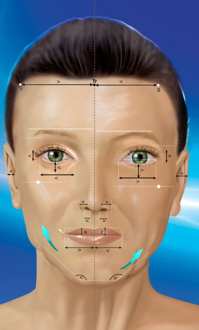
To adapt the procedure to each patient’s submental morphology, cases were secondarily classified by lower face morphotype as broad (open facial angle, horizontal mandibular body) or narrow (closed angle, steeper mandibular inclination), based on morphometric criteria. 12 This classification guided the choice of platysmal plasties and digastric resections, anticipating muscular variations and asymmetries. A degree of facial asymmetry—either directional or fluctuating—was always identified and considered essential in tridimensional submental reconstruction. The study complied with the Declaration of Helsinki, and all patients provided consent for the use of data and images (Fig. 1 ).
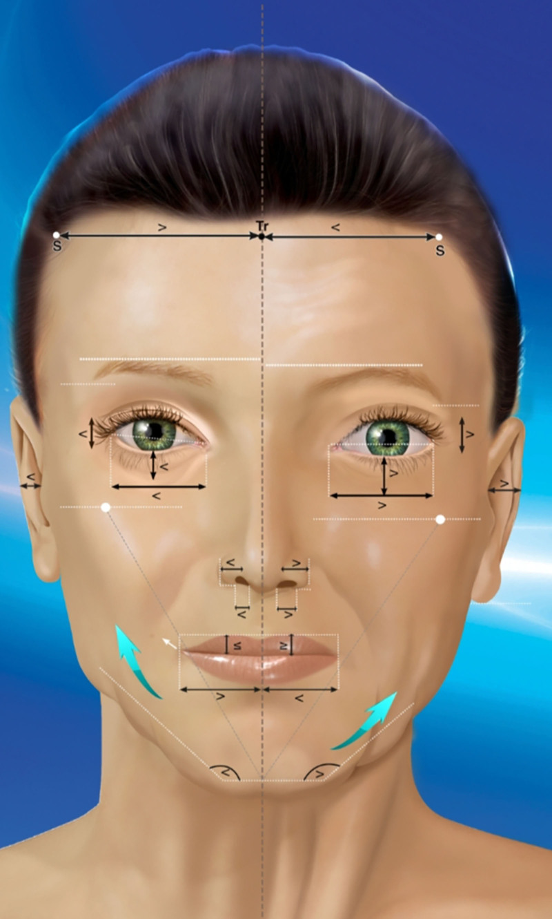
Fig. 1. Frontal morphologic analysis showing consistent lower face asymmetry, with a broader left side (>4-degree difference) and a mandibular branch more parallel to the Frankfurt plane.正面形态分析:左下颌更宽(>4°差异)、下颌支更平行 Frankfurt 平面。说明不对称需个性化设计。Operative technique. ( See Video [online] , which displays how facial asymmetry guides personalized remodeling of the fat compartments and the platysma to create a central fibrous “floor” within the platysmal triangle, restoring a balanced cervicomental architecture beyond surface traction.)
Gentle liposuction with a 3-mm cannula in the subcutaneous plane, extended laterally.
Progressive subcutaneous undermining, leaving a thin superficial fat layer.
Exposure of the platysma after removing supra-platysmal excess.
Delineation of 2 equilateral triangles for excision: right side A–B–apex; left side A′–B′–apex.
Muscular resection: 2.5–3 cm per side. The S–A (or S–A′) distance—from the symphyseal point to the lateral point of the triangle—should ideally match the target symphysis–hyoid height:-too short → the floor cannot unfold;-too long → posterior laxity.
The superior platysmal flap is then elevated, exposing the anterior digastric bellies (Figs. 2 , 3 ).
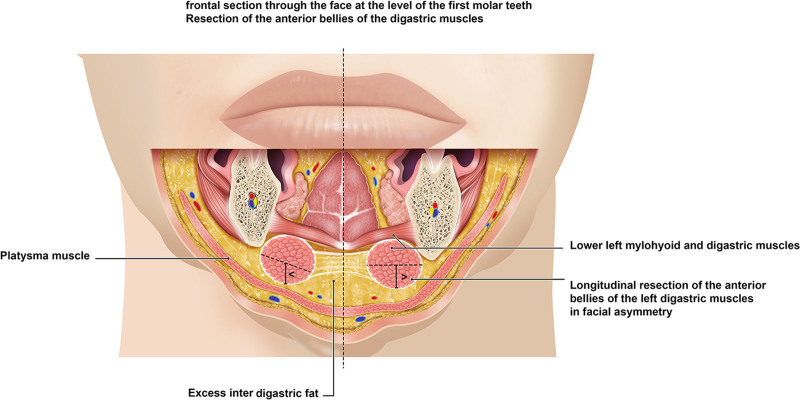
Fig. 2. Anatomical frontal view showing left-side mandibular broadness with a flatter anterior digastric belly. This predisposes to submental bulk, supporting selective resection during a 3D neck lift.解剖正面观:左侧下颌宽、前二腹肌腹扁平,易致颏下膨隆→支持选择性切除。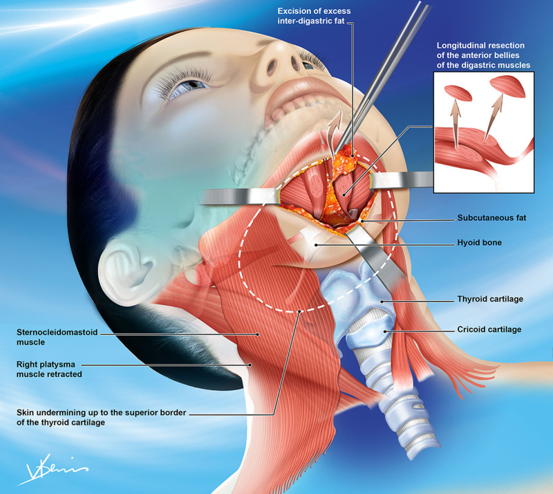
Fig. 3. Longitudinal resection of anterior digastric bellies prevents submental bulging and stabilizes the platysmal flap. This step reinforces fibrotic fixation and ensures durable cervicomental definition.前二腹肌腹纵行切除防止颏下膨出、稳定颈阔肌瓣。关键步骤之一。- 4. Anterior digastric resection and gland control
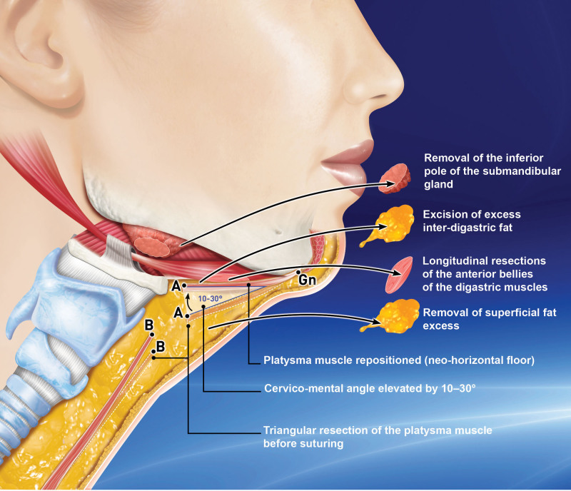
Fig. 4. Diagram illustrating the 3D neck lift combining retroplatysmal fat removal, digastric resection, and vectorized platysma rotation. The method restores an angulated cervicomental profile and may include submandibular gland reduction when needed.3D 颈部提升示意图:retroplatysmal 脂肪切除+二腹肌切除+矢量颈阔肌旋转,恢复颏颈角。- 5. PRP (optional)
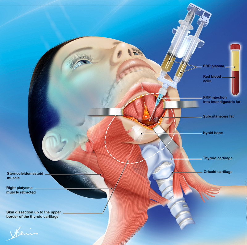
Fig. 5. Occasional PRP injections into interdigastric and retroplatysmal fat were used in men older than 55 years to reduce seromas. PRP may enhance deep-tissue healing, although evidence remains empirical.>55 岁男性偶用 PRP 注射入肌间/retroplatysmal 脂肪以降低血清肿(证据有限)。- 6. Platysma repositioning
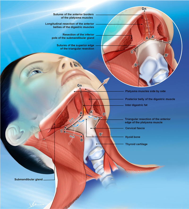
Fig. 6. Triangular platysmal flap (≈3 × 3 cm) rotated 10–30 degrees and anchored to the prehyoid fascia forms a stable support bridge. This restores the cervicomental inflection point and reinforces muscular continuity.三角颈阔肌瓣旋转 10–30° 锚定舌骨前筋膜→稳定支撑桥,重建颏颈转折点。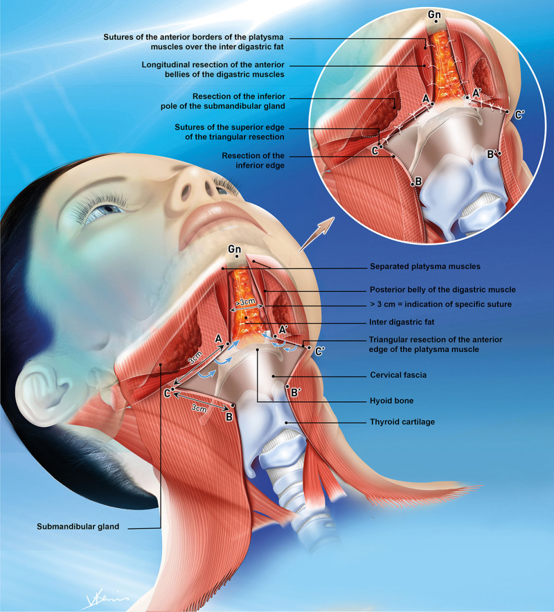
Fig. 7. Median platysmal suture anchored to the prehyoid fascia realigns the musculoaponeurotic plane. This precise fixation redefines submental contour and secures the platysmal triangle.正中颈阔肌缝合锚定舌骨前筋膜,重排肌腱膜平面、固定颈阔肌三角。- 7. Systematic skin undermining and optional radiofrequency
Systematic subcutaneous undermining before closure.
The poorer the skin quality, the farther laterally the undermining is extended to avoid indentations and allow proper redraping.
Exceptionally, percutaneous bipolar radiofrequency may be added in selected moderate stage IV cases.
Optional 7-mm drain placement.
Skin closure.
Application of a molding compressive dressing for 24–48 hours.
补充视频 / 附件(原文未随文提供原图,可于出版方页面查看)gox-14-e7463-s001.mp4Video 1. This video demonstrates how facial asymmetry guides a personalized remodeling of fat compartments and platysma, creating a central fibrous “floor” from the platysmal triangle. This structural approach restores a balanced cervico-mental architecture, rather than simply pulling on surface tissues.
A total of 177 patients were included: 99 women (56%) and 78 men (44%), aged 26–78 years (mean: 59 y). A constant mandibular asymmetry was found, with a left-sided broad face in 93%. Before detailing our clinical series, we present a representative case illustrating this asymmetry and its technical implications (Fig. 8 ).
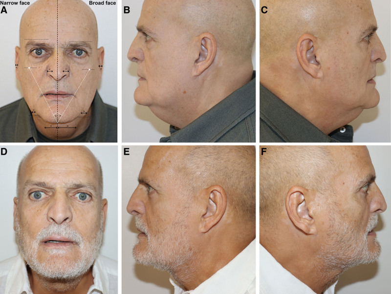
Fig. 8. Male patient, 65 years, Knize stage IV, presenting marked left-sided mandibular broadness and submental laxity. A, Preoperative frontal view. B, Preoperative left lateral view. C, Preoperative right lateral view. Correction was achieved using triangular platysmaplasty and asymmetrical digastric resection. D, One-year postoperative frontal view. E, One-year postoperative left lateral view. F, One-year postoperative right lateral view, showing a stable and symmetrical cervicomental contour.65 岁男性 Knize IV 级:术前正/侧位(A,B)与术后(C,D)。展示重度病例改善。The distribution according to the Knize 8 classification was as follows:
Among these, 16 cases were treated before 2018 using limited skin excision techniques. Since 2018, with the formalization of the 3D neck lift, the technique has been systematically applied to stage II and III cases and progressively extended to 11 moderate stage IV patients, carefully selected according to skin quality and tolerance for limited redraping. Severe stage IV cases with marked skin excess remained excluded from the protocol and were treated by direct excision (Figs. 9 – 11 ). ( See figure, Supplemental Digital Content 1 , which displays a 56-y-old male patient, Knize stage III, who underwent 3D neck lift with liposuction and triangular platysma plication. At 1 y, the L-shaped cervicomental contour remains stable, https://links.lww.com/PRSGO/E631.) ( See figure, Supplemental Digital Content 2 , which displays a 57-y-old male patient, Knize stage IV [moderate], who underwent 3D neck lift with radiofrequency, digastric resection, and interdigastric fat removal. A clear, stable submental break was maintained at 1 y, https://links.lww.com/PRSGO/E632.) ( See figure, Supplemental Digital Content 3 , which displays a 58-y-old male patient, Knize stage IV, who underwent 3D neck lift with gland reduction and a 5-mm chin implant. At 1 y, the patient exhibited a refined profile and harmonious cervicomental definition with full satisfaction, https://links.lww.com/PRSGO/E633.)
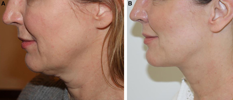
Fig. 9. Female patient, 51 years, Knize stage II, presenting a poor cervicomental angle. A, Preoperative left lateral view. B, One-year postoperative left lateral view following a 3D neck lift with mild liposuction and partial digastric resection, showing restoration of a sharp cervicomental angle and a balanced cervical profile.51 岁女性 Knize II 级:术前与术后 1 年侧位。颏颈角改善。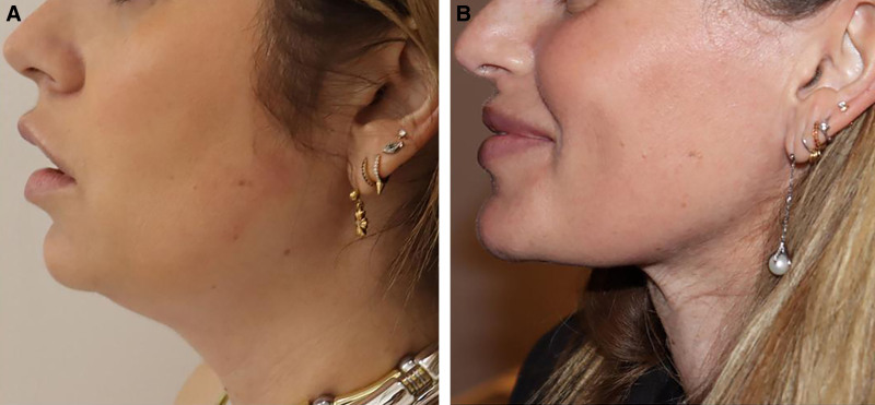
Fig. 10. Female patient, 35 years, Knize stage II. A, Preoperative left lateral view. B, One-year postoperative left lateral view following triangular platysmaplasty with partial myectomy of the anterior belly of the digastric muscle, combined with limited submental liposuction (4 cm³) and partial excision of the left inferior submandibular pole, showing a refined cervical contour and a naturally defined cervicomental inflection.35 岁女性 Knize II 级:三角颈阔肌成形+部分肌切除术前/术后 1 年。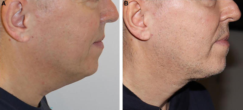
Fig. 11. Male patient, 54 years, Knize stage III, presenting a wide platysmal diastasis treated with triangular plasty, partial digastric resection, and PRP injection. The one-year result shows a clearly defined submental floor and a redefined cervical profile.54 岁男性 Knize III 级:宽颈阔肌分离,三角成形+二腹肌部分切除+PRP 术后 1 年。
A selection of clinical cases illustrates indications and outcomes.
No major complications were observed. Two patients had transient paresis of the marginal mandibular branch of the facial nerve, with full recovery within 6 weeks.
Submental seromas—chronologic trend:
This reduction suggests a possible role for these adjuncts, without proven causality.
Postoperative assessments were performed at 3, 6, and 12 months, focusing on the following criteria:
Chin implants yielded a marked profile improvement with better cervicomental definition.
For more than 4 decades, cervical rejuvenation techniques have been extensively described, particularly in the American literature. Authors such as Connell and Shamoun, 13 Jacono, 14 Rohrich et al, 15 Kaplan et al, 16 and Zins and Menon 17 – 19 established a solid technical doctrine grounded in precise anatomy and proven operative concepts such as the platysmal corset and median plasties. These approaches are effective and foundational. They nevertheless tend to seek an ideal symmetry, whereas clinical experience shows that anatomical variability and structural asymmetries are the rule rather than the exception—especially involving the anterior digastric belly, the submandibular gland, and platysma insertions. 20
A harmonious neck profile depends not on a single angle, but on 2 distinct anatomical angles: the submental angle, immediately beneath the mandibular symphysis, and the cervicomental angle, marking the chin-to-neck transition. These must be differentiated, analyzed, and reconstructed complementarily.
A key foundation of this approach is a subtle yet constant facial asymmetry present from embryogenesis, classifiable as directional (more marked on 1 side) or fluctuating (milder). Asymmetry was observed in 100% of cases. In 93%, the left horizontal mandibular branch lay slightly lower and more parallel to the Frankfurt plane (opening angle on average greater by 4 degrees); in 7%, the reverse was true. This subtle asymmetry is most often characterized by a slightly lower oral floor on the left side, together with marginally lower suprahyoid insertions. This tendency usually accounts for the mild left-sided submental fullness. In most cases, the mandibular platysmal insertions are higher on the right, whereas the anterior borders are never symmetrical in length or shape, being more often looser on the left side. This constant dissociation provides clear clinical evidence of asymmetry.
Accordingly, the initial maneuver is the triangular platysma myectomy, the foundational element of the 3D neck lift. This myectomy induces directed fibrosis that is less contractile than muscle, stabilizes the submental region, and reduces the likelihood of band recurrence. The asymmetrical configuration justifies a side-differentiated triangular plasty with an adapted vector redirection.
After the plasty, the platysmal flap is elevated, exposing the anterior digastric bellies. The resection is tailored to the degree of muscular protrusion. Here, protrusion refers to digastric bulging, not bony projection. When the protrusion is marked—most often on the left side—a tangential reduction is performed. When the protrusion is more limited, the resection is finer and more conservative, carried out as a shaving or partial longitudinal excision. Connell 1 showed that up to 80% of the anterior belly can be resected without functional consequence owing to the synergy of the mylohyoid and contralateral digastric.
The ascended platysmal flap is then fixed anterior to the prehyoid groove at points A–A′:
These fixations create a line of stability that redesigns the cervicomental angle and consolidates the suprahyoid region. The goal is not to elevate the hyoid, but to convert the ascended platysmal flap into a functional support bridge. At this interface, between the fresh bed of the digastric resection and the elevated flap, a directed fibrosis develops, acting as a stabilizer and limiting band recurrence. This phenomenon takes on particular importance in the context of aging, where the hyoid bone tends to descend under the influence of sex, body mass index, and mandibular resorption. 21 Prehyoid fixation of the platysma then helps to maintain a clear and stable inflection point.
By analogy, in orthopedics, several authors have shown that scar fibrosis of tendons and ligaments can provide joint stabilization, partially compensating for muscular loss. 22 – 27 In plastic surgery, the same principle applies to abdominoplasty: rectus plication with absorbable sutures induces a fibrotic reaction that maintains abdominal wall tightening beyond suture resorption. 28 This parallel helps explain the interest—already noted by Connell 1 and Rohrich et al 29 —in selective platysma maneuvers capable of inducing controlled, useful fibrosis. The triangular myectomy shifts the platysma from expression to support, echoing facial reanimation principles.
The interdigastric fat is another key lever. When excessive, its resection deepens and lengthens the submental inflection, refines the chin–neck junction, and optimizes the effect of the muscle plasty. Because this fat is more fibrous and vascularized than superficial fat, its management directly contributes to continuity of the neo-submental plane and provides a valuable fixation bed for prehyoid anchoring.
Submandibular glands also shape the contour: their projection, usually asymmetrical and more pronounced on the left, increases crowding. The triangular platysma plasty often masks this volume, but a partial reduction of the inferior pole was performed regularly (~35%), most often unilateral on the left; bilateral reduction is less frequent but not exceptional. This well-described maneuver effectively reduces glandular fullness without impairing function.
Since 2018, the 3D neck lift has been systematically applied in stage II–III cases and cautiously extended to 11 selected moderate stage IV patients. This gradual extension highlights the prudence of indications, as advanced stage IV cases with marked skin excess remain excluded and continue to be managed by direct excision.
Finally, adjuncts may be used sparingly. PRP, although reported by multiple teams, 23 , 29 , 30 shows encouraging effects on healing and elasticity but lacks robust randomized trials and standardized protocols. An experimental study on abdominal skin-fat samples 30 showed a favorable effect on the lymphatic network, suggesting a possible reduction in seromas, but in neck surgery the use remains empirical. Bipolar radiofrequency was used only very occasionally in selected moderate stage IV cases. 31 These adjuncts mainly helped to cautiously widen indications toward more advanced stages after 2018; they are also the contexts in which outcomes were the most uncertain.
In thin necks with fragile skin, skin retraction remains less predictable and improvement may be incomplete. Platysmal bands, although attenuated in most cases, do not always disappear completely, reminding us that the objective is not to abolish a functional muscle but to restore morphologic balance. Marked bony asymmetries or unrealistic expectations can also limit postoperative satisfaction. The fundamental contribution of the technique is therefore not to promise permanent elimination of cervical cords—whose partial persistence reflects function—but to rebuild a stable architecture perceived by patients as a longer, elegant neck with a harmonious “C-to-L” curve. The occasional use of a chin implant further enhances angle restoration and should be considered within the armamentarium.
The 3D neck lift is a tridimensional, personalized approach to rejuvenation. Its central element is a triangular platysma myectomy creating a stable neo-floor, systematically associated with targeted resection of the anterior digastric belly and, when necessary, selective management of fat or the submandibular gland. The skin redrapes naturally over this restored musculo-fatty framework, supported by its intrinsic elastic properties. As Kaplan et al 16 emphasized, “when the underlying structure is correct, the skin follows—without tension, without artifice.” Thus, the 3D neck lift distinguishes itself by embracing asymmetries, tailoring muscular and fatty maneuvers, and leveraging fibrosis as a stabilizer, allowing since 2018 a prudent extension of indications to certain moderate stage IV necks.
The authors have no financial interest to declare in relation to the content of this article.
Patients provided written consent for the use of their images.
声明:中文精读 · 仅供学术参考。内容来自公开文献检索,不代表本人观点,不构成诊疗建议。 医疗美容需在正规医疗机构由执业医师实施。
基于相同主题推荐 · 继续深挖
背景:传统颈部年轻化(吸脂、颈阔肌成形、皮肤再铺)忽略了下颌-颈 intrinsic 不对称、二腹肌插入、颈阔肌不连续、颌下腺下垂与年龄相关舌骨后移——这些共同决定颏下轮廓,需要三
单中心、单臂前瞻性病例系列(证据等级 III),纳入 2024 年河北医科大学第一医院 786 例男性乳腺发育内镜腺体切除术患者(773 例双侧、13 例单侧),平均随访 96.5

回顾性病例系列(单术者),50 例连续患者(平均年龄 62.1 岁,范围 30–74 岁;女性 98%),2022 年 1 月至 2024 年 12 月行内窥镜额部提升,术前眉峰不