腹壁整形内束腰技术腹直肌折叠联合外斜肌推进
2026年9月5日13 min read
Plastic and Reconstructive Surgery Global Open
结论与实践要点
腹壁整形不止于『切皮抽脂』: 腹直肌分离(diastasis)与侧方肌筋膜松弛是腰线不明显的关键。『内束腰』通过外斜肌内侧推进在筋膜层重建腰线,本组腹围平均减少约 10 cm,并减少侧方残留膨出。
证据怎么读: 属观察性病例系列(无对照、样本小),结论为『该技术可显著缩小腹围、提供腰线定义』,但仍需随机对照与长期随访确认。临床上可作为技术选项之一纳入考量。
安全提醒: 腹壁整形属较大手术,围术期血栓预防、穿支保护与引流管理不可忽视;本组未见血栓事件不代表低风险,须按规范执行 VTE 预防。
Background:Abdominal contour deformities frequently result from skin laxity, localized adiposity, and multidirectional musculoaponeurotic laxity. Although median rectus plication effectively corrects diastasis, residual abdominal bulging and limited waist definition may persist. This study presents a technique combining median rectus plication with medial advancement of the external oblique muscles.
Methods:A prospective observational case series was performed, including 22 female patients (body mass index < 28 kg/m 2 ) undergoing abdominoplasty with median rectus plication and external oblique muscle advancement combined with liposuction. The primary endpoint was reduction in abdominal circumference at the umbilical level at 3 months. Measurements were obtained with the patient relaxed and standing. Paired t test analysis was performed after verification of normality using the Shapiro–Wilk test.
Results:Abdominal circumference decreased from 85.7 ± 7.7 cm preoperatively to 75.4 ± 6.4 cm at 3 months, corresponding to a mean reduction of 10.3 ± 3.5 cm (95% confidence interval, 8.8–11.9 cm; P < 0.001). Mean follow-up was 12 months (maximum 24 mo). No cases of late lateral bulging or thromboembolic events were observed in this limited sample.
Conclusions:The combination of median rectus plication and external oblique muscle advancement demonstrated a significant reduction in abdominal circumference in this observational case series, suggesting that this approach may represent a valuable technical adjunct in contemporary abdominoplasty.
Abdominal contour deformities may result from genetic predisposition and acquired factors, such as pregnancy and weight fluctuations. 1 Modern abdominoplasty requires an integrated approach addressing skin, adipose tissue, and the musculoaponeurotic system. 1 , 2
In addition to rectus diastasis, 2 patients may present multidirectional laxity involving the oblique and transversus abdominis muscles. 3 Isolated correction of rectus diastasis may not fully restore global abdominal wall firmness. 4 , 5
The author previously described the concept of corset plication, which combines a midline plication with additional plications along the semilunar lines. 6 Building upon this principle and the importance of waist definition, 7 , 8 the present study described the association of median rectus plication with medial advancement of the external oblique muscles. 9
The external oblique muscle originates from ribs 5 to 12 and inserts into the rectus sheath and iliac crest. 10 , 11 Its vascular supply follows a Mathes and Nahai type IV segmental pattern. 12
The objective of this study is to describe a technique combining median rectus plication with medial advancement of the external oblique muscles and to evaluate its aesthetic outcomes and safety profile in an observational case series.
A prospective observational case series was performed, including 22 consecutive female patients operated on between April 2023 and November 2024.
All patients were female.
Primary endpoint: reduction in abdominal circumference at 3 months.
Circumference was measured by a single trained nurse at the umbilical level with the patient standing and relaxed, preoperatively and at 3 months.
Statistical analysis was performed using a paired t test. Normality was verified using the Shapiro–Wilk test. Analyses were conducted using standard statistical software. A significance level of 5% was adopted.
The technique followed principles described in contemporary abdominoplasty approaches. 13 , 14 All procedures were performed by the senior author.
Under general anesthesia, initial liposuction was performed in right and left lateral decubitus positions, followed by repositioning to the supine position, requiring 3 patient positions and 2 repositionings. Gluteal lipofilling was performed as indicated.
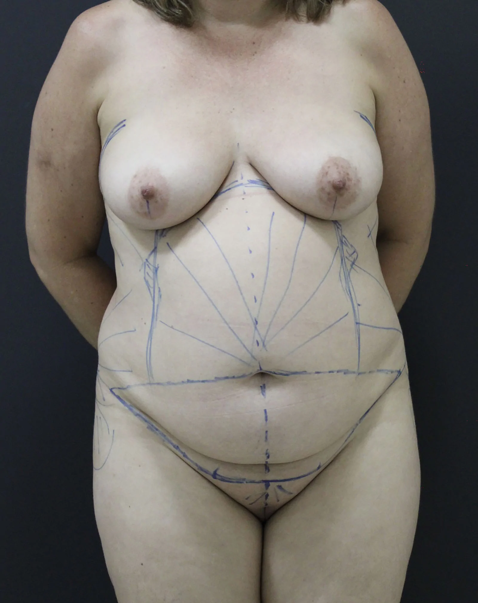
In the supine position, lower abdominal and periumbilical incisions were made. The incision design was planned to minimize discrepancy between the upper and lower incision lengths during closure, facilitating more balanced tissue approximation and reducing the need for excessive tension redistribution or dog-ear correction. (Fig. 1 ).
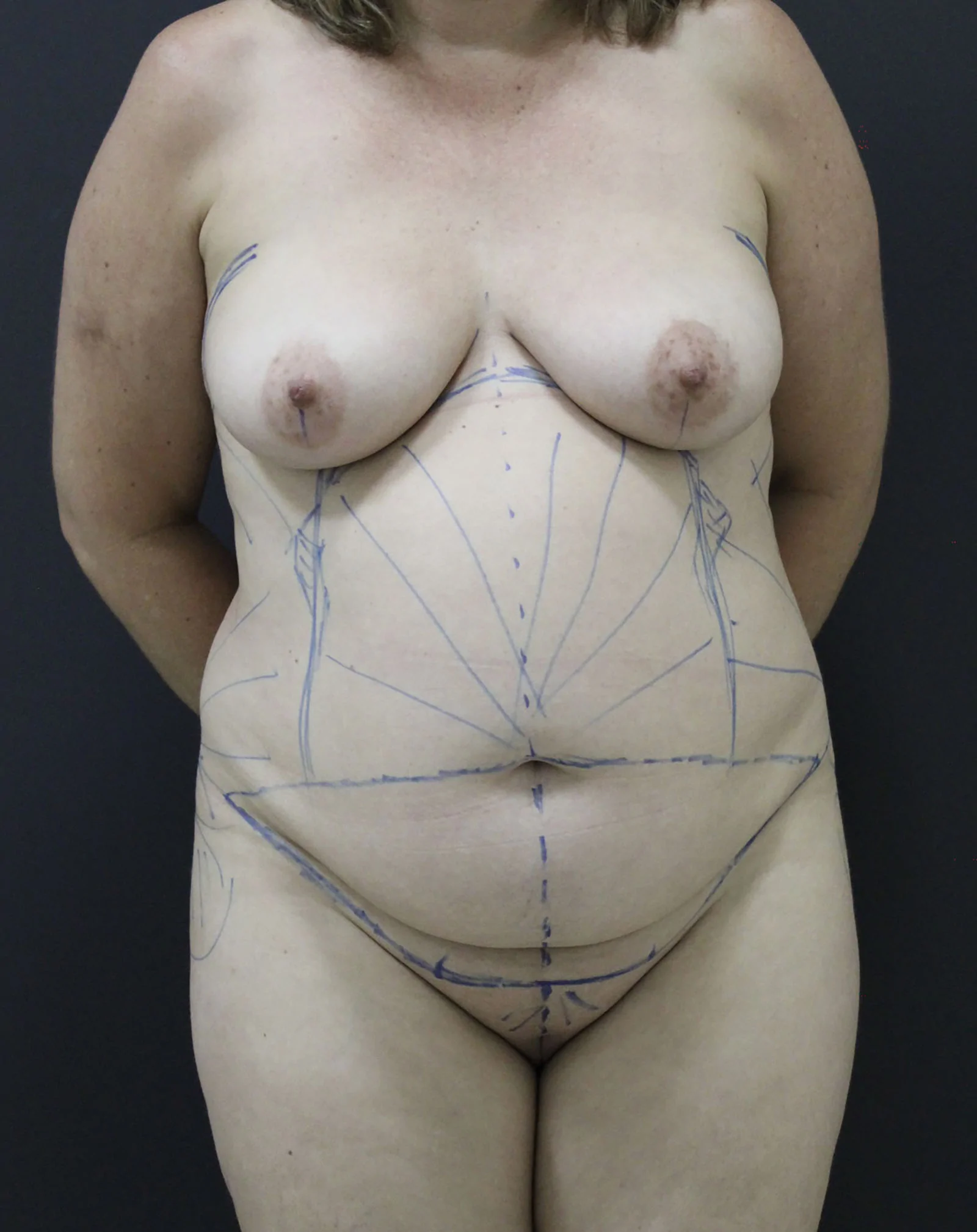
Fig. 1. Preoperative marking demonstrating flap design intended to minimize flap length discrepancy during closure.术前标记示意,设计以减少关闭时皮瓣长度差异。Pre-aponeurotic dissection was carried out up to the umbilicus. A selective supraumbilical tunnel was then created, wide enough to allow correction of rectus diastasis while preserving vascular supply.
Continuous median rectus plication was performed in both supra- and infraumbilical segments using polypropylene sutures, reinforced with barbed sutures. Lateral tunnels were subsequently developed along the semilunar lines up to the costal margins, as previously described. 15 This limited dissection preserves rectus perforators essential for flap vascularity 16 (Fig. 2 ).
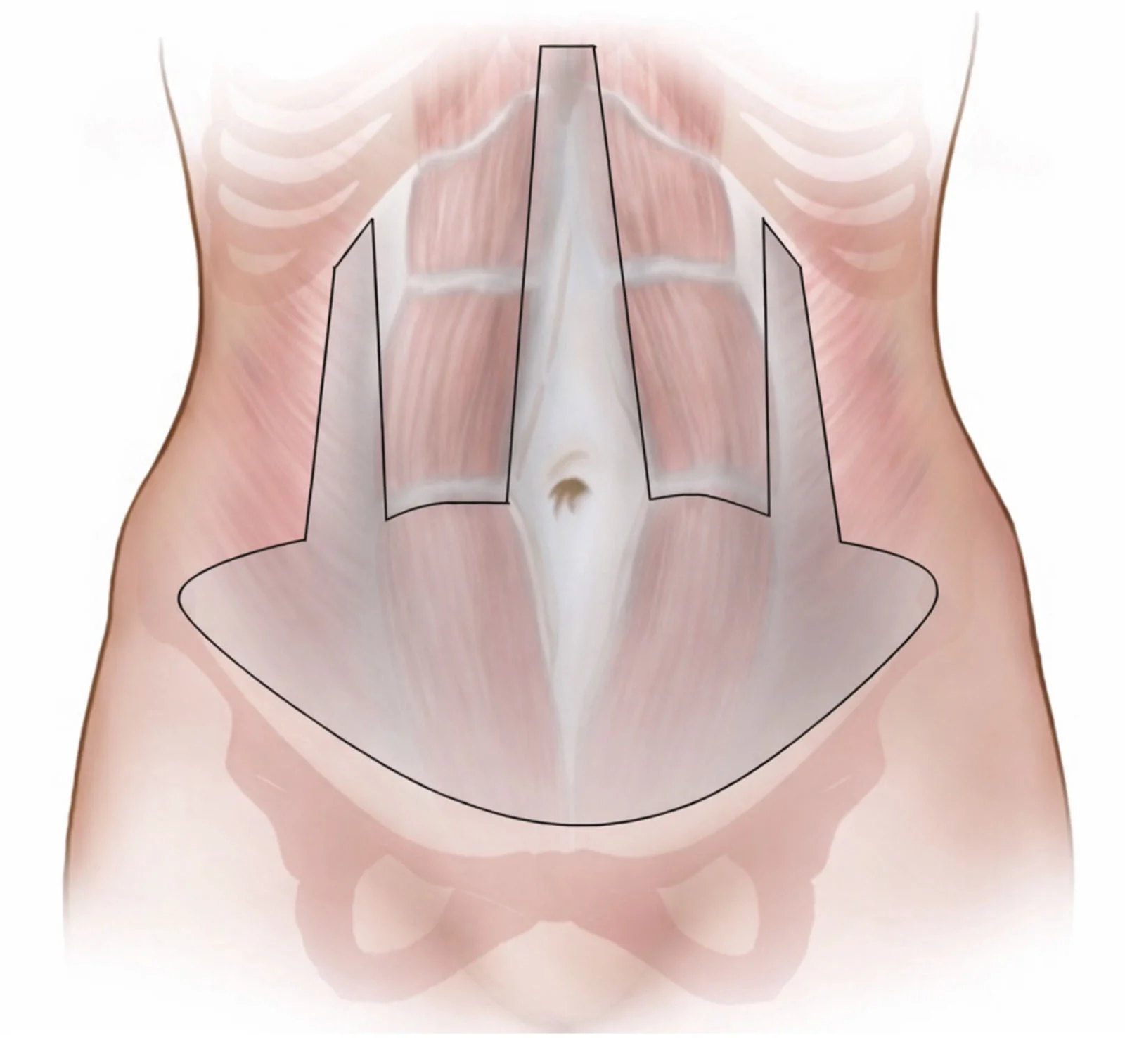
Fig. 2. Area of undermining. Selective supraumbilical dissection preserves rectus abdominis perforators.剥离范围;选择性脐上解剖以保留腹直肌穿支。The external oblique insertions were released while preserving a 1-cm aponeurotic strip for secure fixation. In our experience, external oblique fascia tearing is uncommon when this aponeurotic strip is preserved, allowing stable advancement and fixation of the flap. The muscles were advanced medially and fixed using absorbable X-shaped sutures reinforced with nonabsorbable running sutures. Mean medial advancement was 3.5 cm per side (maximum 5.5 cm to the midline), limited to the midline to avoid overlap with contralateral advancement (Fig. 3 ).
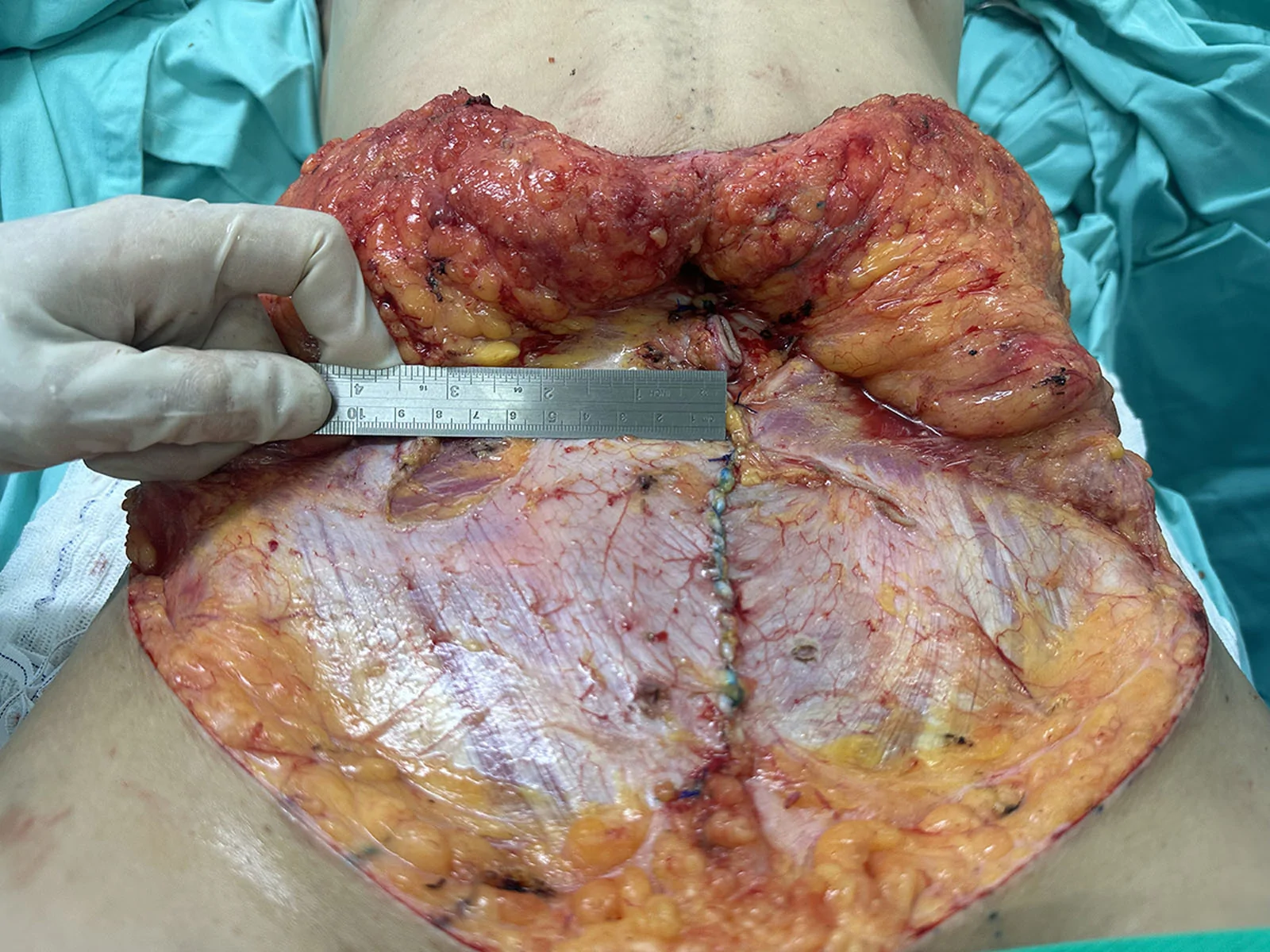
Fig. 3. Left-sided external oblique advancement. A ruler on the right demonstrates 5.2 cm of medial advancement.左侧外斜肌推进,标尺示内侧推进约 5.2 cm。The dermo-fatty flap was resected under appropriate tension, and the umbilicus was repositioned. Closure was performed in layers. Liposuction of the abdominal flap and pubic region was then performed, followed by placement of a closed-suction drain.
Mean liposuction volume was 3300 mL, and mean operative time was 5.5 hours. Thromboprophylaxis consisted of enoxaparin 40 mg initiated 6 hours postoperatively and maintained for 20 days. ( See Video [online] , which displays the overview of key steps in abdominoplasty with median rectus plication and external oblique muscle advancement.)
补充视频 / 附件(原文未随文提供原图,可于出版方页面查看)gox-14-e8068-s001.mp4Video 1. This video provides an overview of the key operative steps of the Internal Corset Technique in abdominoplasty, including median rectus plication, medial advancement of the external oblique muscles, and abdominal wall contouring. The video demonstrates the sequence of the procedure and highlights the surgical maneuvers used to improve waist definition and abdominal contour.
Abdominal circumference decreased from 85.7 ± 7.7 to 75.4 ± 6.4 cm at 3 months.
The mean reduction was 10.3 ± 3.5 cm (95% confidence interval, 8.8–11.9 cm; P < 0.001). Mean follow-up was 12 months (maximum, 24 mo).
Complications included 1 intraoperative external oblique tear (repaired), 2 cases of epidermolysis, and 1 mild infection.
No thromboembolic events were observed in this limited sample. Additionally, no cases of late lateral bulging, functional complaints, or genitourinary complaints were identified (Figs. 4 – 7 ).
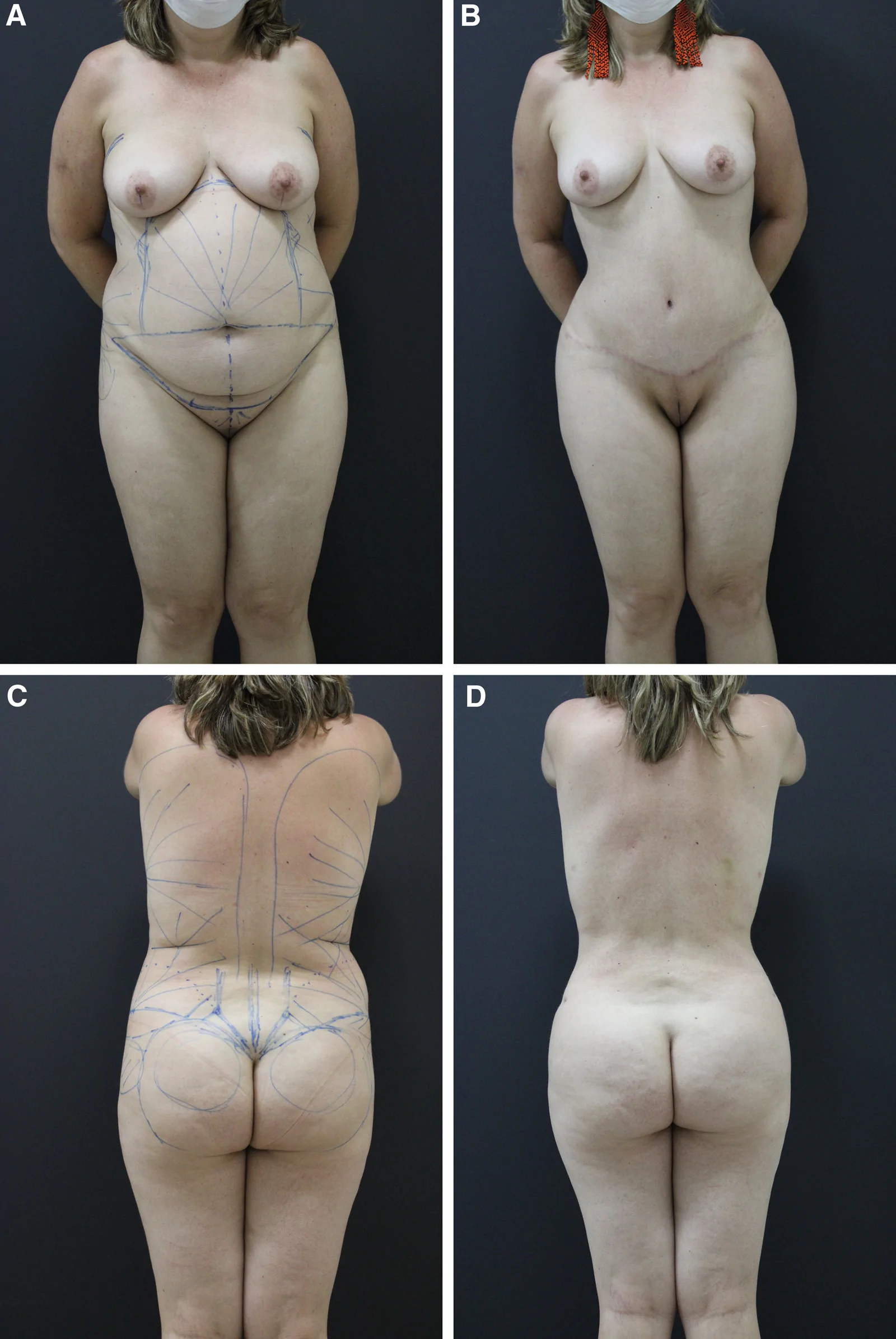
Fig. 4. A, Preoperative anterior view. B, Three-month postoperative anterior view. C, Preoperative posterior view. D, Three-month postoperative posterior view.病例:术前 / 术后 3 月前后位与后位对比。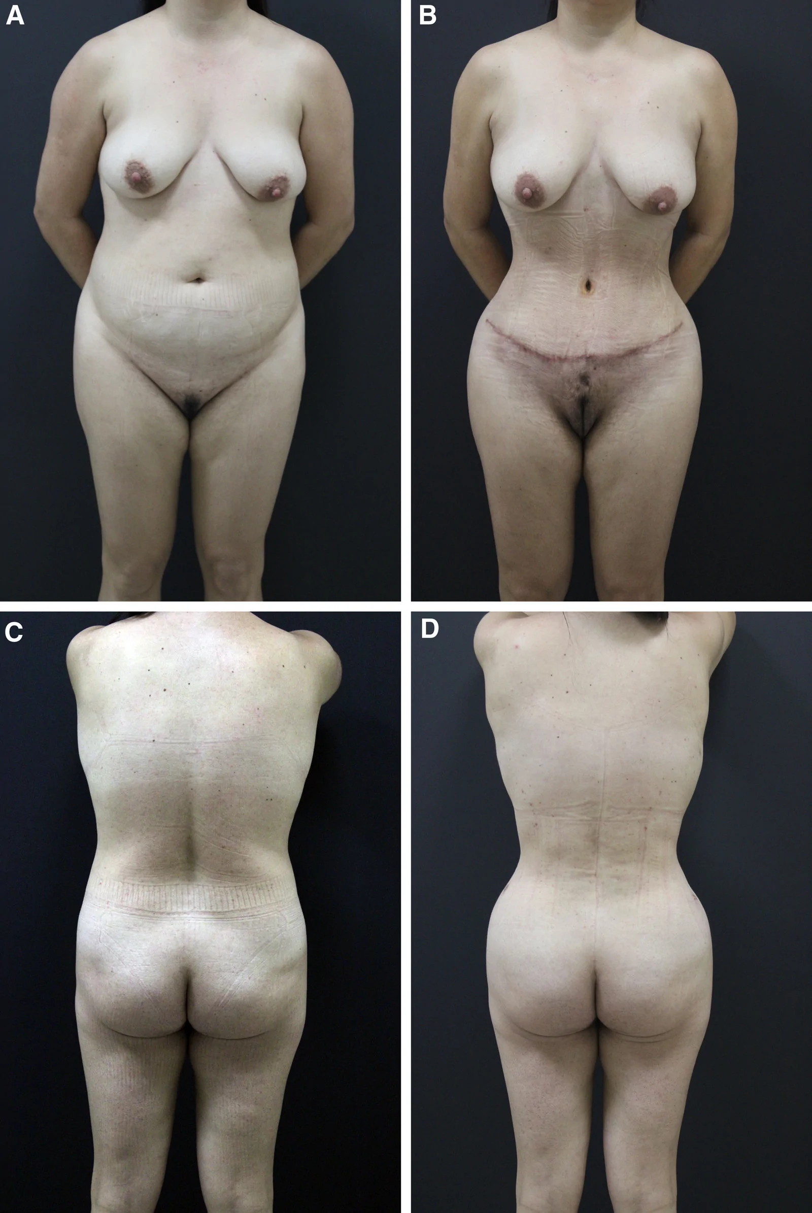
Fig. 5. A, Preoperative anterior view. B, Three-month postoperative anterior view. C, Preoperative posterior view. D, Three-month postoperative posterior view.病例:术前 / 术后 3 月对比。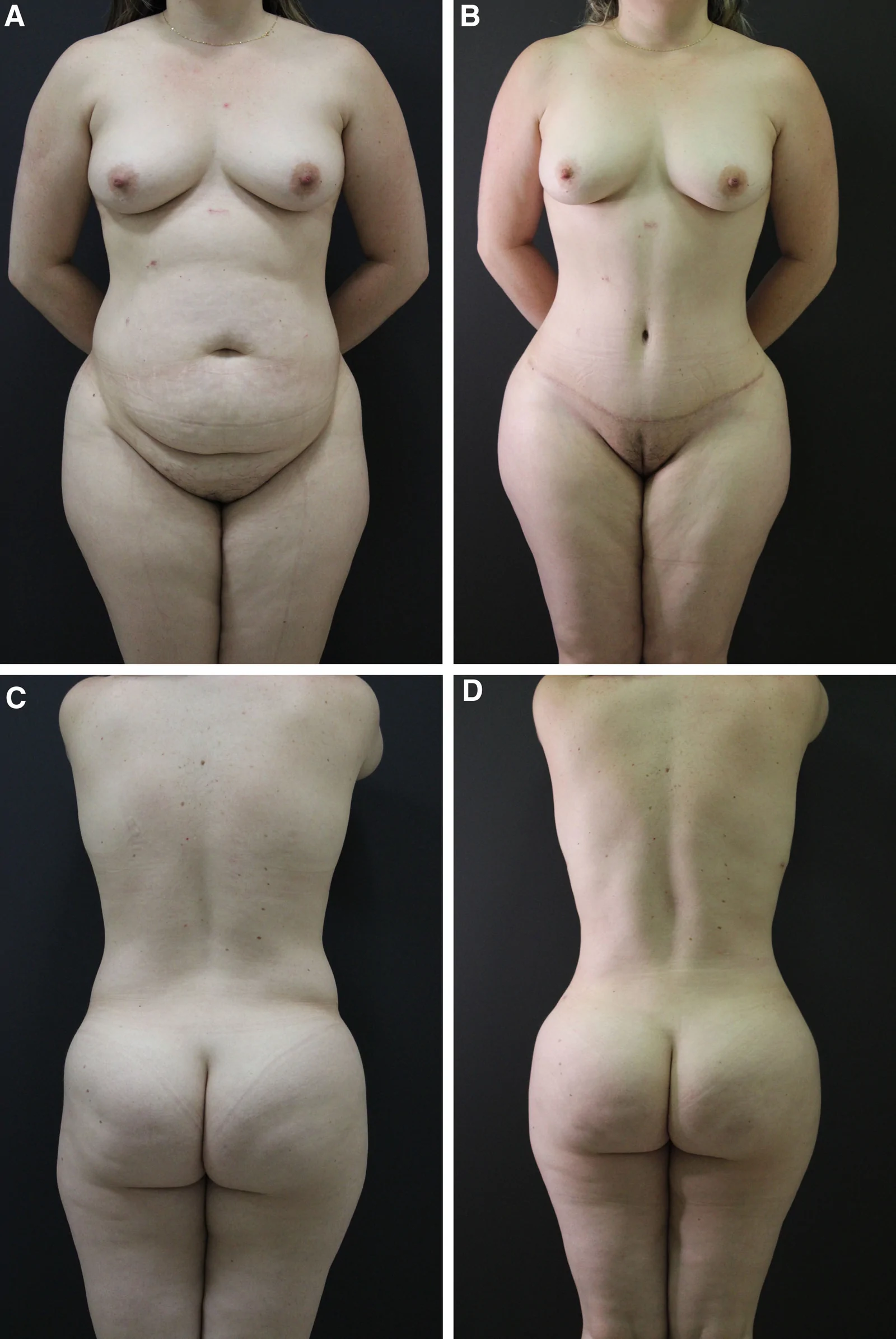
Fig. 6. A, Preoperative anterior view. B, Three-month postoperative anterior view. C, Preoperative posterior view. D, Three-month postoperative posterior view.病例:术前 / 术后 3 月对比。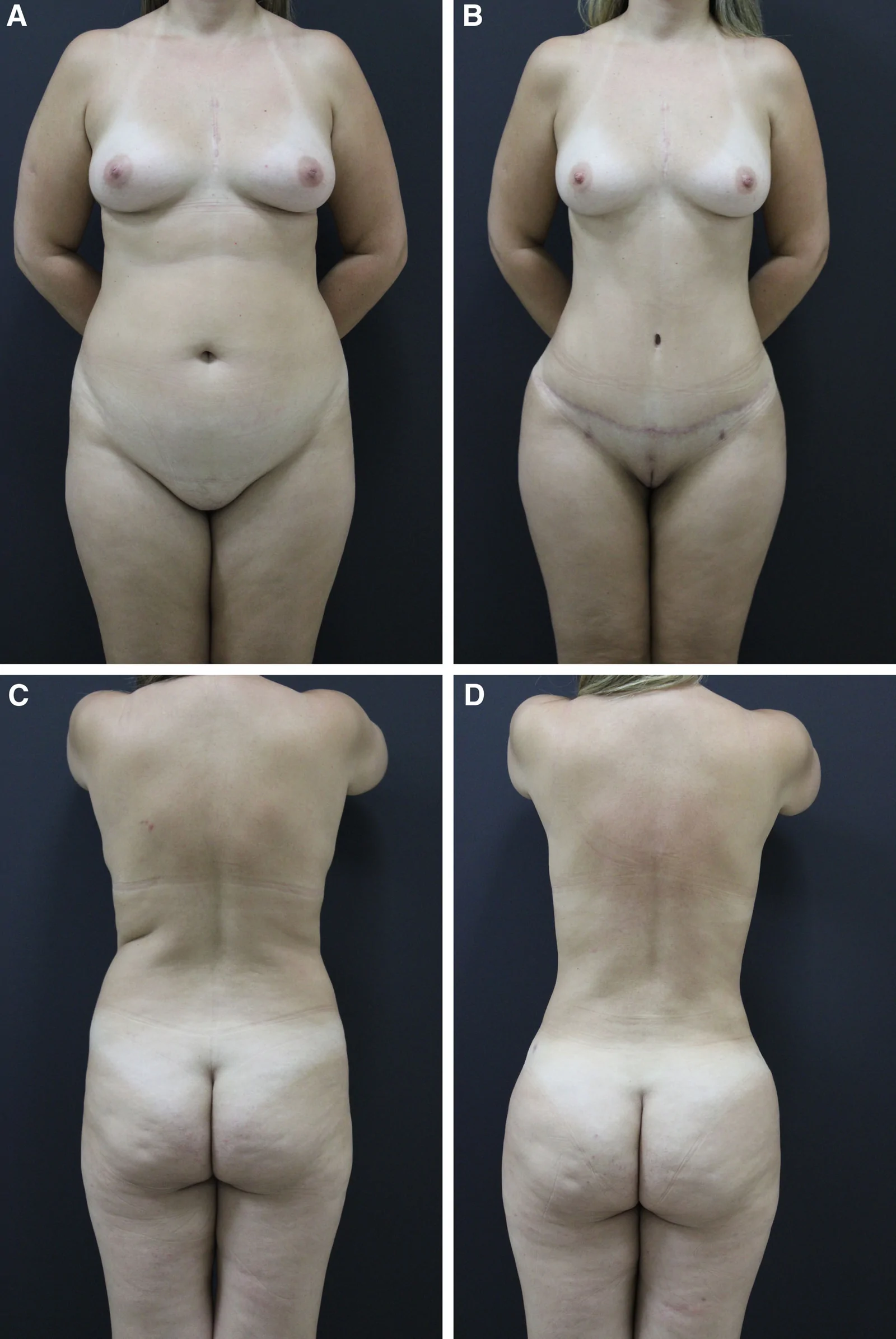
Fig. 7. A, Preoperative anterior view. B, Three-month postoperative anterior view. C, Preoperative posterior view. D, Three-month postoperative posterior view.病例:术前 / 术后 3 月对比。
Anatomic studies have demonstrated the feasibility of external oblique advancement for waistline improvement. 15 The dissection plane for external oblique advancement is developed lateral to the lateral border of the rectus abdominis muscle, along the semilunar line. For this reason, clinically significant rectus abdominis perforators are typically not encountered during this portion of the dissection. 16 , 17 In our experience, only small subcutaneous vessels may occasionally be identified and are routinely controlled with cautery, supporting this plane as a safe and reproducible area for dissection.
Waist contour is influenced not only by soft tissue distribution but also by the relationship between the costal margin and iliac crest. 18
This technique builds upon previously described concepts of abdominal wall plication and lateral contouring by emphasizing a lateral-to-medial vector of tension, which may enhance waist definition beyond midline correction alone. 9 This conceptual approach may help explain the magnitude of circumferential reduction observed in the present series.
The previously described external oblique plication technique improved abdominal wall tension and correction of musculoaponeurotic laxity, 6 particularly in patients with increased abdominal wall flaccidity. However, its ability to produce substantial waistline narrowing was limited. In contrast, external oblique advancement, when combined with median rectus plication, not only enhances abdominal wall tension but also allows a more pronounced and consistent reduction in waist circumference by mobilizing the lateral abdominal wall components.
Medial advancement of the external oblique muscles may increase intra-abdominal pressure. Previous studies have shown measurable increases in intra-abdominal pressure following abdominal wall plication; however, these changes were not associated with significant systemic repercussions. 19 In the present study, no clinical manifestations suggestive of abdominal compartment syndrome or respiratory compromise were observed.
The reduction observed in this study most likely results from the combined effects of median plication, muscular advancement, and liposuction. The isolated contribution of each component cannot be determined in this observational case series. Nevertheless, the magnitude of circumferential reduction, together with qualitative assessment of postoperative photographs, suggests a clinically meaningful improvement in waist definition and lateral contour.
Contemporary rib remodeling techniques have also been described to enhance waist definition, 20 although they may be associated with increased morbidity in selected cases. 21 In contrast, external oblique advancement preserves the structural integrity of the rib cage while still allowing meaningful waistline refinement through mobilization of the lateral abdominal wall components, representing a potentially valuable alternative for selected patients.
This study has important limitations, including small sample size, absence of a control group, single-surgeon design, inclusion exclusively of female patients with body mass index < 28 kg/m 2 , inability to isolate the effect of liposuction, and absence of objective functional assessment.
The primary endpoint was assessed at 3 months postoperatively, which may not fully capture continued improvement occurring between 4 and 6 months as edema resolves and tissue remodeling progresses. Longer follow-up may therefore provide additional refinement of the final aesthetic outcome.
Prospective comparative studies are required to better define the isolated role of external oblique advancement.
The combination of median rectus plication and external oblique muscle advancement demonstrated a significant reduction in abdominal circumference in this observational case series, suggesting that this approach may represent a valuable technical adjunct in contemporary abdominoplasty.
The author has no financial interests to declare in relation to the content of this article.
Written informed consent was obtained from all patients for participation and publication of clinical photographs.
This study was conducted in accordance with the principles of the Declaration of Helsinki. According to local institutional regulations, formal institutional review board approval was not required for this type of observational case series.
声明:中文精读 · 仅供学术参考。内容来自公开文献检索,不代表本人观点,不构成诊疗建议。 医疗美容需在正规医疗机构由执业医师实施。
基于相同主题推荐 · 继续深挖
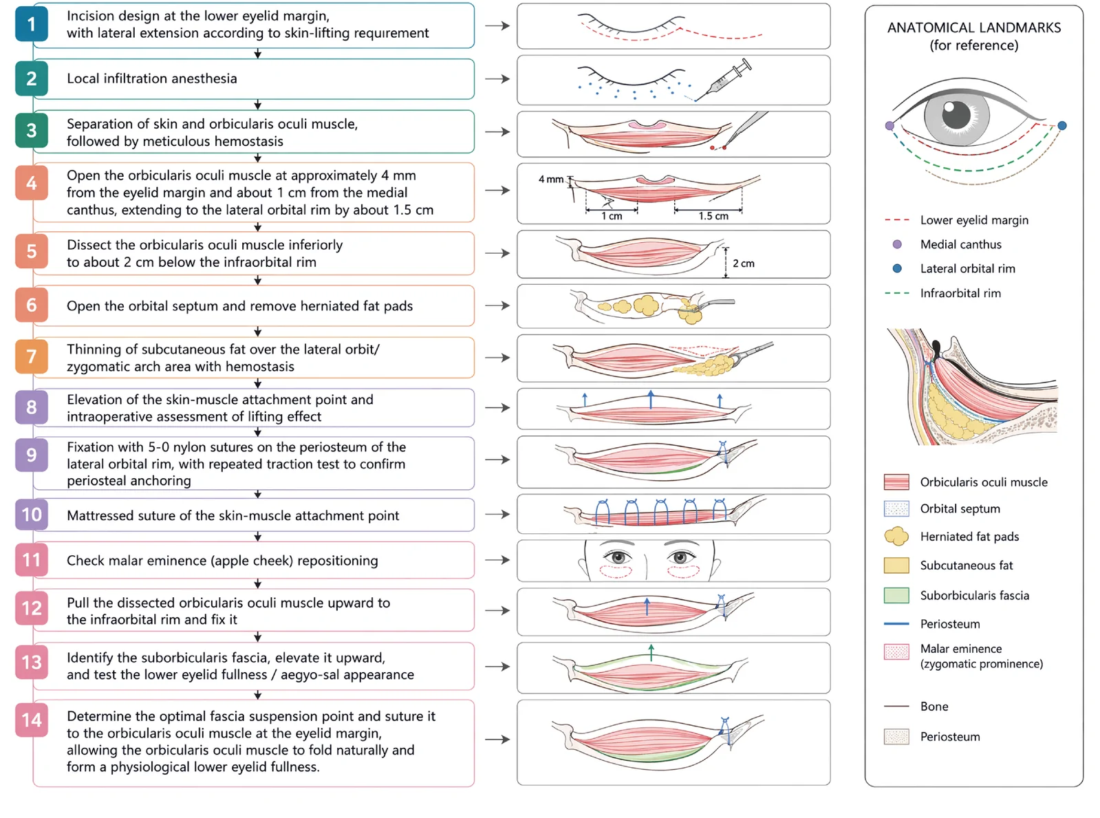
V3中面部提升经睫毛下切口,沿内眦线、外眦线、颧脂肪垫下界三个向量悬吊,可同时改善中面部下垂、下睑袋及睑颊轮廓不连续。
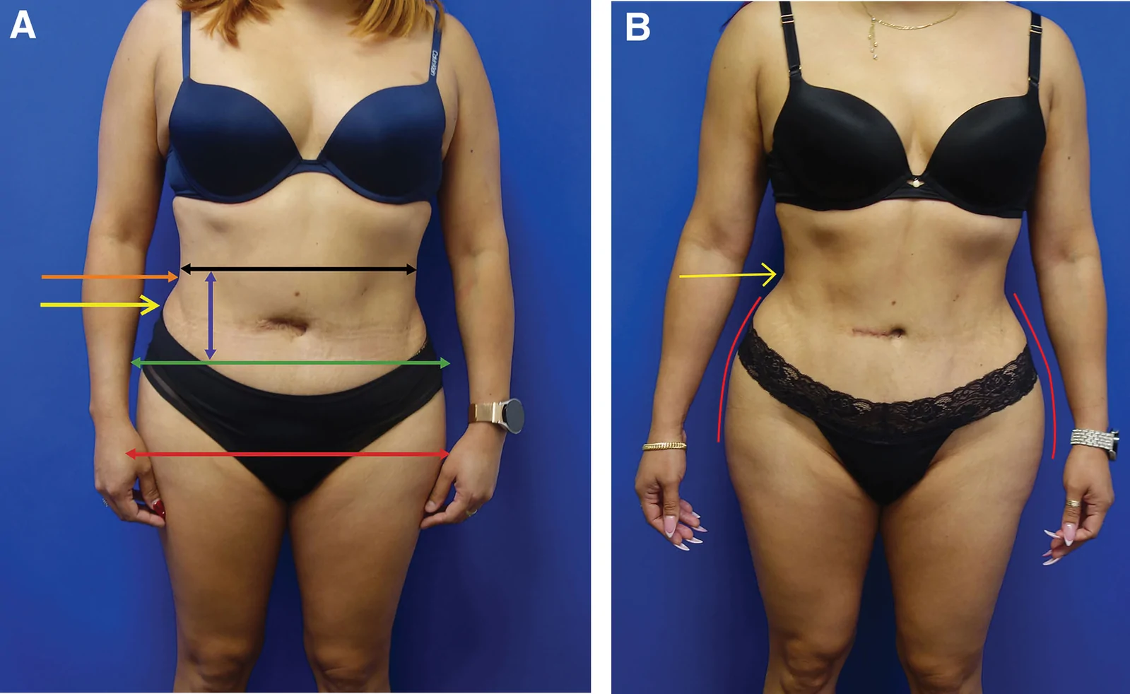
回顾性大样本病例系列(2016年11月至2025年1月),共1999例连续手术,中位年龄36岁(IQR 31至42),中位BMI 29(IQR 26至32),最短随访6个月。
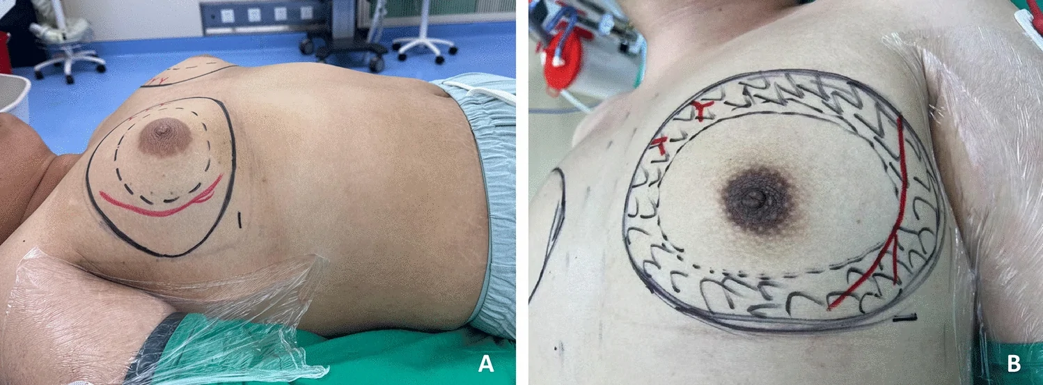
单中心、单臂前瞻性病例系列(证据等级 III),纳入 2024 年河北医科大学第一医院 786 例男性乳腺发育内镜腺体切除术患者(773 例双侧、13 例单侧),平均随访 96.5