眉下切口上睑成形辅助矫正上睑下垂
2026年9月15日23 min read
Plastic and Reconstructive Surgery Global Open
结论与实践要点
该研究为单中心回顾性队列,证据等级中等偏低,但在东亚肥厚型上睑人群中提供了较明确的功能与美学改善信号:MRD1 平均提升约 2.35 mm、睑裂闭合不全可忽略、患者主观满意度较高(中位 8/10)。需意识到其设计局限——缺乏对照组、术者与中心单一,结论外推应保守。
临床落地提示:对皮肤松弛、上睑饱满且合并腱膜性下垂的东亚患者,单纯双眼皮切口可能“去脂不足”而残留厚重感,联合眉下切口去除冗余皮肤软组织有助于获得更自然的轻盈轮廓。操作中应保护提上睑肌腱膜前鞘与 Müller 复合体以维持稳定抬升,并常规评估术后睑裂闭合情况以防暴露性角膜病变。可将“患者主观满意度高但客观评分偏高、两者一致性低”作为知情同意中点明预期差异的依据。
Background:This study analyzed the outcome of combined ptosis repair with adjunctive subbrow blepharoplasty in a predominantly East Asian population that is frequently characterized by bulky upper eyelid tissue.
Methods:A retrospective study of 160 eyelids of 80 patients who underwent bilateral aponeurotic ptosis repair with subbrow blepharoplasty at 1 center by 1 surgeon (A.L.) from March 2021 to December 2023 was performed.
Results:The mean age of the patients included in the study was 56.5 ± 12.5 years; 95% of the patients were women, and 97.5% were of Chinese ethnicity. There was a statistically significant increase in marginal reflex distance 1 of 2.35 ± 1.15 mm postoperatively ( P < 0.001). Postoperatively, the mean lagophthalmos measurement at 3 months was 0.025 ± 0.16 mm. None of the patients had exposure keratopathy or postoperative complications. Three eyelids underwent revision surgery for residual ptosis. The mean subjective grading by patients was 8.6 ± 1.5, and the median score was 8 (interquartile range, 8–10). The mean objective score by the blinded independent observer was 2.6 ± 0.6, with 67.5% and 27.5% achieving an excellent score of 3 and a good score of 2, respectively. The median objective score was 3 (interquartile range, 2–3).
Conclusions:Subbrow blepharoplasty is a useful adjunct to ptosis surgery in the East Asian population. It achieves greater tissue debulking and yields a “lighter” aesthetic outcome with good functional results. This technique achieves good subjective patient response and objective observer grading and has low revision and complication rates.
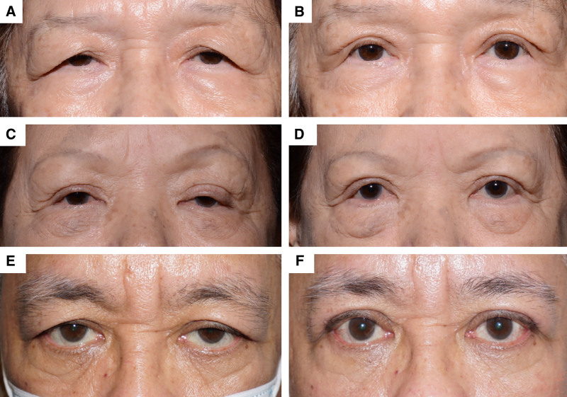
In the older, more mature East Asian patient, aponeurotic ptosis is often coupled with some degree of excess upper eyelid skin and soft tissue. In the absence of significant brow droop, ptosis repair with excision of excess skin only above the skin incision often leads to a persistent “heavy” or full look despite sufficient skin excision and preaponeurotic fat debulking (Fig. 1 ). This results essentially from differential skin and soft tissue thickness in the area extending from the lash-bearing eyelid margin to the eyebrow, with the skin closer to the eyebrow being of greater thickness than the skin closer to the eyelid margin. 1 , 2 To address this, the authors advocate the addition of subbrow blepharoplasty as a useful adjunctive procedure that allows for a “lighter” and more aesthetic outcome following ptosis repair.
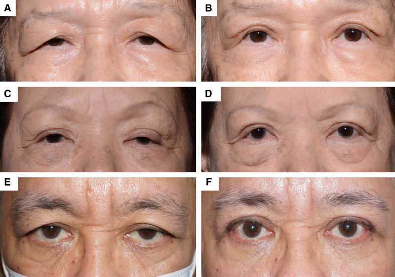
Fig. 1. Pre and postoperative photographs of patients who underwent blepharoplasty and ptosis repair through the traditional eyelid crease incision, showing fullness of the upper eyelid and thick skin above the eyelid crease. A and B, Preoperative and postoperative photographs for patient 1. C and D, Preoperative and postoperative photographs for patient 2. E and F, Preoperative and postoperative photographs for patient 3.传统双眼皮切口患者的术前术后照片,显示上睑饱满、睑缘上方皮肤偏厚。The subbrow approach was first described by Parkes et al in 1976 3 and has since proven to be an effective surgical technique to address excess upper eyelid skin and soft tissue, particularly with lateral hooding of the upper eyelids. 4 This involves an incision just inferior to the eyebrow, allowing for the removal of excess soft tissue whilst hiding the scar at the inferior border of the brow. By itself, this technique offers several advantages, including superior aesthetic outcomes due to scar concealment, 5 a reduced risk of postoperative asymmetry or lid crease issues, 6 and preservation of the natural eyelid crease, especially in patients with a low or absent eyelid crease. 7 It is often preferred by conservative patients who do not desire a significant change in appearance or the creation of prominent upper lid creases. 8 Additionally, subbrow blepharoplasty has been combined with browpexy or ptosis repair to further enhance functional and cosmetic results. 9 Concerns remain, however, regarding the safety of the combined procedure as excision of additional soft tissue may potentially lead to lagophthalmos and corneal exposure issues.
This retrospective review explores our experience with subbrow blepharoplasty used as an adjunct to classic ptosis repair in a predominantly East Asian population.
This was a retrospective study of 80 patients who underwent bilateral aponeurotic ptosis repair with subbrow blepharoplasty at 1 center by 1 surgeon (A.L.) from March 2021 to December 2023.
Patients who underwent this surgery had aponeurotic ptosis with bulky upper eyelid soft tissue and lateral hooding. None had significant brow droop, with the eyebrows, in their entirety, above the superior orbital rim. Patients who only had ptosis repair alone or subbrow blepharoplasty alone were excluded. All patients had eyelid measurements performed pre- and postoperatively by the surgeon. In particular, the marginal reflex distance 1 (MRD1) from the light reflex to upper eyelid preoperatively and 6–8 weeks postoperatively, and amount of lagophthalmos preoperatively and 2–3 months postoperatively were noted. All patients except 10 were followed up for at least 2 months postoperatively. Pre- and postoperative photographs were collected retrospectively and de-identified (Fig. 2 ). These were evaluated independently by a blinded independent observer for the cosmetic outcome (L.T.). Each patient was also asked to provide a subjective grading of their surgical outcome.
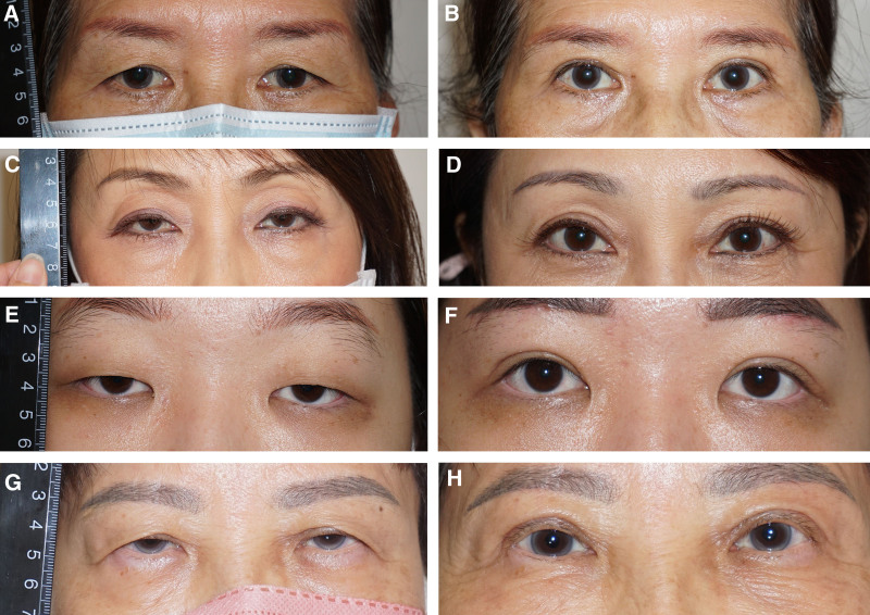
Fig. 2. Pre- and postoperative photographs of patients who underwent ptosis repair and subbrow blepharoplasty, showing a “lighter” and less bulky upper eyelid appearance above the lid crease. A and B, Preoperative and postoperative photographs for patient 1. C and D, Preoperative and postoperative photographs for patient 2. E and F, Preoperative and postoperative photographs for patient 3. G and H, Preoperative and postoperative photographs for patient 4.上睑下垂修复联合眉下切口的术前术后照片,呈现更“轻盈”、减容后的上睑外观。The subjective score by the patients was ranked on a Likert-type scale from 1 to 10, with 5 being satisfactory and 10 being excellent. The objective score was given by a blinded independent observer grading anonymized postoperative photographs. This score was ranked on a three-point scale from 1 to 3. The objective grade of 3 indicated excellent results with the eyelids achieving good height with an MRD1 of at least 2 mm (1 point), good eyelid contour and a well-formed, defined crease (1 point), and MRD1 symmetry within 1 mm (1 point). An objective grade of 2 indicated that results were not satisfactory in one of these parameters. An objective grade of 1 was assigned if 2 or more of these conditions were not achieved.
Skin markings were made with the patient lying supine before injection of local anesthetic (Fig. 3 ). The amount of excess subbrow skin resected varied depending on the severity of the patient’s dermatochalasis. This ranged from 4 to 6 mm wide and 2.5 to 4 cm long. Once the subbrow markings were made, care was taken to elevate the subbrow tissue manually to simulate the elevation of the eyelid skin with subsequent closure of the subbrow wound. The eyelid crease markings were then made with the pinch technique while the patient is instructed to keep eyes gently closed, ensuring that there would be sufficient skin for eye closure postsurgery. All patients underwent bilateral upper eyelid surgery under local anesthesia (lidocaine 2% with 1:80,000 epinephrine admixed with bupivacaine 0.5% with 1:200,000 epinephrine in 1:1 proportion) with or without intravenous sedation in a day surgery setting. A strip of subbrow skin and underlying soft tissue including part of the orbital orbicularis oculi was excised from the infra-brow region with a beveled 15G blade, down to but sparing the periosteum, with the upper excision border just along the lower border of the eyebrow. Following hemostasis with bipolar diathermy, the wound was closed in layers with interrupted 6/0 Vicryl in the deeper tissue and 5/0 prolene in a continuous subcuticular manner. The aim was to ensure good apposition of the deeper layers so that the wound edges came into close apposition even before the skin closure was performed. ( See Video 1 [online] , which shows the crucial steps in subbrow blepharoplasty.) Upper eyelid ptosis repair was then performed in the usual traditional fashion through an upper eyelid crease incision with appropriate excision of residual excess skin and orbicularis. Care was taken to elevate the preaponeurotic fat from the levator without disturbing the anterior fascial sheath. 10 , 11 Effort was also made to elevate the levator-Muller complex from the conjunctiva until the white line that represented the underbelly of the levator aponeurosis was visualized. 12 The advancement of the levator palpebrae superioris onto the tarsal plate was then secured with placement of 2, occasionally 3, 6/0 Vicryl sutures. The first suture was placed just medial to the medial corneal limbus, ensuring as long a bite of the tarsal plate as possible, and the second was placed just lateral to this. The third 6/0 Vicryl suture was applied laterally if needed to address any lateral droop and improve the overall contour. Following this, the skin was closed with 7/0 silk suture in interrupted fashion, with care taken to catch the terminal end of the levator to form a well-defined lid crease. ( See Video 2 [online] , which demonstrates the blepharoptosis repair technique with emphasis on protecting the anterior fascial sheath of the levator aponeurosis and isolating the levator-Muller complex to ensure a strong lift.)
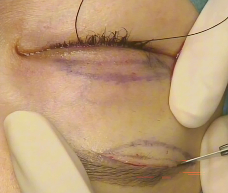
Fig. 3. Intraoperative photograph showing subbrow and eyelid crease incision markings.术中照片,显示眉下与双眼皮切口的标记线。补充视频 / 附件(原文未随文提供原图,可于出版方页面查看)gox-14-e7967-s001.mp4Video 1. This is a video demonstrating the surgical technique of sub-brow blepharoplasty that can be used together with ptosis repair to achieve a more aesthetic “lighter” outcome in the more mature East Asian patient with bulky upper lid soft tissue.补充视频 / 附件(原文未随文提供原图,可于出版方页面查看)gox-14-e7967-s002.mp4Video 2. This is a video demonstrating the crucial steps in blepharoptosis repair in the East Asian patient. Emphasis is placed on protecting the anterior fascial sheath of the levator aponeurosis and isolating the levator-Muller complex to ensure a strong lift. Judicious pre-aponeurotic fat debulking is recommended to avoid a sunken look that is particularly aging in the East Asian eyelid.
Clinical data were collected by the surgeon during the course of patient care and recorded retrospectively in this study.
Where appropriate, continuous variables were compared pre and postoperatively using the t test. Statistical analysis was performed using Stata 15 (StataCorp. 2017. Stata Statistical Software: Release 15;StataCorp LLC, College Station, TX).
All study procedures were performed in accordance with the tenets of the Declaration of Helsinki as revised in 1989. A waiver for written consent from retrospectively recruited participants was obtained from the institutional review board overseeing the hospital where the procedures were performed.
A total of 80 patients (160 eyelids) underwent combined bilateral ptosis repair with subbrow blepharoplasty. The mean age of the patients included in the study was 56.5 ± 12.5 years. Of the 80 patients, 76 (95%) were female and 4 (5.13%) were male. Seventy-eight (97.5%) patients were of Chinese ethnicity.
Mean duration of surgery was 140.9 ± 35.0 minutes, and the mean duration of follow-up was 4.9 ± 4.5 months (Table 1 ). Five patients were lost to follow-up after 1 month, 4 were followed up for 2 weeks postoperatively, and 1 patient was lost to follow-up after 3 weeks. Overall, preoperatively, mean MRD1 was 0.67 ± 1.27 mm. The mean postoperative MRD1 was 2.35 ± 1.15 mm. There was a statistically significant increase in MRD1 of 2.35 ± 1.15 mm postoperatively.
Table 1. Patient Characteristics, Operative Time, and Follow-up Duration
| P | N = 80 |
|---|---|
| Age, mean (SD) | 56.6 (12.5) |
| Sex (%) | |
| Female | 74 (94.5) |
| Male | 4 (5.13) |
| Operative time (min), mean (SD) | 140.9 (35.0) |
| Follow-up duration (mo), mean (SD) | 4.9 (4.5) |
None of the patients had preoperative lagophthalmos. One day postoperatively, the mean lagophthalmos measurement was 0.92 ± 0.9 mm. Three months postoperatively, 5 eyes had lagophthalmos ranging from 0.5 to 1 mm, and the mean lagophthalmos measurement was 0.025 ± 0.16 mm. None of the patients had exposure keratopathy secondary to the minimal lagophthalmos.
Three patients underwent revision surgery for residual ptosis in 1 eye. Of these 3 patients, 2 gave postoperative subjective scores of 9 and 8.5 and were objectively graded with a final score of 3, whereas the third patient declined to respond and was objectively graded with a score of 2.
None of the patients experienced complications of infection, hematoma, wound dehiscence, hypertrophic scars, or bothersome sensory changes. 51 patients responded retrospectively with a mean subjective grading of 8.6 ± 1.5 and a median score of 8 (interquartile range [IQR] 8–10). The mean objective score by the blinded independent observer grading anonymized postoperative photographs was 2.6 ± 0.6 with 67.5% and 27.5% achieving an excellent score of 3 and a good score of 2 respectively (Fig. 4 ). The median objective score was 3 (IQR 2–3) (Table 2 ). Figure 5 shows the distribution of the subjective scores graded by the patients. A kappa analysis of the subjective and objective scores showed poor agreement, with a kappa score of 0.069 ( P = 0.24)
Table 2. Postoperative Measurements and Gradings
| Variable | N = 160 | P |
|---|---|---|
| Preoperative MRD1 (mm), mean (SD) | +0.67 (1.27) | |
| Postoperative MRD1 (mm), mean (SD) | +3.02 (0.46) | |
| MRD1 change (mm), mean (SD) | 2.35 (1.15) | <0.001 |
| Preoperative lagophthalmos (mm), mean (SD) | 0 | |
| 1-day postoperative lagophthalmos (mm), mean (SD) | 0.92 (0.9) | |
| 3-month postoperative lagophthalmos (mm), mean (SD) | 0.025 (0.16) | |
| N = 80 | ||
| Patient subjective grading (1–10) | ||
| Mean (SD) | 8.6 (1.5) | |
| Median (IQR) | 8 (8–10) | |
| Blinded independent observer objective grading (%) | ||
| 1 | 4 (5.0%) | |
| 2 | 22 (27.5%) | |
| 3 | 54 (67.5%) | |
| Median (SD) | 2.6 (0.6) | |
| Mean (IQR) | 3 (2–3) |
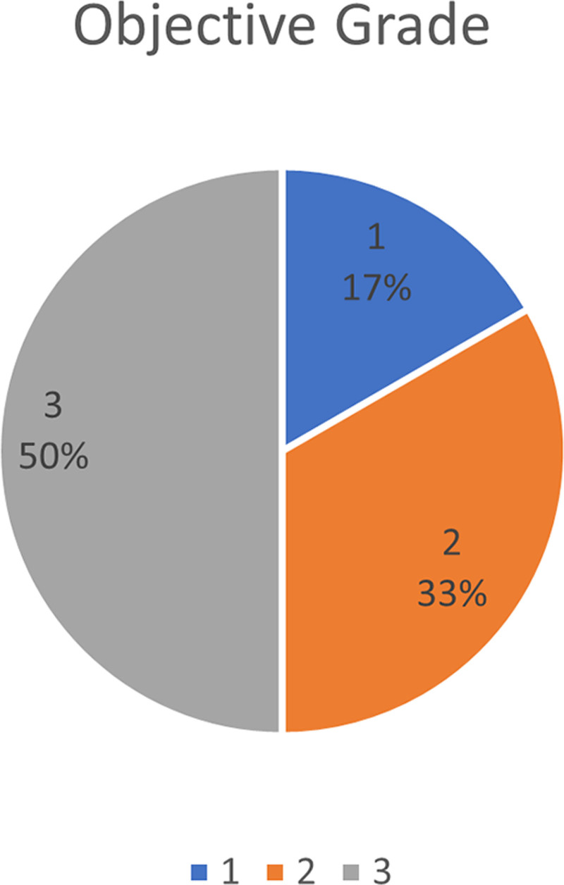
Fig. 4. Objective grading by independent blinded observer.盲法独立观察者的客观分级结果汇总。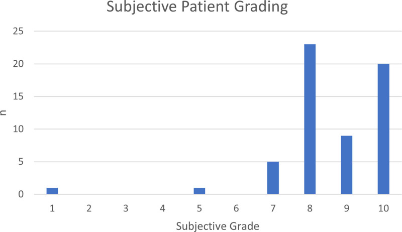
Fig. 5. Subjective grading by patients on a 10-point Likert scale.患者 10 分 Likert 量表的主观评分分布。Of the 10 patients who were only followed up for 1 month or less, only 1 had postoperative lagophthalmos of 1 mm at 1 month postoperatively, but no exposure keratopathy was noted. The median objective grading of their postoperative photographs was 3 (IQR 2–3).
Our study found good results with utilizing the subbrow blepharoplasty as an adjunct to ptosis surgery in an East Asian population, showing that the combined surgery was both effective and safe in addressing blepharoptosis while achieving satisfactory subjective patient reported outcomes and objective observer gradings.
There was a statistically significant increase in MRD1 of 2.35 mm although this was more an indication of good levator palpebrae superioris repair alone. Even though 10 patients were only followed up for 1 month or less, they had an excellent median objective score of 3, and it was likely that they did not return for follow-up reviews due to nonsurgical outcome concerns such as cost or convenience.
Although there have been concerns about the potential to cause worrisome lagophthalmos, this study has demonstrated the safety of the combined approach of ptosis repair with subbrow blepharoplasty. Lagophthalmos tended to be present only in the early weeks after surgery and none presented a long-term problem of exposure. Only 5 eyes (3%) developed lagophthalmos postsurgery, and the degree of lagophthalmos was mild, ranging from 0.5 to 1 mm. There was no statistically significant increase in lagophthalmos postoperatively compared to preoperatively. The onus, however, is still on the surgeon to start with more conservative skin and soft tissue excision first and develop a good judgment of maximal amount of soft tissue excision over time.
All 3 patients who underwent revision surgery had unilateral revision ptosis repair 5–7 months postoperatively and achieved excellent results of mean MRD of +3.17 mm and a mean objective score of 2.67. The requirement for revision ptosis surgery was unlikely to be related to the choice of a combined approach. Other known complications specific to subbrow blepharoplasty include wound dehiscence, hypertrophic scarring, and trauma to the supraorbital arteries and nerves, which none of our patients experienced.
A large center-based review led by our surgeon (A.L.) in 2013 found a strong correlation between patient satisfaction scores and MRD1 scores and symmetry. 13 In the pursuit of good patient satisfaction, it is therefore important to also focus on other facets of surgical outcome aside from MRD1 scores. Achieving symmetry is one area. Achieving a “lightness” in the final result is arguably another, as patient satisfaction can be diminished by a perception of a persistent heavy or full appearance of the upper eyelids due to the thicker skin and soft tissue that come to lie above the newly formed crease with a traditional eyelid crease surgical approach; even when MRD1 improves postsurgically. Eliminating the thicker skin and soft tissue that lie closer to the eyebrow, rather than the thinner and more delicate skin nearer the eyelash allows for the final eye appearance to be “lighter” and less full.
Previous authors, including Osaki et al, expressed similar sentiment in combining subbrow blepharoplasty with the traditional upper blepharoplasty. 14 Applying this combined approach to ptosis repair refines the overall outcome of ptosis repair and helps improve patient satisfaction further.
Our results show good patient satisfaction with the cosmetic outcome of this surgery, albeit with 64% of patient response to a retrospective request for grading. Those who responded gave an average grade of 8.6 out of 10, indicative of a high level of patient satisfaction. The favorable cosmetic result was further evidenced by the good mean objective score of 2.6 out of a maximum score of 3.
There exist varying surgical options for rejuvenation of the mature ptotic upper eyelid. Patient selection is of key importance in determining which technique is optimal. Where the skin and soft tissue bulk is not significant, the traditional ptosis repair is an excellent technique to lift up the droopy eyelid and eliminate excess skin and soft tissue. Where the skin and soft tissue bulk is significant, with prominent lateral hooding but no significant brow droop as yet, as is often seen in East Asian patients, adding on subbrow blepharoplasty to the classic ptosis repair allows for a “lighter,” more cosmetically pleasing upper eyelid. This is particularly so because the lateral hooding beyond the lateral canthus is better addressed by the longer subbrow incision that extends to the area lateral to the lateral canthal angle. 15
It has been said that endoscopic brow lift may be a better adjunct procedure to manage the lateral hooding but the counter-argument is that the lateral brow has not drooped significantly in this group of patients and an endoscopic browlift could lead to overelevation of the brow, whereas subbrow blepharoplasty results in a more natural appearance. 16 The additional subbrow procedure has a short learning curve and can be readily adopted into one’s surgical armamentarium. It also helps that the wounds heal remarkably well with good surgical technique and good-postoperative care and attention. 8 , 17 In patients with scant brow hairs, the scar may be a little more obvious but has not been shown to be bothersome to patients. It may also be readily masked by having the patient undergo a brow embroidery/tattoo procedure. Indeed, the ideal patient is one who has ptosis with bulky upper lid soft tissue and a preexisting brow tattoo or embroidery.
One limitation of this study is that it is conducted retrospectively and patients do not respond fully to a retrospective request for a grading. Only 64% of patients responded with a grading. Furthermore, the objective and subjective scores were graded on different scales, which may account for the poor correlation in the kappa analyses and make it challenging to interpret the kappa score meaningfully. Additionally, our study represents the experience of a single, experienced oculoplastic surgeon and does not consider the potential learning curve for this surgical technique. As all the patients were of Chinese ethnicity except 2 who were of Indian and Malay ethnicity respectively, ethnic variations may render this surgical technique less predictable in Occidental populations. Eid and Tsirbas described the use of subbrow blepharoplasty alone in selected White patients with severe lateral hooding and combined this in some cases with internal browpexy. 18 To a large extent, their focus was on functional improvement, and it remains to be seen whether this combined ptosis repair and subbrow blepharoplasty has a role to play in select White patients with ptosis and heavy upper eyelid tissue. The main concern lies in the thinner and less bulky orbicularis in the Occidental eyelid which may lead to a greater degree of lagophthalmos for the same degree of work done. In this article, there is no aim to lift the brow at all, and we emphasize care to be taken to apply this combined technique only in the ptotic patient with heavy, bulky upper eyelid soft tissue in the absence of significant brow ptosis.
Ptosis repair with subbrow blepharoplasty allows one to achieve a “lighter” and more cosmetic outcome in the more mature, East Asian ptotic patient even as a functional result is secured. This surgical approach directly addresses the differential soft tissue thickness in the East Asian upper eyelid and is sound as evidenced by both the subjective patient response and independent objective observer grading as well as the low revision and complication rates. It is a technique that can readily be added to the arsenal of the oculoplastic surgeon in rejuvenating the periorbital area.
The authors have no financial interest to declare in relation to the content of this article.
Patients provided written consent for the use of their images.
声明:中文精读 · 仅供学术参考。内容来自公开文献检索,不代表本人观点,不构成诊疗建议。 医疗美容需在正规医疗机构由执业医师实施。
基于相同主题推荐 · 继续深挖
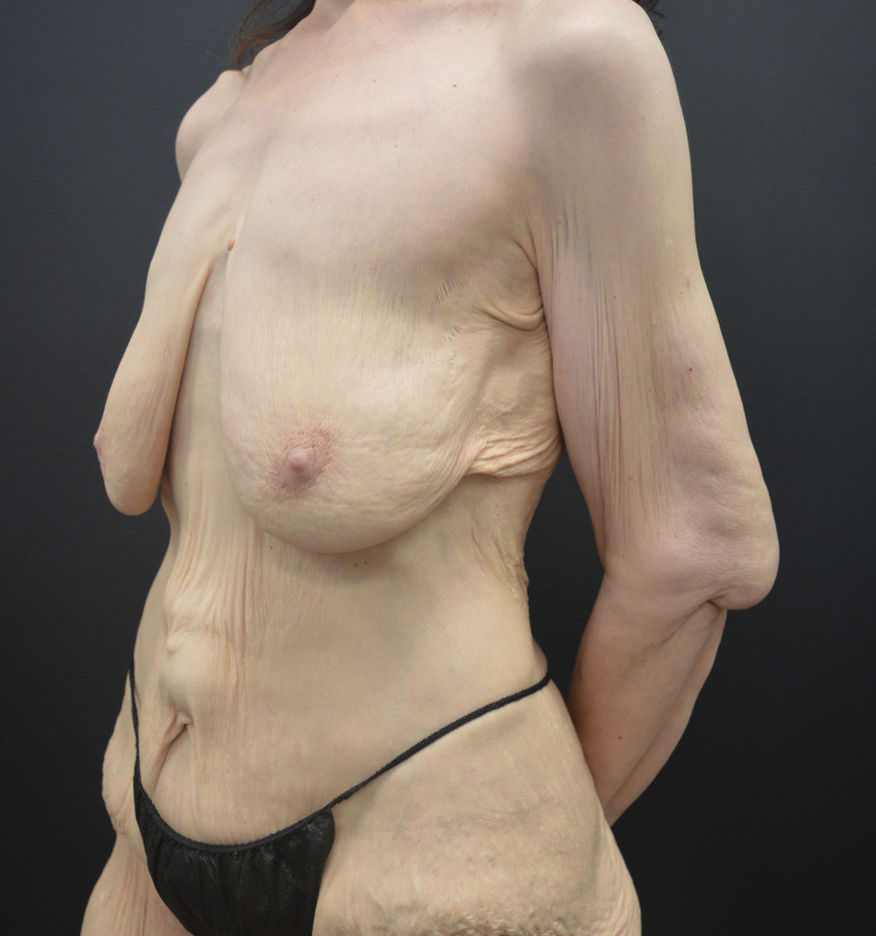
文章对躯干与四肢提出美学单元(AU)与粘连分区(ZA)的系统分类,作为身体塑形手术(BCS)切口规划的解剖基础。
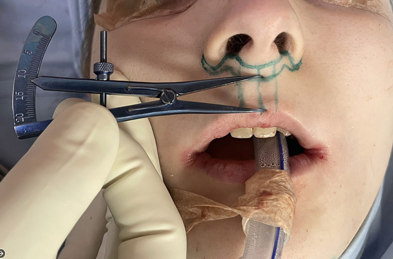
1997-2023年共988例出生分配为男性的性别焦虑者接受唇女性化与年轻化,累计1789例次手术,年龄22-75岁(平均39.5岁),79.8%小于50岁。
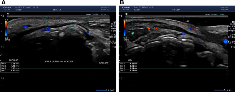
前瞻性研究纳入 25 例 21–65 岁女性,接受 Cleviel Volume 透明质酸(1.0 mL,22 mg/mL)的 11 点注射(11-PIT)并联合高频超声血管定位进