乳房假体取出同期基于解剖的乳房塑形重建
2026年9月16日27 min read
Plastic and Reconstructive Surgery Global Open
结论与实践要点
循证与证据等级。 本项为单中心回顾性队列(n=204 例 / 408 侧,2021–2025),证据等级中等偏低、描述性为主;报告了腔隙分布、人群参数与并发症发生率,但缺乏对照组与标准化患者报告结局(PRO)量化。
局限。 回顾性设计存在选择偏倚与信息偏倚,未提供详细的并发症分级、再手术率或长期(>1 年)轮廓维持数据;文中并发症以“例数/百分比”表述,分母在部分指标上以患者或侧计,跨研究比较须注意口径;且 BST 为术者依赖型技术,可重复性有待多中心验证。
可落地提示。 对计划行假体取出者,术前站立位、配合上肢动态活动标记 footprint 并据腔隙类型规划缩窄/组织募集策略,有助于在取出同期获得更自然的轮廓;存在既往瘢痕、多次手术或 tubular/瀑布畸形等复杂因素时,应提前设定瘢痕不可完全去除等预期,并可将脂肪移植作为后续补充手段。
Background:Over time, there has been a significant rise in the number of patients who require and/or desire breast explantation. From implant complication issues to more contemporary reasons, such as breast implant disease, the causes are varied.
Methods:This retrospective study analyzed 204 patients (408 breasts) who underwent explantation between 2021 and 2025 using the “breast sculpting” technique. The approach incorporates strategies such as tissue recruitment, muscle repositioning, and reshaping of the breast footprint, with the objective of restoring contour and achieving optimal aesthetic balance.
Results:About 78.6% of the implants were in the retroglandular plane, 11.5% in the submuscular plane, and 9.8% in the dual plane. Two (0.98%) patients developed deep vein thrombosis, 1 (0.49%) patient experienced a minor pulmonary embolism, and 5 (1.22%) patients experienced unilateral breast hematomas.
Conclusions:The breast sculpting technique promotes a tailored approach to breast tissue management. It facilitates multiple strategies for reconstructing breast shape following implant removal, with a particular emphasis on restoring natural breast contour—thereby differentiating it from previously described methods in the literature. This technique demonstrates a low risk of complications and does not preclude the use of lipofilling as a subsequent procedure.
In recent years, the number of breast augmentation procedures has increased due to their popularity, and there has been a notable rise in the number of explantations. 1 The main causes for explantation are patient health concerns, lifestyle changes, or aesthetic preferences. 2 In this context, breast explantation and reconstruction pose unique challenges due to anatomical distortions caused by implants over time. 3 However, much of the previous research and traditional surgical techniques have not adequately addressed the substantial anatomical distortions resulting from long-term implants, which vary according to implant placement (retroglandular, retropectoral, or dual-plane). 4 , 5
Most techniques focus on volume restoration and fat augmentation. 3 Some authors choose to perform routine fat grafting associated with mastopexy after breast explantation. 4 The use of the chest wall–based flap may be considered as an alternative in cases with significant breast volume in the lower pole. 4 The “breast sculpting” technique (BST), presented in this study, can be applied to all breast explantation situations, with implants placed in the dual plane, retroglandular, and submuscular planes.
The objective of this study is to demonstrate the various possibilities for breast reconstruction without fat grafting following breast explantation and to emphasize the importance of using different strategies depending on the implant placement plane. It also highlights a new technique that allows surgeons to correct implant-related anatomical distortions and restore breast shape without routinely using fat grafting, although fat grafting can still be considered later if additional volume is desired.
A retrospective review was conducted of 204 patients consecutively treated by the senior author from July 2021 to October 2025. This study was conducted in accordance with the ethical standards of the Declaration of Helsinki.
All patients who underwent breast explantation combined with any form of skin adjustment, performed by the senior author at various São Paulo hospitals during the study period, were included, irrespective of age. Exclusion criteria comprised breast explantation without concomitant skin adjustment, procedures performed outside the designated study period, and explantation with implant replacement. Preoperative magnetic resonance imaging (MRI) was requested for all patients with the intention of assisting in the patient’s decision to undergo explantation.
When indicated, areolas were mobilized through a superior pedicle with complete dermal release, enabling areolar transposition and skin redraping. For retromuscular and dual-plane explantation (with or without mastopexy), as well as retroglandular explantation without vertical scar mastopexy, suction drains were placed in initial cases and removed on postoperative day 3. Penrose drains were used initially for retroglandular explantation involving vertical scar mastopexy, with their removal occurring on postoperative day 2. Since 2024, drains have no longer been used routinely.
Tranexamic acid was administered only intravenously: 1 g at the start of the surgery, 500 mg in the evening, and 500 mg in the morning before hospital discharge. Upon completion of the surgical procedure, a nonadhering dressing composed of knitted cellulose acetate fabric impregnated with petrolatum emulsion was applied directly to the wounds. Micropore tape was used over the incisions to minimize tension. Additionally, micropore tape was placed over the entire breast, strategically applied to provide a taping effect for contour stabilization and edema control. The thoracic compression bandage was removed on either postoperative day 1 or 2. Following the removal of the bandage, patients were instructed to wear a nonstretch, back-closing support bra.
Breast implants cause anatomical changes that differ based on their placement. MRI of the breasts is crucial for a comprehensive anatomical analysis, directly informing and refining surgical planning.
Retroglandular implants induce global, yet nonuniform, expansion of the mammary tissue and frequently migrate inferolaterally, potentially exposing the lateral and inferior thoracic musculature.
Retromuscular implants contribute to breast enlargement not via global mammary tissue expansion, but by distending the pectoralis major muscle.
Dual-plane implants lead to a hybrid effect: the overlying breast tissue in the superior pole is typically unaltered due to muscle coverage, whereas the inferior pole, similar to retroglandular placement, exhibits thinning and expansion of the glandular tissue.
Regardless of implant positioning, the guiding principle of the BST is to narrow the implant-expanded breast footprint. The ideal postexplant breast should exhibit appropriate projection on the patient’s thorax, a uniform and ripple-free contour, and an anatomical morphology characterized by a gently sloping upper pole transitioning to a fuller, well-defined lower pole.
Markings are made with the patient awake and in the standing position, allowing for accurate delineation of the anatomical footprint of the breast. ( See Video 1 [online] , which displays the preoperative markings.)
Initially, the chest midline and the line connecting the beginning of both axillae are marked. The markings are performed with dynamic arm movement: with the arms down to mark the upper contour of the footprint, which is typically located about 2–4 cm below the intersection between the hemithorax line and the line connecting the axillae (the “finish line”). To accurately define the lateral breast contour, the patient’s arms should be partially elevated, and a maneuver involving medial displacement of the implant is performed. This maneuver facilitates precise identification of the lateral outline, which is typically situated approximately 4–5 cm medial to the midaxillary line. Then, the arms should be fully raised to determine the lower contour (not considering any previous scars).
The new point “A” of the areola is calculated based on the distance from the areola to the upper contour of the breast footprint. Since there is often significant skin stretching in the upper pole of the breasts with implants, caution is advised to avoid elevating the areola excessively. Point A is typically calculated as 7–8 cm below the dotted line. Not infrequently, the areolas are already located at this level or even higher. In these cases, the incision is made flush with the areola, with particular attention given to attempting to lower the position of the areola when planning the new location of the inframammary fold. Skin resection may be marked before the removal of the implants using the Le Jour 6 maneuver (lateralization and medialization of the breast, aligning it with the thoracic meridian to mark the area for deepithelialization).
First, the areolas are marked with a cookie cutter because, after the removal of the implants and breast reconstruction using the described technique, there will not be enough internal space to place supports (compresses or implants) for the areolar incision or deepithelialization. When mastopexy is performed, access is achieved through a vertical incision along the breast meridian for the removal of implants and capsules, regardless of the current position of the areola, which may be significantly lateralized by the implants.
A small horizontal incision may be made to assist with capsulectomy, but the final horizontal scar should only be defined after reconstructing the breast contour. This usually allows for a smaller and better-positioned scar. The intention is to position the inframammary fold at the theoretically ideal anatomical height of the footprint lower contour (sixth or seventh rib). 6 , 7
Preoperative counseling is essential to set expectations that existing scars may not be removed, as excision could unnecessarily enlarge the explantation scar. In contrast to breast reduction, large scars after explantation can more strongly compromise aesthetics because the breasts are typically smaller postexplant; prominent scars may look disproportionate and can make the breasts appear even smaller.
补充视频 / 附件(原文未随文提供原图,可于出版方页面查看)gox-14-e8027-s001.mp4Video 1. This video displays preoperative markings.
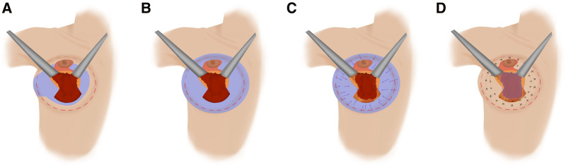
Retroglandular ExplantationsIn retroglandular explantations (Fig. 1 ), we usually observe a breast with a broad base, a large explantation defect, and a retroareolar region lacking projection (Fig. 2 , right breast). For reconstruction, we perform extensive release of the parenchyma beyond the boundaries of the footprint, allowing for greater tissue mobility. 8 Subsequently, separate centripetal traction sutures are placed 360 degrees around the breast using 2-0 Vicryl, aiming to recruit the breast tissue back into the footprint, narrow the defect resulting from explantation, and redefine the entire breast contour 8 (Fig. 2 , left breast).
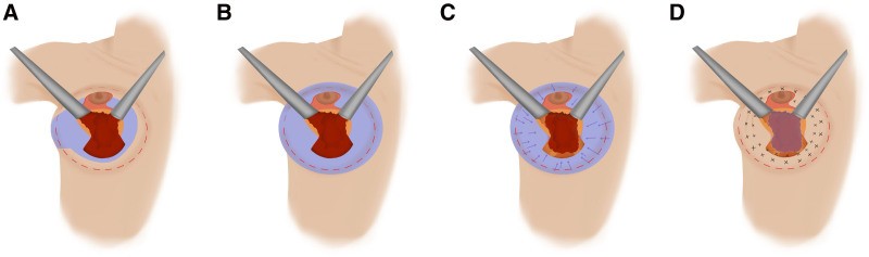
Fig. 1. Footprint narrowing in retroglandular explantations. A, The original defect (purple) resulting from explantation with total capsulectomy can be seen. B, The defect is then enlarged and homogenized, extending beyond the desired footprint contour. C, Interrupted recruitment sutures are placed with 2-0 Vicryl to draw tissue from outside the footprint and reestablish the rounded breast contour. D, After recruitment, the explantation defect becomes smaller, facilitating more uniform and predictable filling of the retroareolar region and the lower pole, thereby improving symmetry.腺体后间隙取出中的 footprint 缩窄流程:原缺损、扩大均质化、褥式缝合收拢等步骤示意。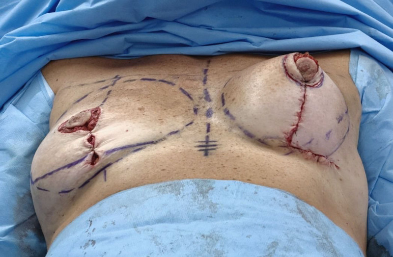
Fig. 2. A 320-cm3 retroglandular explantation. Compare the right breast, treated with skin removal only, with the left breast, where the BST produced greater reshaping and a more defined contour.320 cm³ 腺体后间隙取出病例:右侧仅行去皮,左侧行乳房塑形(BST)重建,后者轮廓更清晰、塑形更明显。Dual-plane ExplantationsFootprint reduction is initiated by specifically addressing 3 anatomical changes: first, the pectoralis major muscle, which is compromised by detachment of its inferior insertion—a hallmark of the dual-plane technique; second, the upper pole mammary tissue, which remains unexpanded and anchored to the pectoralis major; and third, the lower pole tissue, which, having lost its attachment to the muscle, is left expanded and thinned by the presence of the implant (Fig. 3 ). ( See Video 2 [online] , which is a short animation that demonstrates muscular imbrication for cases of retromuscular and dual-plane explantation.)
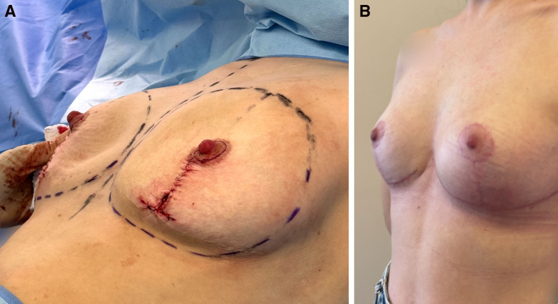
Fig. 3. Intraoperative and postoperative outcomes of explantation with dual-plane implant removal and BST reconstruction. A, Intraoperative appearance after explantation of 325-cm3 dual-plane implants. Note the thinning of the inferomedial quadrant. B, Five months after reconstruction with strategic muscle repositioning and tissue redistribution inside the footprint.双平面假体取出 + BST 重建的术中与术后:325 cm³ 双平面假体取出后下内侧象限变薄,5 月后轮廓恢复。In dual-plane cases, surgical correction is centered on the reconstruction of the pectoralis major muscle by reattaching its upper and lateral portions and, when anatomically feasible, the inferior portion, which may not always be present. ( See Video 3 [online] , which displays the approach to repositioning the pectoralis major muscle for breast contour reconstruction in a 400-cm 3 dual-plane explantation.) The pectoralis major muscle, often divided and elongated from its native anatomy, requires not only reconnection of its segments but also muscular imbrication to effectively narrow the breast base. ( See Video 4 [online] , which displays strategies for correcting tissue irregularities without fat grafting in a dual-plane explantation.) Strategically suturing the pectoralis major muscle with imbrication sutures using 2-0 Vicryl brings both the muscle and its adherent mammary tissue back toward the retroareolar region and to the lower pole of the breast.
It is not always possible to reinsert the muscle into its original anatomical position; however, this is not the primary goal. The goal is to narrow the breast base while establishing a fixation point for upper-abdominal tissue that is more reliable than the intercostal musculature or the costal periosteum. The next step is to recruit the tissue of the upper abdomen by anchoring it to the reconstructed pectoralis major muscle (Fig. 4 ). This comprehensive approach restores the natural breast contour (Fig. 3 ) and aids in correcting deformities, such as bottoming out or window shading, frequently found with dual-plane implants.
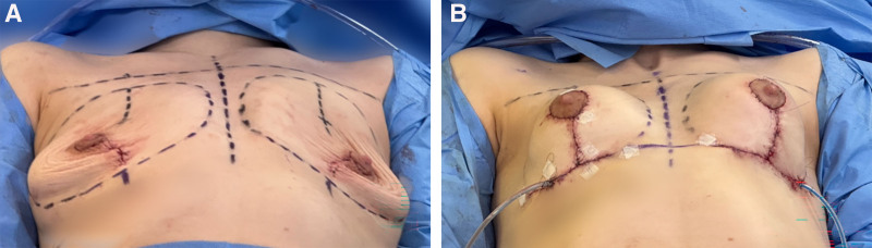
Fig. 4. Dual plane explantation and BST reconstruction in a patient with low BMI and thin soft tissue coverage. A, Explantation of 325-cm3 dual-plane implants. The BMI of the patient was 18 kg/m2. B, After reconstruction with strategic muscle repositioning and tissue redistribution inside the footprint. No fat grafting or pedicles were used.低 BMI、软组织覆盖薄者的双平面取出 + BST 重建(BMI 18 kg/m²),经肌肉复位与组织重塑改善轮廓。补充视频 / 附件(原文未随文提供原图,可于出版方页面查看)gox-14-e8027-s002.mp4Video 2. This video displays the surgical technique.补充视频 / 附件(原文未随文提供原图,可于出版方页面查看)gox-14-e8027-s003.mp4Video 3. This video displays tissue redistribuiton inside the footprint to address irregularities without fat grafting.补充视频 / 附件(原文未随文提供原图,可于出版方页面查看)gox-14-e8027-s004.mp4Video 4. This video details in “footprint narrowing” and “valley-and-mountain” strategies in immediate (left) and delayed (right) reconstruction in a 450 cc retroglandular explantation.Retromuscular ExplantationsInitially, dissection is performed in the retroglandular plane to expose the central area of the pectoralis major muscle. Total capsulectomy is carried out through careful incision of the muscle. Next, muscular imbrication sutures using 2-0 Vicryl are placed, aiming to narrow the breast footprint by horizontally shortening the muscle, which had previously been expanded by the implant. In these cases, muscle narrowing alone is usually sufficient to restore an aesthetically pleasing breast contour, making extensive retroglandular dissection unnecessary. Glandular dissection is therefore limited, performed only to allow placement of a few centripetal traction sutures that further enhance and define the narrow breast contour (Fig. 5 ).
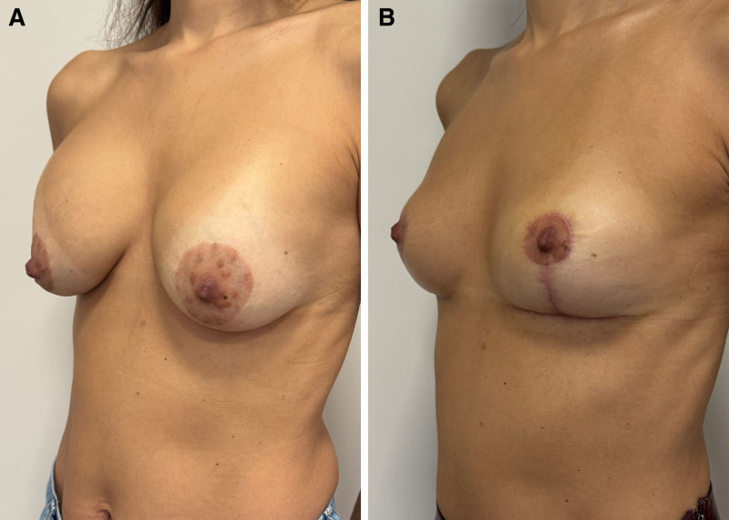
Fig. 5. Preoperative waterfall deformity and postoperative result after retromuscular explantation with BST reconstruction. A, A 41-year-old patient with 250-cm3 total retromuscular implants. Waterfall deformity. B, Three months after explantation with reconstruction.肌肉后间隙取出 + BST 重建矫治 waterfall 畸形:250 cm³ 完全肌后假体,术后 3 月改善。
After footprint narrowing, lax breast tissue is typically observed centrally within the breast. This laxity often exposes tissue irregularities from long-term implant presence, especially in retroglandular and dual-plane cases.
In these situations, tissue laxity allows the tissue to “walk” within the breast footprint using the “valley and mountain” strategy: visual or palpable depressions (valleys) surrounded by more robust tissue (mountains) are identified; then, “mountain” peaks are sutured to conceal the “valleys”, and the expanded tissue is internally shortened ( see Video 4 [online] ). ( See Video 5 [online] , which details strategies used for immediate reconstruction of the left breast and delayed reconstruction of the right breast after 450-cm 3 retroglandular explantation.) With this approach, areas at risk for retractions and adhesions (valleys) do not require lipofilling. Over the years, we have increasingly relied on additional “valley-and-mountain” sutures rather than pedicle creation (Fig. 6 ). Classic pedicle creation becomes necessary only if the pedicle is too long for the desired footprint or if rotation is required to address distant regions—most often to fill severely empty inferomedial quadrants.
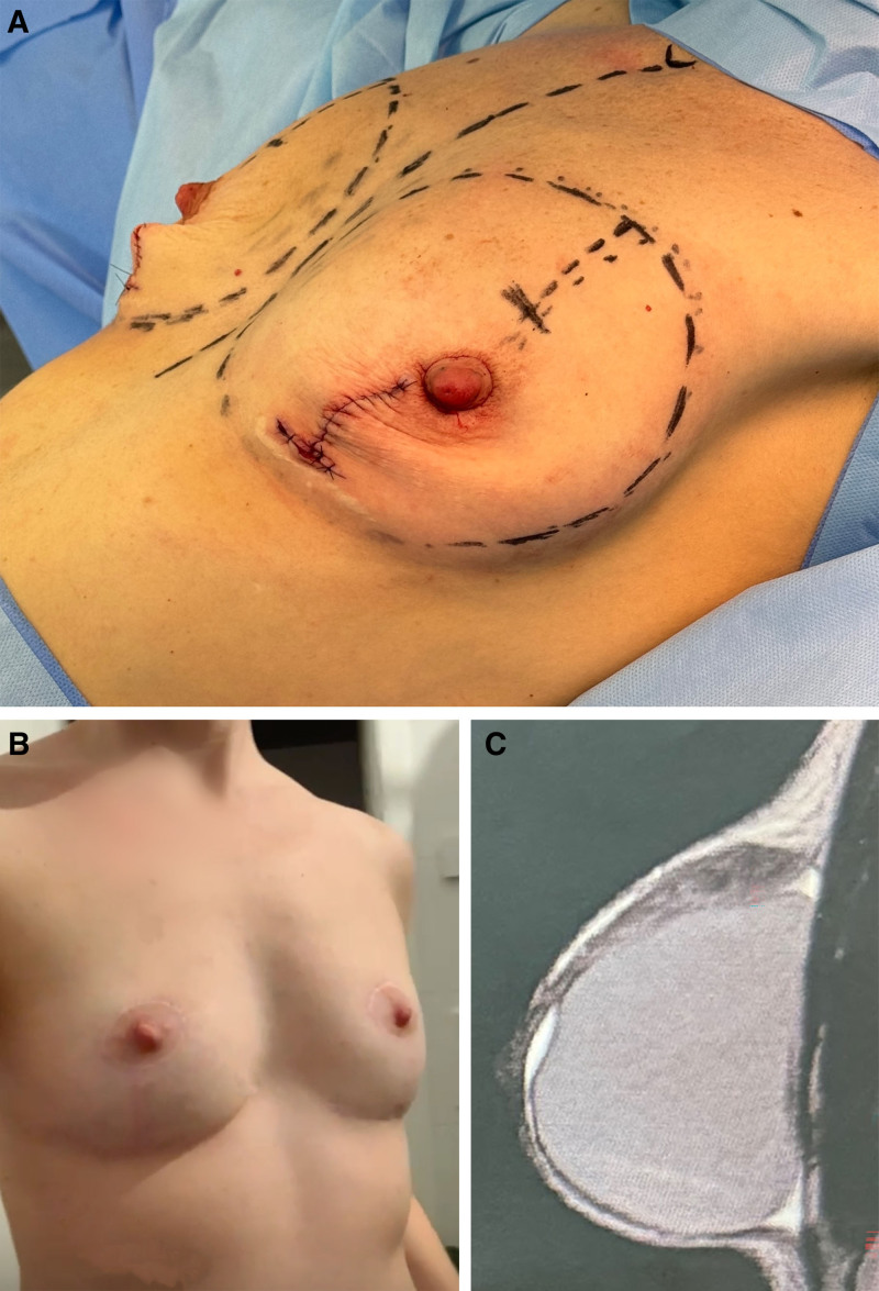
Fig. 6. Preoperative, postoperative, and imaging findings in a patient undergoing retroglandular explantation with BST reconstruction. A, Intraoperative image immediately after explantation, before reconstruction with the described technique. B, Image 10 months after explantation with retroglandular reconstruction strategies described in this article. C, Breast MRI of a 33-year-old patient with 325-cm3 retroglandular implants (BMI, 18 kg/m2).腺体后间隙取出 + BST 重建的术前、术后与影像:即刻与术后 10 月对比。补充视频 / 附件(原文未随文提供原图,可于出版方页面查看)gox-14-e8027-s005.mp4Video 5. This video demonstrates muscular imbrication for cases of retromuscular and dual plane explantation.
A total of 408 breast implants were explanted from 204 patients, all through total capsulectomy. Implant volumes ranged from 80 to 560 mL, with a mean of 282.5 ± 68.6 mL. All devices were silicone-based. Patients’ ages ranged from 25 to 78 years, with a mean of 46 ± 10.3 years. Body mass index (BMI) ranged from 16.80 to 32.32 kg/m 2 , with a mean of 23.83 ± 2.95 kg/m 2 . The interval between implantation and explantation ranged from 2 to 39 years, with a mean of 12 ± 5.9 years.
With respect to implant placement, the majority of cases (n = 321, 78.67%) involved retroglandular positioning. Total submuscular placement was identified in 47 breasts (11.51%), whereas the dual-plane technique was observed in 40 breasts (9.80%). Three patients had implants placed in different planes, with one side in the retroglandular plane and the other in the total retromuscular plane.
Postoperative complications were uncommon. Two (0.98%) patients developed deep vein thrombosis, 1 (0.49%) patient experienced minor pulmonary embolism, and 5 (1.22%) patients experienced hematomas—3 in the retroglandular plane, 1 in the retromuscular plane, and 1 in the dual plane. Minor revisions, such as scar positioning correction, were performed in 2 patients, and 3 patients underwent scar correction for keloids. No cases of infection or skin or nipple necrosis were observed.
Breast explantation surgery has experienced increasing demand and involves challenges that go beyond the simple removal of the implant. Between 2008 and 2021, breast explantation surgery cases increased by 653%. 9 According to the Food and Drug Administration, the risks of complications of undergoing breast implant surgery are even more concerning and may include capsular contracture (51.7%), rupture or leaking (31.2%), and the need for reoperation (59.7%). 10
As demonstrated in our previously published article, 11 MRI not only allows identification of the implant plane but also enables evaluation of tissue thickness and glandular symmetry. MRI-based breast volumetry may prove to be a helpful technique for providing more objective information regarding breast reconstruction. 12
A decision must also be made regarding capsule management. Retroglandular capsulectomy is less invasive and technically easier, 13 whereas retropectoral capsulectomy carries higher risks. 3 Total capsulectomy may be considered for complex capsular contracture, breast implant illness, asymptomatic textured implants, breast implant-associated anaplastic large cell lymphoma, or at the patient’s request. 6
To enable proper anatomical reconstruction, anterior capsulectomy is essential for retroglandular explantation, as the capsule maintains tissue position, creates defects, and hinders both tissue mobilization and the detection of implant-related irregularities. 13 In response to patient concerns, in our practice, we routinely perform total capsulectomy, even for retromuscular capsules. For safety, when there is significant adherence, minimal segments of the capsule may not be removed but are always cauterized. After silicone implant insertion, distinct anatomical changes occur 14 depending on the plane: in the retroglandular plane, there is a 360-degree widening of the footprint due to centrifugal displacement of breast tissue, whereas in the retropectoral plane, expansion happens by stretching the pectoralis major muscle.
Despite these differences, most articles describing explantation techniques do not account for anatomical variations when presenting the available reconstruction options. Pompei et al 1 report that simple implant removal often results in internal tissue reorganization, including quilting with barbed absorbable sutures and cutaneous scarring, and that mastopexy is required in patients with grade III ptosis or greater.
For patients requiring additional aesthetic correction after implant removal, mastopexy is frequently the best option for contouring the breast. 15 In cases where there is little remaining breast volume, another option is to use auto-augmentation flaps. 15
Another technique published in the literature is the use of a vertical bipedicle. This technique may be useful in cases where several previous procedures have been performed. The major difficulty in using this technique is in patients with very ptotic breasts, where the pedicle becomes very long, and it can be difficult to prevent folding and thus avoid compromising its vascularization. 16
To narrow the footprint in retroglandular explantations, we adopted the innovative technical approach that we have proposed. 8 This technique requires additional internal sutures to reposition the parenchyma and enables better breast volumization by using breast tissue within the footprint.
Because this technique allows us to freely redesign the breast footprint, encouraging explantation results can be achieved in tuberous breasts (Fig. 7 ). In these cases, the footprint is already excessively narrow, but it can be widened using reasoning similar to that described in Figure 1 .
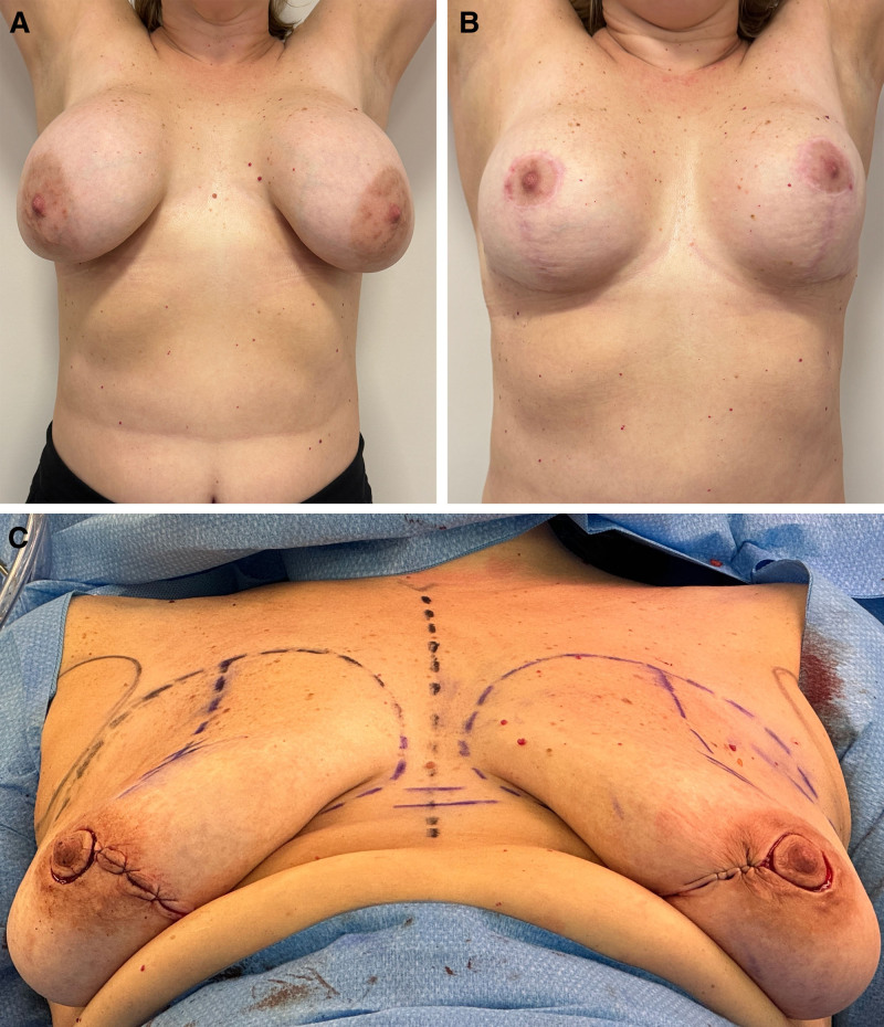
Fig. 7. Tuberous breasts undergoing retroglandular explantation and BST reconstruction. A, A 46-year-old patient with tuberous breasts with 375-cm3 retroglandular implants. B, Nine months after reconstruction, focusing on restoring breast contour. Note the improvement in the tuberous appearance. C, Intraoperative appearance.tubular 乳房腺体后间隙取出 + BST 重建:375 cm³ 假体,9 月后重点恢复乳房轮廓。In retromuscular explantation, broad breast tissue dissection is unnecessary because strategic plication stitches on the muscle already effectively improve the breast contour. Another option for explantation described in the literature, mainly for retromuscular implants, is the mammary imbrication lift and fixation technique. 17 This technique is different from ours because it treats the more superficial tissues. In our technique, we perform deeper muscle imbrications to restore breast shape.
In 3 patients in our study, the implants were found in 2 separate planes: total retromuscular on one side and retroglandular on the contralateral side. The BST allowed for a tailored approach on each side, ensuring symmetry—an outcome that alternative procedures may not have been able to achieve (Fig. 8 ).
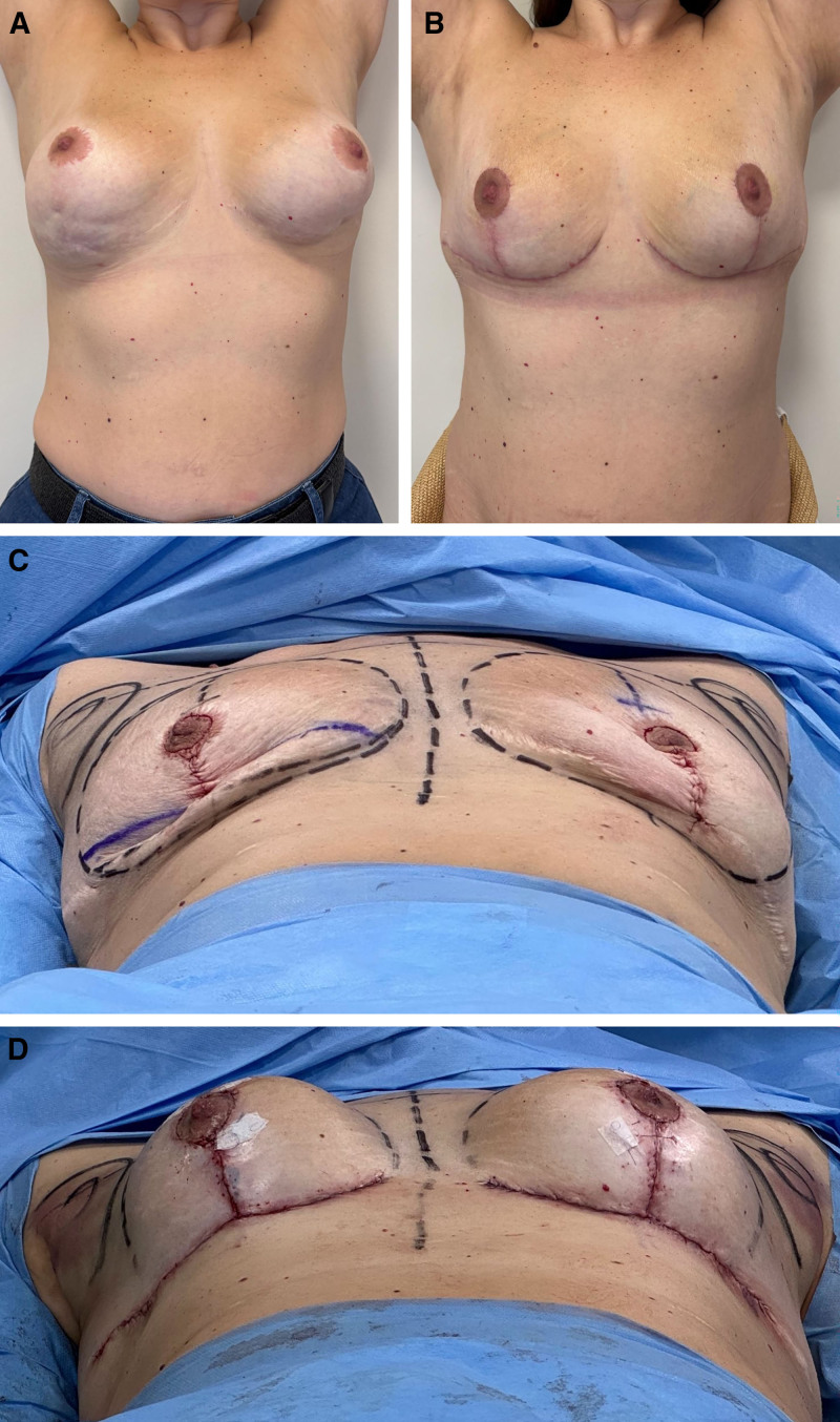
Fig. 8. Distinct implant pocket locations and BST reconstruction. A, After 4 previous surgical procedures, the patient presented with 300-mL implants placed retroglandularly on the left and in a dual-plane pocket on the right. B, At 45 days, the breasts exhibit adequate contour and satisfactory symmetry. C, Intraoperative findings: typical retroareolar depression on the left breast (retroglandular explantation) and better retroareolar support on the right (dual-plane explantation). D, Immediate postoperative results show reconstruction using different BST strategies tailored to each deformity.不同假体腔隙位置的 BST 重建:左侧腺体后、右侧双平面,经 4 次既往手术后 45 天。Simple explantations may heal unpredictably, revealing implant-induced contour irregularities that can markedly compromise the aesthetic outcome (Fig. 9 ). Although improvement has been reported over time, this expectant approach often imposes substantial patient anxiety. The BST enables immediate, controlled contour restoration at the time of explantation by internal parenchymal redistribution and directed tissue healing, yielding a predictable rounded shape with uniform fullness, adequate retroareolar support, nonretracted nipples, and short horizontal scars (Fig. 9 ).
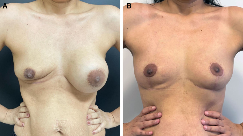
Fig. 9. Comparative dynamic evaluation of simple retroglandular explantation versus immediate reconstruction with the BST. A, Dynamic evaluation of the right breast at 5 months after simple explantation of a 450-cm3 retroglandular implant. B, The left breast shows stable contour and volume 5 months after reconstruction with the BST, with no deformities or need for fat grafting.单纯腺体后取出与即刻 BST 重建的动态对比:右侧单纯取出 5 月,左侧即刻重建。Fat grafting is performed in most cases of breast explantation. 15 Pompei et al 1 claim that prior implant capsule can be preserved when designing a fat graft because it provides a better plane for adipose tissue infiltration.
Although autologous fat grafting is a safe and effective option for breast volumization, 18 the BST minimizes reliance on it by using individualized approaches that leverage immediate postexplantation skin laxity and breast tissue looseness to narrow the footprint and uses the “walking breast” strategy to address volume-deficient areas, thereby reducing the need for lipofilling ( see Video 5 [online] ). Fat grafting remains available as an optional adjunct for additional volume at a subsequent stage.
With regard to complications, a related article that examined the prevalence of explantation complications in 64 patients revealed minor cellulitis in 1 (1.6%) patient and late-onset hematoma with infection in 1 (1.6%) patient. In our study, 2 (1.2%) patients developed deep vein thrombosis, and 5 (1.22%) patients experienced hematomas. No cases of infection or skin or areola necrosis were observed. This technique is quite safe for the areola because it remains attached to its original site, as no rotational pedicles or repositioning are necessary.
This retrospective study is limited by the absence of validated metrics (BREAST-Q). Future prospective research incorporating patient-reported outcomes and complication analyses is essential to further establish the technique’s clinical standing.
When performing silicone breast implant explantation, our method—known as the BST—promotes a customized approach to breast tissue management. It can be applied to explantation in the retroglandular, submuscular, and dual-plane pockets, enabling predictable outcomes with a rounded breast shape and adequate retroareolar filling, without the need for fat grafting. Its low rate of complications makes it a procedure that can also be regarded as safe.
The authors have no financial interest to declare in relation to the content of this article.
声明:中文精读 · 仅供学术参考。内容来自公开文献检索,不代表本人观点,不构成诊疗建议。 医疗美容需在正规医疗机构由执业医师实施。
基于相同主题推荐 · 继续深挖
背景:因假体并发症、乳腺植入物相关症状等需求行假体取出者增多;如何在取出同期重建自然轮廓、避免“空瘪”外观是临床难点。
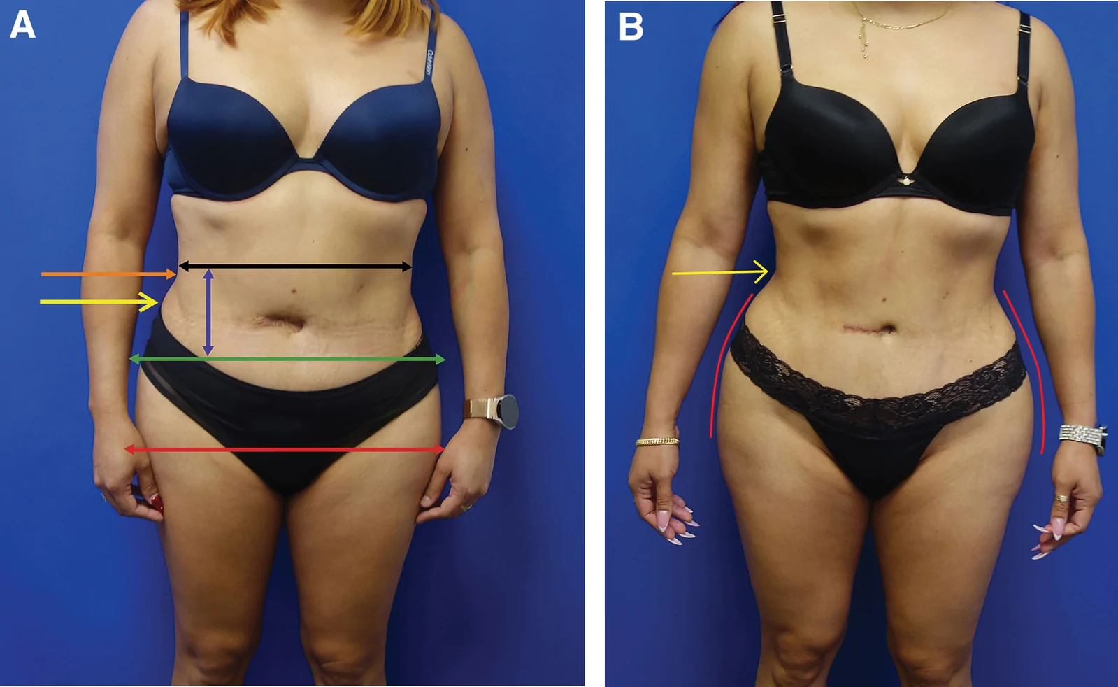
研究设计:PRS Global Open 回顾性研究,2016-11 至 2025-01 共 1999 例连续手术,旨在界定并塑形『腰腹过渡美学单元(waist transitio
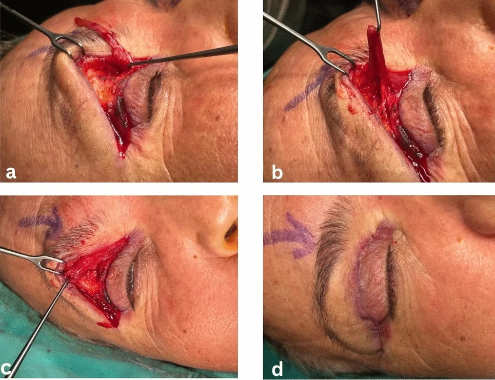
背景:眶上区(上睑)凹陷可由衰老、创伤或上睑成形术过度去脂引起,传统脂肪移植存在移植萎缩与眼眶内栓塞等风险。