耳轮缘复合移植片鼻部重建
2026年9月20日22 min read
Plastic and Reconstructive Surgery Global Open
结论与实践要点
循证与证据等级。 本项为单中心、单术者回顾性队列(n=65,2010–2024),属低至中等证据等级的观察性研究;但样本量在鼻部复合移植文献中较可观,且与历史报道(失败率可达 70%)相比表现良好,提供一定实践参考。
局限。 回顾性设计、未使用术中冰冻切片、缺损/移植物尺寸以病理测量为代理(甲醛固定后收缩,可能低估体内实际大小);缺乏客观的长期美学评分;术者经验集中,结论外推需谨慎。文中“5% 不完全切除率”与已发表数据相当。
可落地提示。 对累及鼻翼缘、最大约 20 mm 的全层缺损,该术式可一期完成、供区隐蔽且可一期闭合,是值得考虑的重建选项;应强调术前戒烟(≥2 周)、优化受区血管床,对吸烟或存在放疗/瘢痕史者需充分告知较高失败风险并准备备选方案(如额部皮瓣)。
Background:The use of composite grafts for reconstruction of nasal defects involving the alar rim has long been established. The standard donor site is the root of the helix, which limits the size of the graft and can distort the upper pole of the ear. An alternative approach using the mid-helical rim, by contrast, allows a larger graft size to be harvested and eliminates the need for additional flap closure of the donor site. We detail our 1-stage technique and outcomes of using a mid-helical rim composite graft for nasal defects involving the alar rim.
Methods:This retrospective cohort outlines a single surgeon’s use of the mid-helical rim composite graft for reconstruction of full-thickness nasal alar defects.
Results:A total of 65 patients underwent the mid-helical rim composite graft procedure from 2010 to 2024. The procedure was performed under local anesthetic with intravenous sedation in 63 (97%) patients following excision of a basal cell carcinoma. The median defect size along the alar rim was 14 mm (range, 9–20 mm), as measured by the pathologist following formalin fixation. The excision of the tumor and the reconstruction were performed in a single procedure. Four grafts were lost, with 3 losses occurring in patients with a history of smoking.
Conclusions:The mid-helical rim composite graft can be used for 1-stage reconstruction of nasal defects involving up to 20 mm of the alar rim, with reliable outcomes. Graft take is improved by creating a defect that increases the size of the recipient vascular bed.
Defects of the alar, when full-thickness and/or involving the alar rim, soft triangle, and/or nasal tip, are difficult to repair. The goals for reconstruction are to maintain the contour of the nose and preserve nasal function. The use of composite grafts (combined cartilage, skin, and soft tissue) harvested from the helix of the ear is an effective way of achieving a good aesthetic and functional reconstruction. The “standard” donor site is the root of the helix. However, closure of this donor defect often requires a separate preauricular skin flap to avoid distorting the upper pole of the ear. 1 This adds complexity and further scarring to the reconstruction. In comparison, a helical rim donor site can be closed with a wedge and allows for larger grafts to be harvested. Increasing the size of the recipient defect to include a vascular bed for the graft helps improve graft take. We report here the technique and results of using mid-helical rim composite grafts for nasal reconstruction by a single surgeon.
This retrospective cohort examined 65 patients who underwent surgery between 2010 and 2024, performed by a single surgeon (G.H.). Their clinical and histopathologic reports were retrieved and analyzed. The specimen size was obtained from histopathology measurements. The study adhered to the principles outlined in the Declaration of Helsinki.
The indication for the use of mid-helical composite grafts was a nasal alar and/or soft triangle defect involving or extending to within 2–3 mm of the alar rim, with a maximum defect of approximately 20 mm. In all cases, the alar rim was included in the excision to maximize pathological clearance. Patients who smoked were advised to discontinue tobacco use at least 2 weeks before and after surgery, and anticoagulants were stopped if deemed safe.
The technique is shown in Figures 1 and 2 and Videos 1–4. ( See Video 1 [online] , which displays the harvesting of a mid-helical rim composite graft from the left ear.) ( See Video 2 [online] , which shows the marking of the site for deepithelialization of the composite graft.) ( See Video 3 [online] , which demonstrates the trimming of the composite graft with an extended dermal pedicle.) ( See Video 4 [online] , which displays the insetting of the trimmed composite graft.)
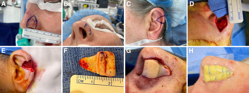
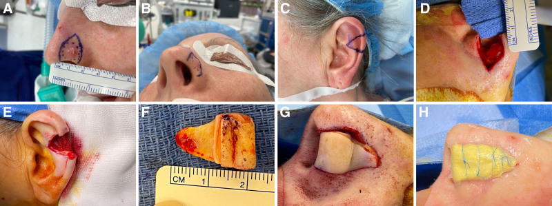
Fig. 1. Key steps in planning, harvesting, and insetting a mid‑helical rim composite graft for 1‑stage reconstruction of nasal alar rim defects. A–C, The nasal lesion is marked with an extended triangular-shaped wound bed proximal to the defect. This marking is copied 1:1 onto either ear, depending on patient preference, using the helix and antihelix. Markings are made only anteriorly, as the graft is harvested at full thickness with a 15-blade. D, Nasal defect showing the extended superior triangular wound bed and the lower 3–5 mm of the defect at full thickness along the alar rim. E and F, The mid-helical rim composite graft with the triangular end of the anterior side of the ear trimmed of skin and cartilage. This part matches the defect, including the triangular-shaped wound bed created above the defect on the nose. G, The graft is placed with the postauricular skin externally and the anterior surface of the ear internally within the nostril. The inset is performed with 5/0 nonabsorbable monofilament, Novafil (Covidien, MA) on the skin and 4/0 absorbable monofilament, Monocryl (Ethicon, GA) within the nostril. H, A bolster of occlusive gauze strip with 3% bismuth tribromophenate in petrolatum blend (Xeroform; Covidien, MA) secured with the 6/0 Novafil as a tie-over bolster, with 1% chloramphenicol ointment to the edges. This is typically left in place for 5–7 days.耳轮缘复合移植片规划、切取与植入的关键步骤示意(A–H),含扩大三角形血管化创面床的标记与 1:1 移植片设计。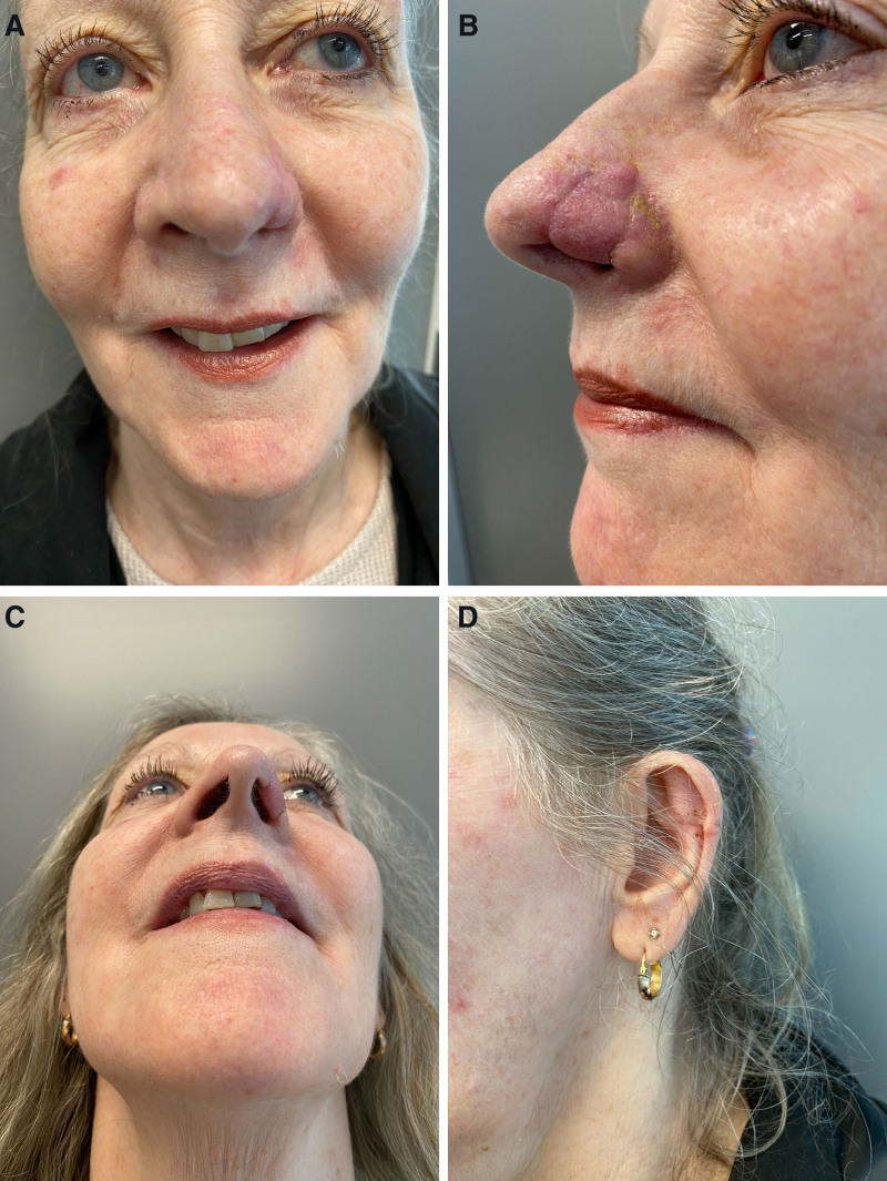
Fig. 2. Six weeks postoperatively, showing mild swelling, as expected, with continued refinement of contour. A, Front view. B, Lateral view. C, View from below. D, Donor site.术后 6 周外观(A 正面、B 侧面、C 仰视、D 供区),可见预期内轻度肿胀并逐步轮廓细化。The tumor and excision margin, including a triangle superior to the defect, are marked out preoperatively (Figs. 1 A, B). This triangle, representing the extended dermal pedicle, provides a vascularized wound bed to improve graft take. The width of the defect is drawn perpendicular to the alar rim (Fig. 1 C). The composite graft is marked out preoperatively 1:1 on the mid-helical rim, using the contra- or ipsilateral ear, depending on patient preference and the availability of good-quality skin.
Infiltration is performed with 1% ropivacaine with adrenaline (1:100.000). The excision is performed, starting with the superior triangle, which involves excising skin and soft tissue while preserving at least the mucosa of the nasal vestibule (Fig. 1 D). Approximately 3–5 mm above the alar rim, the excision is performed full-thickness to include the vestibular mucosa.
These steps, excising a skin-only triangle superior to the defect and excising the alar rim full–thickness, are performed to provide a well-vascularized wound bed, to match the donor site, and to provide structural support of the composite graft. Careful hemostasis is performed, ensuring the wound bed remains as vascular as possible.
The composite graft is harvested as a wedge with the anterior side of the graft turned over to line the vestibulum and become the inside of the nostril (Fig. 1 E). Part of the anterior side of the composite graft is trimmed of skin and cartilage on table to match the recipient site wound bed (Fig. 1 F). This part of the composite graft will act as a full-thickness skin graft.
The graft is inset with the postauricular skin externally and secured in place with 5/0 nonabsorbable monofilament, Novafil (Covidien, MA), externally and 4/0 absorbable monofilament, Monocryl (Ethicon, GA), in the nostril (Fig. 1 G). A bolster of an occlusive gauze strip with 3% bismuth tribromophenate in a petrolatum blend, Xeroform (Covidien, MA), is carefully secured as a tie-over and left intact for 5–7 days, with minimal disruption to blood flow at the margin (Fig. 1 H). The donor site is closed in a standard fashion. No systemic antibiotics are given postoperatively, but the patient is advised to apply topical chloramphenicol 1% ointment to the edges of the bolster twice daily until the first follow-up. Figures 2 and 3 – 4 show the composite graft and donor site at 1 and 6 weeks postoperatively. Figure 5 presents further examples of preoperative markings and 6-week postoperative outcomes.
Fig. 3. Postoperative images showing composite graft at 1 week (A) and 6 weeks (B).复合移植片术后 1 周(A)与 6 周(B)图像对比。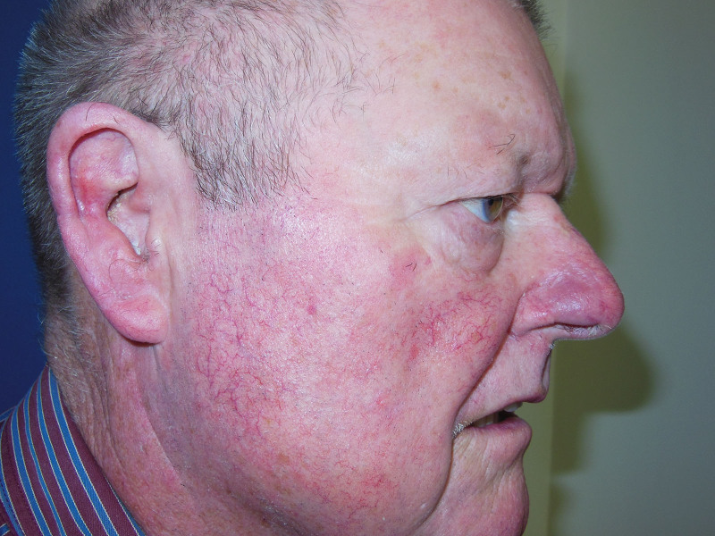
Fig. 4. Composite graft 6 weeks postoperatively.复合移植片术后 6 周外观。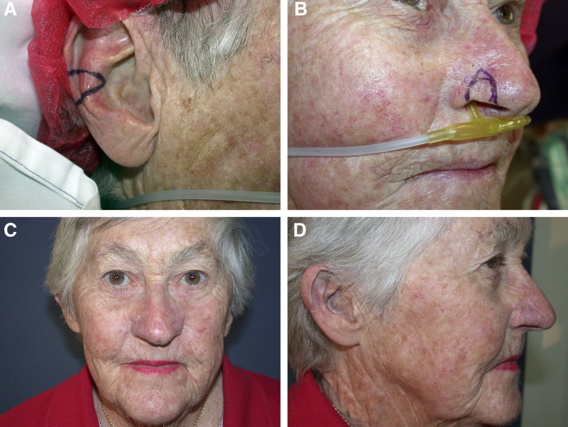
Fig. 5. Composite graft with preoperative markings and results 6 weeks postoperatively. A, Donor site. B, Planned excision. C, Front view 6 weeks postoperatively. D, Lateral view showing both the mid helical rim donor site and the composite graft.含术前标记与术后 6 周结果的更多示例(A 供区、B 规划切除、C 正面、D 侧面示供区与移植物)。补充视频 / 附件(原文未随文提供原图,可于出版方页面查看)gox-14-e7733-s001.mp4Video 1. This video displays the raising of an ipsilateral mid helical rim composite graft and inset to the alar rim defect.补充视频 / 附件(原文未随文提供原图,可于出版方页面查看)gox-14-e7733-s002.mp4Video 2. This video displays the trimming of the mid helical composite graft.补充视频 / 附件(原文未随文提供原图,可于出版方页面查看)gox-14-e7733-s003.mp4Video 3. This video displays the inset of an ipsilateral mid helical rim composite graft and inset to the alar rim defect. The composite graft is turned over and inset to the alar rim defect.补充视频 / 附件(原文未随文提供原图,可于出版方页面查看)gox-14-e7733-s004.mp4Video 4. This video displays the trimmed ipsilateral mid helical rim composite graft turned over and inset to the alar rim defect.
From 2010 to 2024, a total of 65 mid-helical rim composite graft reconstructions of the alar were performed (Table 1 ). The majority were men (n = 36, 55%), with 29 (45%) women. The median age was 74 years (range, 48–96 y). Seven (11%) actively smoked, and 44 (68%) had at least 1 comorbidity. Fifty-seven (88%) underwent reconstruction following excision of a basal cell carcinoma (BCC), 3 (5%) following excision of squamous cell carcinoma, and the remaining 5 (8%) were 2 patients with defects created by failed local flaps by a referring surgeon, 1 patient with a pyogenic granuloma, 1 with bowenoid actinic keratosis, and 1 with basosquamous carcinoma. The median defect size along the alar rim was 14 mm (range, 9–20 mm), as measured by the pathologist following formalin fixation. The procedure was performed in 63 cases (97%) under local anesthetic with intravenous sedation. Clear margins were obtained in 62 (95%) cases, with 3 cases (5%) showing involved margins. Of these cases, 1 was treated with topical imiquimod for residual superficial BCC, and 2 patients declined further surgery and elected to undergo clinical surveillance instead. Frozen sections were not used in our series. Frozen section does have a false-negative and false-positive rate, and it adds additional time and cost to the procedure. Our incomplete excision rate of 5% is in keeping with established and published incomplete excision rates. In 53 (81%) cases, there were no adverse events following surgery. Twelve (17%) patients experienced adverse events following the procedure. There was 1 case of minor bleeding treated conservatively, superficial epidermolysis in 6 (9%) patients, partial loss of graft in 1 case resulting in notching (2%), and total graft failure in 4 (6%) cases. Notching was noted in 2 patients. Of these, 1 was due to partial graft loss. This patient was offered a revision composite graft but declined.
Table 1. Demographics, Nasal Defect and Composite Graft Characteristics, and Results
| Age at the time of surgery, median (range) | 74 (48–96) |
|---|---|
| Sex, n (%) | |
| Men | 36 (55) |
| Women | 29 (45) |
| Smoking status, n (%) | |
| Nonsmoking | 58 (89) |
| Smoking | 7 (11) |
| Comorbidity, n (%) | |
| None | 21 (32) |
| ≥1 | 44 (68) |
| Etiology, n (%) | |
| BCC | 57 (88) |
| SCC | 3 (5) |
| Other * | 5 (8) |
| Complete excision † | 62 (95) |
| Anesthesia, n (%) | |
| Local anesthesia with sedation | 63 (97) |
| General anesthesia | 2 (3) |
| Nasal defect characteristics | |
| Width of specimen along alar rim, mm, ‡ median (range) | 14 (9–20) |
| Composite graft characteristics | |
| Donor site, n | |
| Helical rim | 63 |
| Ear lobule | 2 |
| Postoperative characteristics within 30 d, n (%) | |
| No complications | 53 (81) |
| Recipient site (nasal alar) | |
| Postoperative bleeding | 1 (2) |
| Superficial skin loss | 6 (9) |
| Lost parts of the graft | 1 (2) |
| Complete loss of tissue | 4 (6) |
| Infection | 0 |
| Minor notch | 2 (3) |
| Temporary alar collapse | 1 (2) |
| Donor site (mid-helical rim) | |
| Wound healing issues | 1 (2) |
| Follow-up in months, median (range) | 1 (1–100) |
Superficial epidermolyses were all managed conservatively with acceptable outcomes. Of the 4 complete graft losses, 3 occurred in patients who smoked. The defect sizes of these graft failures ranged from 10 to 14 mm, with smoking as well as comorbidity for 1 patient, being the only registered variable that stood out. Only 1 patient who did not smoke (1 of 58 = 2%) experienced graft failure. This patient, who had no other comorbidities, had undergone Mohs surgery elsewhere, followed by a failed local flap reconstruction. It is likely that poor blood supply and scarred tissue as a result of the previous failed local flap, in combination with a large full-thickness defect of 6 × 10 mm, were factors that led to graft failure. This patient proceeded with a staged forehead flap reconstruction and was the only subject in our cohort requiring secondary flap reconstruction. No graft infections were encountered. One patient had temporary alar collapse, which did not impact nasal airflow, and no patients recorded alar collapse with functional nasal obstruction at the time of last follow-up. Most patients were discharged to follow-up at 6 weeks, with a median follow-up of 1 month by the treating surgeon (range, 1–100 mo).
The nose and nasal tip are frequent locations for skin cancer, especially BCC, and the reconstruction of resultant excisional defects is therefore frequently required. 2 The challenge faced by the reconstructive surgeon is to provide the best aesthetics and preserve nasal airflow and prevent nostril collapse on inspiration. Reconstruction should aim to provide adequate structural support, especially if the excisional defect involves and/or is near the alar rim. Skin flaps or grafts that encroach upon the alar rim or just above it will frequently result in notching of the rim or collapse of the nostril. The use of composite grafts for these defects has the advantage of providing structural support using the cartilage of the ear. These grafts are, however, limited by the need for adequate vascularization of both the soft tissues and the underlying cartilage. This has traditionally limited the size of the grafts to no more than 10–20 mm, so that no part of the graft is more than 1 cm from a free edge. 3 Increasing the size of the wound to include a superior wound bed, and removing skin and cartilage from the anterior skin of the composite graft at its apex, provides a full-thickness graft component to the composite graft, which increases neovascularization. This allows for larger composite grafts to take more reliably. In our series of 65 mid-helical rim composite graft reconstructions of the alar, the median (range) size of the free alar rim edge was 14 mm (9–20 mm), as measured by the pathologist after formalin fixation. With the expected postexcision shrinkage from formalin fixation, these measured defects would be 10%–20% larger if measured in vivo. 4 A 20% increase in specimen size would mean that the average size of our defects, and hence the grafts, would be 17 mm, and the largest would have been up to 24 mm along the alar rim.
The “standard” donor site for composite grafts is the root of the helix. 1 These composite grafts are limited in size and also cause distortion of the upper pole of the ear, often requiring additional preauricular flaps and resulting in scarring on the face to close the donor site. The use of a mid-helical rim composite graft allows a larger graft to be harvested with primary closure of the donor site, away from the face.
In our series, all defects were converted to full-thickness defects for the inferior 3–5 mm to allow the helical rim to reconstruct the alar rim full thickness. This approach provides a better aesthetic and functional result, with the shape of the helical rim cartilage maintaining the curve of the nostril and, hence, the airway. Creating a full-thickness alar defect allows for a stable and aesthetically pleasing composite graft design. This uses a well-established plastic surgery principle of matching the defect to the reconstruction.
To create a well-vascularized wound bed, the triangular superior skin excision led to a 94% graft take. This triangle can be placed within the alar, respecting the alar crease. For larger defects, the alar crease can be transversed.
In our series, all patients except 1 who did not use tobacco achieved complete success with the mid-helical rim composite graft. Three of the 4 cases of graft failure occurred in patients who used tobacco, with 1 patient undergoing a second composite graft, which also failed. The remaining failure occurred in a patient with no history of tobacco use who had previously had a failed local flap and a large defect. This patient required a subsequent staged forehead flap reconstruction.
In patients who use tobacco, composite grafts should be performed with caution. In our series, 7 patients with a history of tobacco use underwent composite graft reconstruction, with graft failure occurring in 3 (43%) patients.
Notching at the alar rim, as observed in 2 (3%) patients, can be caused by poor wound healing, poor inset, movement, or uneven scar contracture. Careful inset and fixation of the graft may reduce this risk.
Our series compares very favorably with previous reports, which have recorded failure rates of up to 70%, even in the absence of adjunctive treatments to improve healing, such as cooling, hyperbaric oxygen, delayed procedure, or pharmacological agents. 5 By contrast, our cohort did not have previous radiation therapy to the wound bed, and only 11% of patients had a history of tobacco use. The extended dermal flap of the composite graft, combined with the triangular-shaped wound bed proximal to the defect, most likely contributed to our relatively low failure rate.
Alternatives to a composite graft for an alar defect include a staged forehead, melolabial flap ± free cartilage graft ± mucosal flap, concomitant lip lifting using contralateral lateral ala, or a free microsurgical composite graft. These complex procedures are often staged and time-consuming, requiring general anesthesia. 6 – 8 By comparison, the mid-helical composite graft is well tolerated by patients, has a relatively shorter operative duration, and can be performed under local anesthetic with intravenous sedation, a further advantage given the age of our patient cohort. At follow–up of around 6 weeks, our grafts were well healed, with good aesthetic outcomes and no obvious impact on nasal inflow and airway (Figs. 2 A and B, 3 A, 4 , 5 C and D).
One potential limitation of the mid-helical rim is the maximum size that can be harvested without compromising the aesthetics of the ear. Although we did not measure the size of each graft directly, we have used histopathological measurements as a proxy to estimate the size of both the defect and graft. We acknowledge that this may have led to underestimation in our study.
In conclusion, mid-helical rim composite grafts provide a simple, stable, predictable option for the reconstruction of nasal alar defects.
Anand K. Deva is a consultant and research coordinator for AbbVie, Johnson & Johnson, 3M, and BellaSeno. The other authors have no financial interest to declare in relation to the content of this article.
Patients provided written consent for the use of their images.
声明:中文精读 · 仅供学术参考。内容来自公开文献检索,不代表本人观点,不构成诊疗建议。 医疗美容需在正规医疗机构由执业医师实施。
基于相同主题推荐 · 继续深挖
回顾性研究纳入23例接受JAMBER(JB)盘绕PDO线鼻部塑形患者(8男15女,平均35.3岁,21–72岁),随访至术后12个月。
回顾性单中心研究纳入59例巴西多族裔患者(20–60岁,平均36.0±10.3岁),由53名鼻部填充经验各异的注射者实施透明质酸(HA)非手术隆鼻。
鼻部塑形疗效与安全性:12个月前瞻性研究/PMC13100342_fig1.jpg)
这项单中心前瞻性研究纳入24名中国女性受试者,接受CaHA凝胶鼻部注射(骨膜上/骨膜浅层,单侧单次总量≤2.0 mL,平均0.93±0.22 mL),全部完成12个月随访,无脱落。