基于 CT 的鼻骨形态与厚度分型用于截骨术:Avellaneda 系统
2026年9月22日28 min read
Plastic and Reconstructive Surgery Global Open
Plastic and Reconstructive Surgery Global Open
结论与实践要点
本研究为前瞻性病例系列(125例,2021—2024),由 Avellaneda 团队提出并验证了一套基于术前鼻部 CT 的鼻骨形态与厚度分型,用于指导鼻整形中的截骨塑形(osteoplasty)。其核心是把"形状"与"厚度"两个独立维度组合:形状分凹(1型)、平(2型)、凸(3型);厚度分薄 A(<2mm)、中 B(2–4mm)、厚 C(>4mm),两两组合形成 9 种亚型。
临床价值在于风险分层与术式个体化:研究将 3A/3B/2A 列为高危(凸+薄、凸+中、平+薄),建议极小范围塑形,必要时转为可控截骨;1A/2B/3C 为中度风险,需谨慎;1C/1B/2C 为低风险,可较充分地减除骨性隆起。所有患者均行开放性鼻整形并以"锉磨雕塑"代替传统截骨,随访中无意外骨折发生,提示该分型在规避医源性骨折方面的潜在价值。
对临床的启示是:术前常规鼻部 CT 并据此分型,可能比凭经验统一操作更利于平衡塑形效果与安全性,尤其在处理薄而凸的鼻骨时。
Background:Successful rhinoplasty relies heavily on understanding the anatomical diversity of the nasal bones. Variability in nasal bone shape and thickness directly influences the technical complexity and outcomes of osteoplasty; the aim of this study was to develop and validate a computed tomography (CT)–based classification system of nasal bone shape and thickness to guide surgical planning in nasal osteoplasty and optimize patient-specific outcomes.
Methods:A prospective study was conducted on 125 patients who underwent preoperative CT scans between 2021 and 2024. Nasal bone shape was categorized as concave, planar, or convex, and thickness as thin (<2 mm), medium (2–4 mm), or thick (>4 mm); these were combined into 9 possible subtypes. Open rhinoplasty with tailored osteoplasty was performed in all patients, without osteotomies. Outcomes were assessed with CT comparisons and the FACE-Q Rhinoplasty Module at 3, 6, and 12 months.
Results:CT analysis revealed substantial anatomical variability: 47.2% of right nasal bones were convex, whereas only 24% were concave; 51.2% had medium thickness, and 45.6% were thick. The most common Avellaneda types were 3B (convex-medium) and 3C (convex-thick). No unintended nasal bone fractures occurred. Patient satisfaction improved significantly postoperatively.
Conclusions:The Avellaneda classification offers a novel, pragmatic framework to individualize nasal osteoplasty based on CT findings; integrating bone shape and thickness allows surgeons to tailor their technique to the patient’s anatomy, avoid intraoperative fractures, and enhance surgical precision. This system may improve outcomes, promote surgical safety, and serve as a reference for future refinements in rhinoplasty.
Nasal bone anatomy is critical to the overall structure and aesthetics of the nasal pyramid, as it forms the upper third of the nasal skeleton and defines the shape of the dorsum, contributing to harmonious dorsal lines and the lateral nasal walls; the nasal bones articulate with the frontal bone superiorly and with the maxilla superolaterally, creating a bony vault whose thickness varies along its length; at the nasofrontal junction, the nasal bone is thicker, whereas it tapers as it approaches the piriform aperture. 1 This gradient of thickness explains why the caudal portion, which is thinner, is prone to fractures, whereas trauma to the cephalic portion, which is more robust, may result in more complex nasal deformities. 1
On average, the nasal bone thickness is 1.5–2 mm, with variations according to sex: women generally have thinner nasal bones than men. These anatomical differences are critical because thicker bones (particularly in male or ethnic groups) may require different surgical approaches and tools compared with thinner bones. 2
Nasal bones may be convex, planar, or concave, influencing the dorsal profile. Anthropometric studies show that most individuals exhibit an S-shaped bony dorsum, whereas a minority present a straight profile. One series reported 88% with an angulated dorsum and hump, and 12% with a linear configuration. Maximum convexity typically occurs at the osteocartilaginous transition (K-point), representing the apex of the preoperative prominence. The overall dorsal contour is determined by the interaction of the radix, bony vault, and osteocartilaginous vault. 3 In contrast, complex 3-dimensional deformities may show contralateral concavity and convexity, with marked asymmetries posing significant surgical challenges. 4 , 5
Nasal bone osteotomy has traditionally been a fundamental step in rhinoplasty to correct dorsal deformities and narrow the nasal pyramid. A careful approach to osteotomies is essential to achieve a smooth contour, but their execution carries risks: an inadequate osteotomy can lead to bony irregularities that compromise the result. This underscores the importance of mastering different osteotomy techniques and selecting the most appropriate one based on each patient’s anatomy, optimizing the nasal contour without functional detriment. 4
In recent years, rhinoplasty has incorporated innovations that complement or replace conventional osteotomies. One example is dorsal preservation rhinoplasty, in which, instead of resecting the hump, the dorsum is preserved intact and undergoes controlled descent. 1 This preservation approach requires a detailed understanding of nasal anatomy and meticulous preoperative planning with computed tomography (CT) to avoid complications, especially if the low position of the K-area necessitates resecting the perpendicular bone of the ethmoid. 1
Moreover, nasal osteoplasty has been enhanced, being understood as direct bony remodeling without complete fractures. Osteoplasty allows for smoothing bony prominences or correcting minor residual irregularities. Koçak and Senturk 3 and Koçak et al 6 reported that in deviated noses, lateral osteoplasty can replace the traditional double osteotomy, with equivalent results, avoiding complications associated with multiple bony fractures. By avoiding uncontrolled fractures, this bony remodeling reduces the risk of long-term instability and maintains the correction of the deviation without significant recurrences. 3
The aim of this article was to propose and validate a novel anatomical classification system for the nasal bones based on preoperative CT analysis, integrating both bone shape and thickness. This classification is intended to guide the surgical decision-making process in nasal osteoplasty, allowing for individualized, anatomy-driven approaches that minimize the risk of complications such as unintended fractures or asymmetries.
This prospective study was conducted between 2021 and 2024 and included patients who underwent preoperative assessment at Nuara Esencia de la Belleza. The objective of the study was to develop and validate a systematic approach to nasal osteoplasty based on anatomical parameters obtained through nasal CT, with the aim of reducing the risk of unexpected fractures caused by excessive thinning of the nasal bone during surgery.
Patients who underwent primary rhinoplasty with an indication for nasal osteoplasty were included in the study. The inclusion criteria were as follows: patients aged 18 years or older, a diagnosis of nasal deformity requiring surgical intervention (dorsal deviations, prominent humps, or bony irregularities), and availability for documented postoperative follow-up according to the study protocol. Patients with a history of nasal surgery, congenital malformations, systemic diseases that could affect healing, or any contraindications to CT were excluded.
Each patient underwent a CT scan of the nasal bones to evaluate the bony morphology of the nose before surgery for surgical planning, prevent possible fractures, and evaluate possible posterior or high septal deviations (all patients accepted the CT scan because it was part of the standard surgical planning protocol). The obtained images were processed and analyzed using specialized software to measure the thickness of the nasal bone at key points along the dorsum and the base of the nose. The following measurements were recorded: nasal bone thickness in the medial, intermediate, and lateral regions of the dorsum; length and shape of the dorsum; and the presence of bony irregularities.
A classification system was used to categorize the shape and thickness of the nasal bone, which was determined from the results of the CT scan. This classification includes 2 main parameters: the shape of the nasal bone and its thickness.
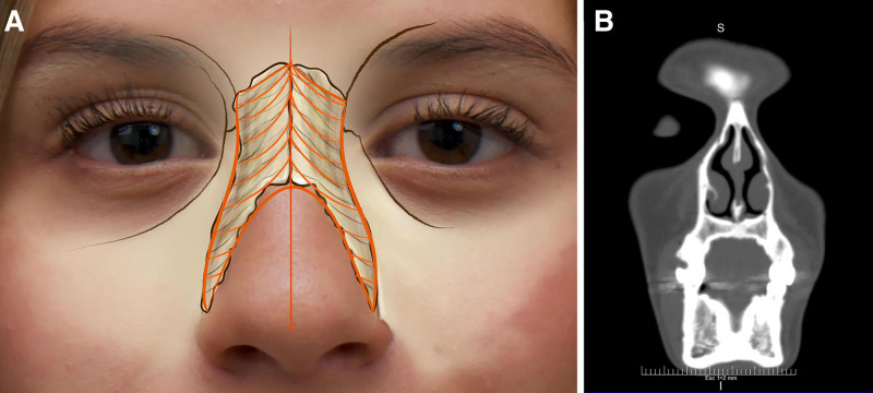
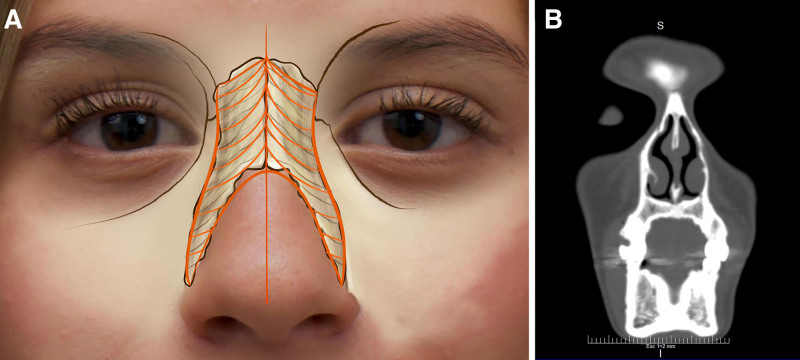
Fig. 1. Concave nasal bone morphology (type 1). A, Clinical illustration with anatomical scheme showing a concave nasal bone morphology (type 1), characterized by an inward curvature of the lateral nasal walls. B, Coronal CT scan demonstrating the concave bony configuration of the nasal dorsum (type 1).凹形鼻骨(1型):侧鼻壁向内凹陷,文中视为女性鼻的美学理想形态;含示意图与冠状CT。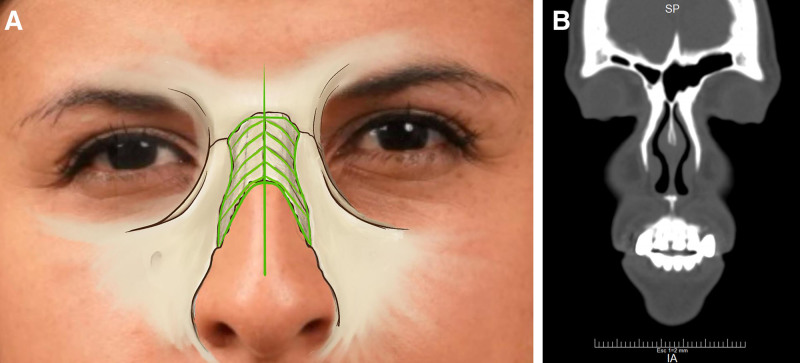
Fig. 2. Planar nasal bone morphology (type 2). A, Clinical illustration with an anatomical scheme depicting a planar nasal bone morphology (type 2), defined by straight lateral walls without significant curvature. B, Coronal CT scan demonstrating a planar bony vault (type 2).平坦鼻骨(2型):侧鼻壁平直无显著弯曲,文中视为男性鼻的美学理想形态;含示意图与冠状CT。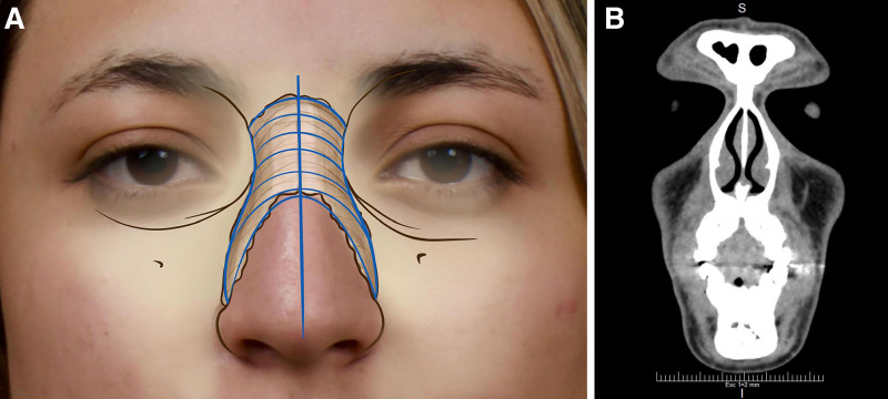
Fig. 3. Convex nasal bone morphology (type 3). A, Clinical illustration with anatomical scheme showing a convex nasal bone morphology (type 3), characterized by outward bulging of the lateral walls. B, Coronal CT scan confirming the convex bony prominence of the dorsum (type 3).凸形鼻骨(3型):侧鼻壁向外隆起形成可见凸起,美学上多不被偏爱;含示意图与冠状CT。
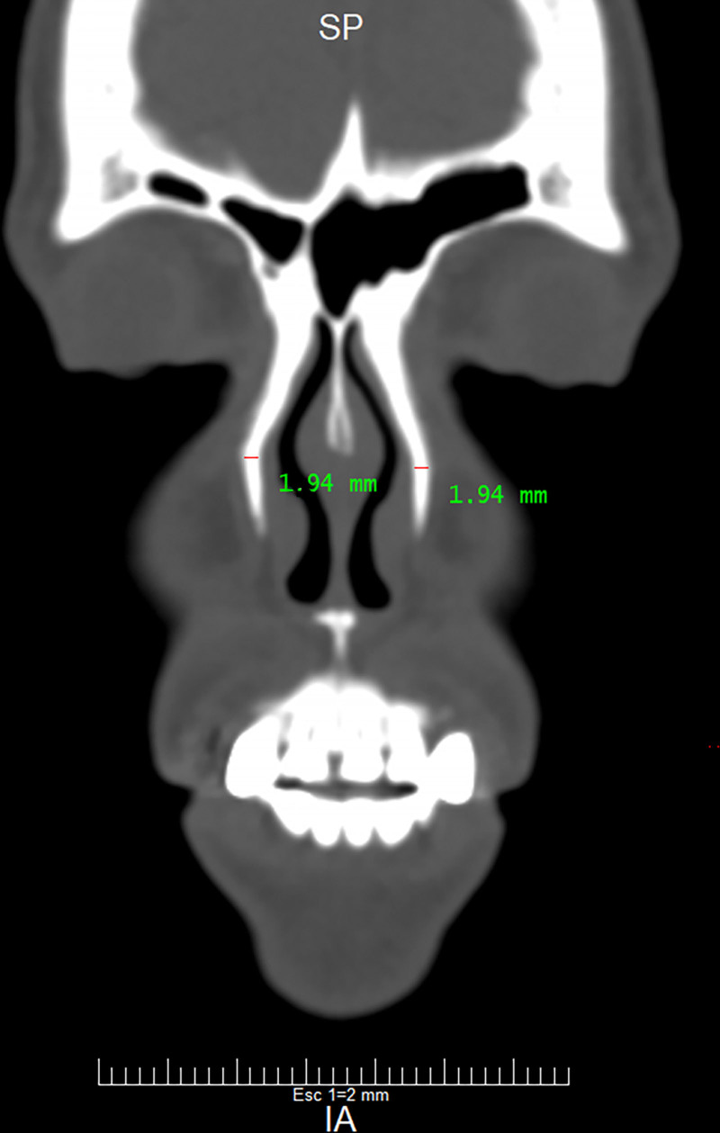
Fig. 4. Thin nasal bone thickness on CT scan (type A). Coronal CT scan of a patient with thin nasal bones (<2 mm, type A). This group requires delicate osteoplasty to avoid intraoperative fractures.薄鼻骨(A型,<2mm):需精细塑形以降低术中骨折风险;冠状CT示例。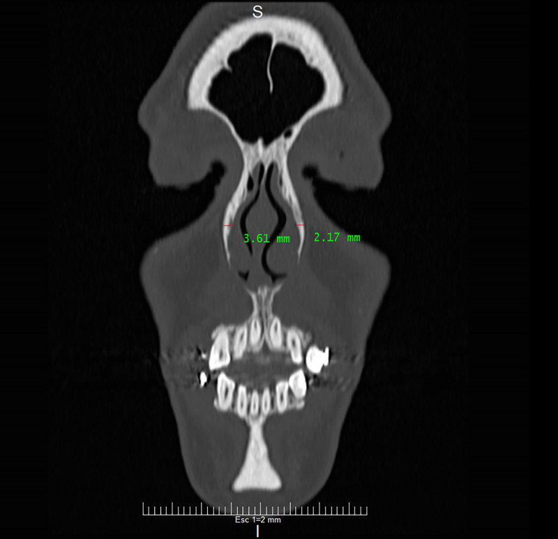
Fig. 5. Medium nasal bone thickness on CT scan (type B). Coronal CT scan of a patient with medium-thick nasal bones (2–4 mm, type B). These bones are suitable for conventional osteoplasty, with a moderate fracture risk.中等厚度鼻骨(B型,2–4mm):适合常规塑形,骨折风险中等;冠状CT示例。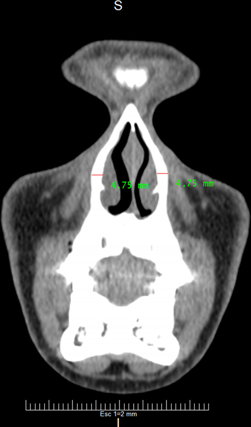
Fig. 6. Thick nasal bone thickness on CT scan (type C). Coronal CT scan of a patient with thick nasal bones (>4 mm, type C). These robust bones allow more aggressive osteoplasty with low fracture risk.厚鼻骨(C型,>4mm):结构稳健、医源性骨折风险低,可较积极塑形;冠状CT示例。Based on this classification, patients were assigned to one of the corresponding anatomical groups, and personalized surgical strategies were developed according to the characteristics of each patient’s nasal bone.
All patients underwent open rhinoplasty with transcolumellar and marginal incisions to provide adequate visualization and access to the nasal dorsum. A transcolumellar incision was made along the lower border of the columella, extending into the marginal incisions along the lateral borders of the nares, allowing complete exposure of the nasal pyramid. Additionally, full exposure of the lateral nasal wall was performed to ensure adequate visualization from its junction with the frontal bone superiorly to the junction with the upper lateral cartilage inferiorly, and laterally to the articulation between the nasal bone and the ascending process of the maxilla.
Unlike other surgical approaches, no osteotomies were performed on any patient. Instead, osteoplasty was exclusively used. The goal was to remodel the nasal bone by filing and sculpting the bone in the affected area, removing prominences or irregularities, and shaping the lateral bony wall to be as concave as possible in female patients and as straight as possible in male patients. This technique allowed for the smoothing of convex or concave areas without performing complete fractures, thereby minimizing the risk of complications associated with traditional osteotomies.
Osteoplasty focused on reducing the prominence of the lateral wall and dorsum in cases of convex bone (classification 3), smoothing irregularities in planar bones (classification 2), and restoring symmetry in cases of asymmetric concave bones (classification 1).
Preoperative CT scans were performed to obtain the nasal bone measurements, which were compared with postoperative images at 3 and 12 months to assess anatomical changes and the stability of the results. Surgical complications and aesthetic outcomes were documented using a visual analog scale for both the surgeon and the patients.
Statistical analysis was performed to provide a descriptive characterization of the study population and the CT-based anatomical findings. Continuous variables, including age, weight, height, body mass index, and FACE-Q scores, were summarized using means and SDs. Categorical variables, including sex, skin thickness, nasal bone shape, nasal bone thickness categories, Avellaneda classification subtypes, and postoperative complications, were described using absolute frequencies and percentages.
For the patient-reported outcome analysis, preoperative and postoperative FACE-Q Rhinoplasty module scores were compared using the Student t test for paired samples, as these measurements were obtained from the same patients before and after surgery. The mean difference, 95% confidence interval (95% CI), and corresponding P value were calculated. All statistical tests were 2-tailed, and a P value less than 0.05 was considered statistically significant.
The study was approved by the ethics committee of the Nuara Institute, and all patients signed an informed consent form before participating in the study. The procedures were conducted in accordance with international ethical standards and the principles of the Declaration of Helsinki.
A total of 125 patients were included in the study between 2021 and 2024. The average age was 30.9 years (SD = 6.99 y). The average weight was 68.2 kg (SD = 12.3 kg), the average height was 165.4 cm (SD = 9.2 cm), and the average body mass index was 24.9 kg/m 2 (SD = 3.1 kg/m 2 ). Of the participants, 78.6% were women and 16.8% were men. Regarding skin thickness, most patients had medium skin (45.6%), followed by thick skin (36%) and thin skin (18.4%) (Table 1 ).
Table 1. Demographic Characteristics
| Variable | Measure of Central Tendency or % | Dispersion Measurement |
|---|---|---|
| Age, y | Mean: 30.9 | SD: 6.99 |
| Sex, n (%) | Total: 125 | NA |
| Female | 103 (78.6) | |
| Male | 22 (16.8) | |
| Weight, kg | Mean: 68.2 | SD: 12.4 |
| Height, m | Mean: 1.65 | SD: 0.09 |
| BMI, kg/m 2 | Mean: 24.9 | SD: 3.1 |
| Skin thickness, n (%) | Total: 125 | NA |
| Thin | 23 (18.4) | |
| Medium | 57 (45.6) | |
| Thick | 45 (36) |
The nasal bone shape, as evaluated through CT imaging, was classified as shown in Table 2 .
Table 2. Nasal Bone Classification
| Variable | n | % |
|---|---|---|
| Shape of the right nasal bone | ||
| Concave | 30 | 24 |
| Planar | 36 | 28.8 |
| Convex | 59 | 47.2 |
| Shape of the left nasal bone | ||
| Concave | 36 | 28.8 |
| Planar | 55 | 44 |
| Convex | 34 | 27.2 |
| Thickness of the right nasal bone | ||
| A (<2 mm) | 4 | 3.2 |
| B (2–4 mm) | 64 | 51.2 |
| C (>4 mm) | 57 | 45.6 |
| Thickness of the left nasal bone | ||
| A (<2 mm) | 20 | 16 |
| B (2–4 mm) | 70 | 56 |
| C (>4 mm) | 35 | 28 |
The Avellaneda classification was based on the combination of the shape and thickness of each side of the nasal bone, determining the surgical approach for each patient. The following are the frequency distributions of the Avellaneda classification (Table 3 ).
Table 3. Frequency Distributions of the Avellaneda Classification
| Variable | n | % |
|---|---|---|
| Right nasal bone | ||
| 1A | 1 | 0.8 |
| 1B | 14 | 11.2 |
| 1C | 15 | 12.0 |
| 2A | 01 | 0.8 |
| 2B | 18 | 14.4 |
| 2C | 17 | 13.6 |
| 3A | 02 | 1.6 |
| 3B | 32 | 25.6 |
| 3C | 25 | 20.0 |
| Left nasal bone | ||
| 1A | 7 | 5.6 |
| 1B | 20 | 16.0 |
| 1C | 09 | 7.2 |
| 2A | 06 | 4.8 |
| 2B | 31 | 24.8 |
| 2C | 18 | 14.4 |
| 3A | 07 | 5.6 |
| 3B | 19 | 15.2 |
| 3C | 08 | 6.4 |
The surgical approach was determined by the classification, combining the shape and thickness of the nasal bones on both sides. The following guidelines were followed for each combination.
In cases classified as 3A (convex and thin), 3B (convex and medium), or 2A (planar and thin), minimal osteoplasty was recommended. Correction was achieved through gentle osteoplasty and light bone remodeling, with conversion to controlled osteotomy when necessary.
For combinations such as 1A (concave and thin), 2B (planar and medium), and 3C (convex and thick), cautious osteoplasty was performed. In these cases, moderate osteoplasty was performed with additional care to avoid unwanted fractures.
For combinations such as 1C (concave and thick), 1B (concave and medium), and 2C (planar and thick), full osteoplasty was performed without restrictions. In these cases, more aggressive osteoplasty was used to effectively reduce bony prominences and achieve a symmetrical and concave correction of the nasal lateral wall.
During follow-up, no unintended nasal bone fractures were observed in any patient. However, 1 case of nasal tip necrosis was reported, which was later attributed to an undiagnosed underlying vasculitis. Additionally, 2 cases of postoperative infection and 24 cases of ecchymosis and edema of the lateral nasal wall were documented. All complications were managed conservatively with antibiotic therapy and cold compresses, without the need for surgical intervention.
Patient satisfaction was evaluated using the FACE-Q Rhinoplasty module. The average preoperative satisfaction score was 23.7 out of 40 points, whereas the postoperative satisfaction score was 36.5 out of 40 points, reflecting a significant improvement in patients’ aesthetic perceptions of their nose (Student t = 39.8; mean difference = 13.8; 95% CI = 13.1–14.5; P < 0.001) (Figs. 7 – 9 ). ( See Video 1 [online] , which displays the preoperative facial assessment—case 1.) ( See Video 2 [online] , which displays postoperative outcome at 3 mo—case 1.) ( See Video 3 [online] , which displays the preoperative facial assessment—case 2.) ( See Video 4 [online] , which displays the postoperative outcome at 3 mo—case 2.)

Fig. 7. Clinical case: preoperative and postoperative views after nasal osteoplasty. A, Preoperative frontal photograph demonstrating nasal dorsum irregularity and lateral wall asymmetry associated with a convex nasal bone morphology. B, Postoperative frontal photograph at 12 months showing improved dorsal symmetry, smooth lateral nasal walls, and harmonious nasal proportions achieved exclusively through osteoplasty without osteotomies.病例1:凸形鼻骨患者截骨塑形前后对比,术后12个月鼻背对称、侧壁平滑。
Fig. 8. Clinical case 2: preoperative and postoperative views after nasal osteoplasty. A, Preoperative oblique view revealing bony dorsolateral prominence and irregular nasal contour corresponding to a moderate-risk Avellaneda classification. B, Postoperative oblique view at 12 months demonstrating effective bony remodeling, preservation of dorsal aesthetic lines, and stable correction without evidence of secondary fractures or relapse.病例2:中度风险Avellaneda分型患者塑形前后斜位对比,骨性重塑稳定。
Fig. 9. Clinical case 3: preoperative and postoperative views after nasal osteoplasty. A, Preoperative lateral photograph showing dorsal convexity related to nasal bone morphology. B, Postoperative lateral photograph at 12 months illustrating a smooth, natural dorsal profile with preservation of nasal support and structural stability, confirming the long-term effectiveness of CT-based osteoplasty planning.病例3:鼻骨凸度相关病例塑形前后侧位对比,术后获得平滑自然的鼻背轮廓。补充视频 / 附件(原文未随文提供原图,可于出版方页面查看)gox-14-e7903-s001.mp4Video 1. Preoperative facial assessment – Case 1. Preoperative video demonstrating standardized facial views, including frontal, lateral, and oblique angles, to evaluate nasal morphology and dorsal profile.补充视频 / 附件(原文未随文提供原图,可于出版方页面查看)gox-14-e7903-s002.mp4Video 2. Postoperative outcome at 3 months – Case 1. Postoperative video at 3 months showing standardized facial views (frontal, lateral, and oblique), demonstrating nasal contour, dorsal profile, and overall aesthetic outcome.补充视频 / 附件(原文未随文提供原图,可于出版方页面查看)gox-14-e7903-s003.mp4Video 3. Preoperative facial assessment – Case 2. Preoperative video displaying standardized facial angles to assess baseline nasal anatomy, symmetry, and dorsal characteristics.补充视频 / 附件(原文未随文提供原图,可于出版方页面查看)gox-14-e7903-s004.mp4Video 4. Postoperative outcome at 3 months – Case 2. Postoperative video at 3 months illustrating facial views in multiple angles, highlighting nasal refinement, symmetry, and dorsal contour following surgery.
This study’s CT-based analysis revealed marked variability in nasal bone anatomy, both in shape and thickness, which has significant implications for osteoplasty. The nasal bones were found to exhibit 3 distinct shape profiles as well as a wide range of bony thicknesses. These findings underscore that nasal bone morphology is far from uniform. The shape variability observed here aligns with prior morphologic classifications of nasal bones in different populations. For instance, Kaplanoglu et al 7 classified nasal bones into 5 types based on suture shape (types A–E) and similarly reported a diversity of forms present within their cohort.
In our series, the simplified classification of concave, convex, and planar patterns captures the geometric spectrum in a more clinically intuitive manner. Bone thickness varied widely; although many patients showed cortical thickness around 2 mm, consistent with reported averages, a notable subset exceeded 4 mm, 8 an anatomical feature rarely described. Previous CT studies reported mean thicknesses of 1.8 mm at lateral osteotomy sites and 2.1 mm medially, with minimal gender variation. The identification of patients with >4 mm nasal bones suggests outliers requiring specific surgical considerations. 8 Together, variability in curvature and thickness underpins our proposed classification system, which we consider novel in nasal osteoplasty.
We propose a combined nasal bone classification integrating shape and thickness to link anatomical assessment with surgical planning. Previous studies have typically evaluated these parameters separately. For instance, Asghar 9 reported type A as the most common shape (45%) in a North Indian population, followed by type C (20%). In contrast, Hwang et al 10 found a predominance of type B (52%) and type A (43%) in a Korean cohort, underscoring the influence of ethnic and genetic factors. These variations support the concept that concave, planar, and convex configurations represent normal anatomical diversity rather than anomalies.
Our classification’s strength lies in coupling such shape descriptors with thickness, which has been largely absent from prior schemas. Although the 5-type classification described by Kaplanoglu et al 7 was morphologically comprehensive, it did not explicitly address bone thickness. 7 Similarly, earlier works focused on linear dimensions, for instance, studies by Karadag et al 8 measured nasal bone lengths and thickness across populations, and Lee et al 11 pioneered computer-aided measurements of the bony pyramid for surgical planning.
However, none of these classifications provided a practical combined framework to guide surgical technique. By integrating shape and thickness, our system addresses this gap, as CT findings indicate that thickness variability is as clinically relevant as shape. Previous radiological studies have shown that nasal bone thickness varies by sex, with males generally presenting greater thickness, and by ethnicity. 12 , 13 Our classification builds on this evidence by explicitly defining anatomical extremes (>4 mm), which had been only implicitly described in prior studies.
The preoperative anatomical assessment may assist surgical planning; by delineating the nasal bone into categories of shape and thickness, it may help surgeons consider anatomical variability when planning osteoplasty. Precise knowledge of bone shape guides surgical strategy: for instance, a markedly convex lateral nasal wall may indicate a pronounced outward bulge and a wide dorsum; in such cases, reduction maneuvers and possibly additional osteotomies might be required to adequately mobilize and reposition the convex segments. 14 Indeed, in cases of severely convex nasal sidewalls, intermediate osteotomies or innovative techniques can be used to straighten the bony vault. 1 , 14 On the other hand, a concave bone (type 1) may correspond to an inward-curving sidewall; surgically, this may not necessitate grafting or out-fracture but rather controlled greenstick breaks to create a smooth contour 1 ; the classification may help identify these anatomical scenarios preoperatively.
Perhaps even more critically, understanding bone thickness is key to selecting the proper instrumentation and force for osteotomy. Thin nasal bones are delicate; surgeons may use fine osteotomes and minimal force to avoid shattering the bone. In fact, the average nasal bone thickness along common osteotomy lines is about 2.5–3 mm, and standard osteotomes of similar width are designed to fit this typical dimension. 15
Bones significantly thinner than this benchmark require a particularly gentle technique. Conversely, thick bones (>4 mm) present distinct challenges: a standard 3-mm osteotome may be insufficient, and forceful fracture increases the risk of uncontrolled cracking. For such cases, alternative approaches have been explored. Demirbilek and Evren 16 compared conventional osteotomes with a powered concave saw for lateral osteotomies, showing that the latter enables more controlled cuts in thick nasal bones, reducing comminution and postoperative irregularities. 16
Likewise, piezoelectric ultrasonic osteotomes have been advocated in recent years for precise cutting of thick or fragile nasal bones with less trauma to surrounding tissue. Our classification system would immediately flag a “3C” patient (convex shape, thick bone) as one likely to benefit from such adjunct technologies or modified techniques. In essence, this classification may contribute to a more anatomy-driven surgical approach and may improve surgical predictability. A recent CT-based study demonstrated that knowing the nasal bone thickness in advance allows the surgeon to choose the optimal osteotomy type and apply appropriate force, thereby preventing uncontrolled fractures and mucosal tears during surgery. 13
This study has limitations: first, the Avellaneda classification was applied internally without comparison with existing anatomical systems, limiting assessment of its relative performance and reproducibility; second, although nasal bone morphology reflects universal parameters, the predominantly Colombian cohort may introduce population-specific craniofacial variations, potentially limiting generalizability to other ethnic or geographic groups; third, the absence of a control group undergoing conventional rhinoplasty with osteotomies precludes direct comparison of surgical approaches and prevents establishing causal relationships between outcomes and the proposed classification. Therefore, external validity should be interpreted cautiously, and further multicenter, comparative studies are needed to evaluate its broader applicability and reproducibility.
In conclusion, precise preoperative assessment of nasal bone anatomy, as facilitated by our novel CT-based classification, may support a more structured approach to surgical planning in rhinoplasty and nasal osteoplasty. This system may help guide individualized decision-making and reduce reliance on uniform surgical strategies in nasal surgery. Within the context of an observational case series, our findings suggest that integrating anatomical parameters such as bone shape and thickness could contribute to improved surgical predictability and a more anatomy-driven approach to nasal osteoplasty, while providing a framework for future clinical investigation and validation.
The authors have no financial interest to declare in relation to the content of this article.
Patients provided written consent for the use of their images.
声明:中文精读 · 仅供学术参考。内容来自公开文献检索,不代表本人观点,不构成诊疗建议。 医疗美容需在正规医疗机构由执业医师实施。
基于相同主题推荐 · 继续深挖
本文报告在正颌手术(Le Fort I截骨)同期以Sticky Bone(颗粒牛骨移植材料+富血小板纤维蛋白)行鼻旁区体积增量这一此前文献中未见描述的术式应用。
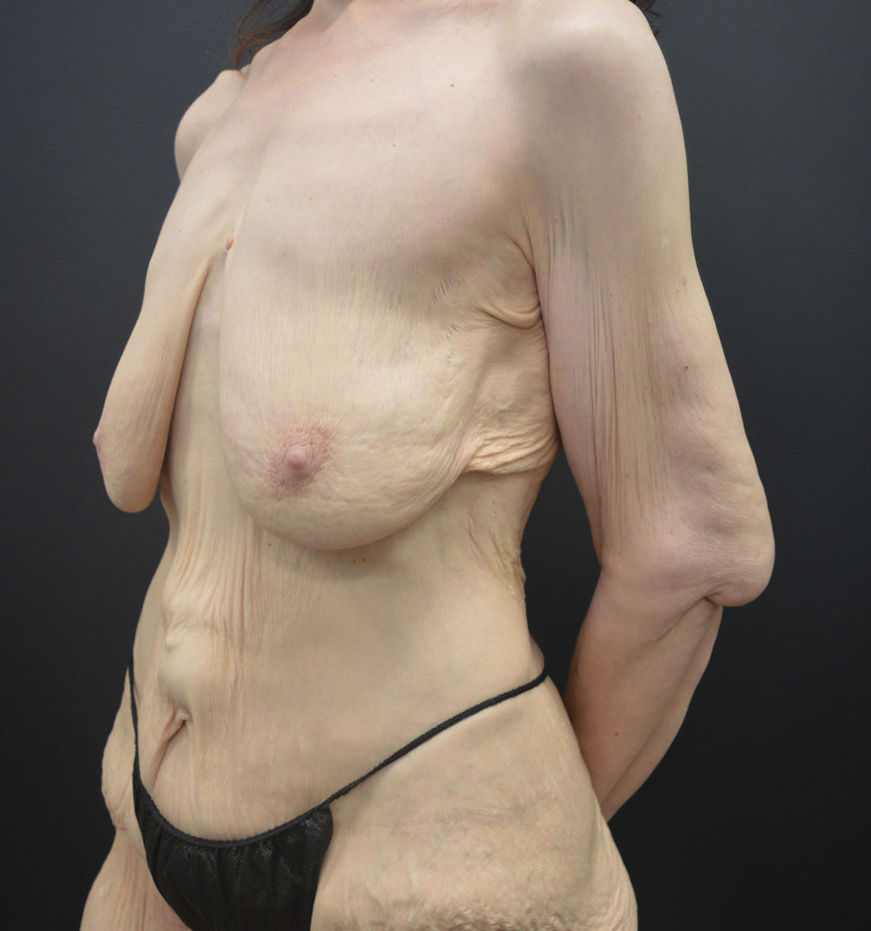
文章对躯干与四肢提出美学单元(AU)与粘连分区(ZA)的系统分类,作为身体塑形手术(BCS)切口规划的解剖基础。
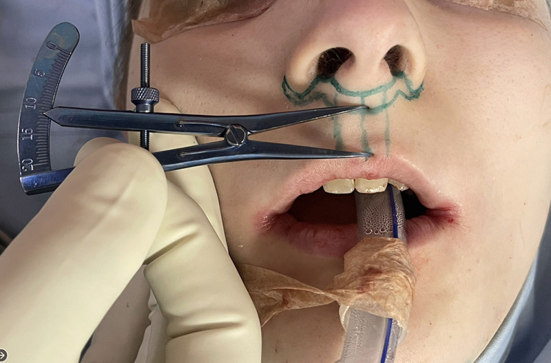
1997-2023年共988例出生分配为男性的性别焦虑者接受唇女性化与年轻化,累计1789例次手术,年龄22-75岁(平均39.5岁),79.8%小于50岁。