睑缘下切口中面部提升联合双环MACS悬吊
2026年10月1日25 min read
Plastic and Reconstructive Surgery Global Open
结论与实践要点
技术要点(可直接迁移): 本文的『经睑缘下切口骨膜上中面部提升 + 双环 MACS 悬吊』把中面部处理并入 MACS 体系,避免传统中面部独立冠状/睑缘长切口的额外瘢痕。本系列已覆盖 MACS 提升联合辅助术式(12957109)与深平面提升(13557784),本文补充『睑缘下小切口路径直达骨膜上』这一低瘢痕入路,适合以中面部轻中度下垂为主、不愿接受大范围剥离者。
与深平面的区分: 深平面/复合提升对重度中面部下垂与颊脂垫移位更强,但创伤与恢复更大;本文路径创伤更小、恢复更快,代价是对重度骨吸收/容量缺失者提升有限。临床应按下垂分级选择:轻中度选 MACS+睑缘下中面部,重度选深平面。
循证边界: 单中心回顾性、无对照、样本有限,结局以照片与满意度为主。结论为『技术可行性支持』,证据等级中等;对我团队的启发:在 MACS 方案中常规纳入睑缘下中面部悬吊作为轻中度下垂的标准模块,并以 pinch test 量化皮瓣冗余。
Background:Minimal access cranial suspension (MACS) lift is a well-established face lift technique commonly included in the facial plastic surgeon’s armamentarium. It has consistently demonstrated safety and efficacy. The midface may also be addressed using various adjunctive maneuvers in combination with the MACS lift. The aim of this study was to evaluate the use of supraperiosteal midface lift via a subciliary incision with a 2-looped MACS lift.
Methods:This retrospective case series included patients who underwent a MACS lift with 2 suspension purse-string loops, combined with upper and lower blepharoplasty and a supraperiosteal midface lift via a subciliary incision. Outcomes were evaluated by comparing the palpebromalar groove position before and after surgery. Patient satisfaction was measured at 6 weeks postoperatively using a 1–10 scale. Complications were documented during follow-up.
Results:The study enrolled 25 patients. The palpebromalar groove position improved from a mean of 54 mm preoperatively to 62 mm postoperatively (mean improvement = 8 mm). Mean patient satisfaction was 8.96 on a 1–10 scale.
Conclusions:Combining a supraperiosteal midface lift via a subciliary incision with a MACS lift proved safe and effective, resulting in a low complication rate and high patient satisfaction.
Question: Does a midface lift performed via subciliary incision in adjunct to a minimal access cranial suspension lift yield a safe and efficient outcome?
Findings: The palpebromalar groove ascended by an average of 8 mm. Mean patient satisfaction was 8.96 on a scale of 1–10.
Meaning: A midface lift performed via a subciliary incision in adjunct to a minimal access cranial suspension lift yielded a safe and efficient outcome with high patient satisfaction.
Face lift surgery remains a cornerstone of facial rejuvenation, delivering long-lasting results that meet patients’ expectations. Although noninvasive procedures have surged in popularity in recent years, face lift surgery remains in high demand, with research showing both increasing patient satisfaction and a rise in the number of procedures performed. In fact, more than 78,000 face lifts were completed in 2023, an 8% increase from 2022, underscoring the enduring appeal of this surgical approach. 1 Furthermore, face lifts often provide superior long-term outcomes compared with noninvasive alternatives, offering more noticeable and lasting improvements. 2
Among the various face lift techniques, the minimal access cranial suspension (MACS) lift has gained popularity due to its effective results, minimal invasiveness, and faster recovery times. Though it is performed via smaller incisions and uses less extensive dissection than the classic superficial musculoaponeurotic system (SMAS) lift, the MACS lift achieves comparable results with an excellent safety profile, shorter operative time, and swifter downtime. Surgeons rely on precise tissue repositioning and a deep understanding of facial anatomy to ensure natural outcomes while minimizing risks. 2 – 4
The midface lift, a specialized procedure targeting the central face, addresses volume loss and sagging around the cheeks and under-eye areas, aiming at restoring a more youthful appearance. When combined with a face lift, it enhances the overall rejuvenation by refining facial contours for a more balanced, harmonious result. 5 , 6
Traditionally, the MACS lift involves using 3 suspension loops to anchor the SMAS layer, providing targeted lifting with minimal scarring. 3 , 7 In this retrospective case series, we used a 2-loop MACS lift, replacing the third loop with a midface lift performed through a lower blepharoplasty (subciliary) incision. This approach offers a streamlined and effective solution with the potential for enhanced outcomes.
This study aimed to evaluate the outcomes of using a 2-loop MACS lift combined with a midface lift via a lower blepharoplasty incision. We assessed the effectiveness, safety, and complications of this technique, with the goal of optimizing midface rejuvenation while minimizing risks and improving patient satisfaction.
This study was a retrospective case series including male and female patients aged 40 years or older who presented to the Plastic Surgery Clinic at Cairo University Hospital from January 2019 to January 2024. The study gained ethical approval from the Faculty of Medicine, Cairo University, as part of an MD thesis. The inclusion criteria consisted of men and women older than 40 years who exhibited signs of facial aging, such as midface and jawline sagging, nasolabial fold creases, marionette lines, facial fat loss or displacement, and jowl formation. Additionally, these patients sought aesthetic procedures, specifically blepharoplasty and face lift surgery. Patients with bleeding disorders, those classified as American Society of Anesthesiologists IV or V, individuals with facial fractures or asymmetry, and those with psychiatric conditions were excluded. Additionally, patients with unrealistic expectations, active autoimmune diseases, ongoing chemotherapy or immunosuppression, and current smoking were also not considered suitable for inclusion in the study or procedure.
The midface lift was evaluated by measuring the distance from the patient’s oral commissure (point A) to a perpendicular point on the palpebral groove (point B) using pre- and postoperative photographs and measurements. The AB distance (oral commissure to palpebromalar groove) was selected for its anatomical relevance, as descent of the malar fat pad lengthens this vector and accentuates midface aging. This measurement has precedent in published midface lift studies and provides a reproducible, objective correlate of surgical lift efficacy. Our goal was to quantify the vertical repositioning of the midface complex after surgery, providing a reproducible and objective measure of surgical lift efficacy. 8 Patient satisfaction was self-administered using a 1–10 Likert-type scale during follow-up visits without physician interference.
Each patient completed a questionnaire covering personal data, smoking, alcohol use, previous facial surgery, and medical history. This was followed by a general physical examination. Careful examination included the patients’ facial skin quality, the severity of facial rhytids, areas of fat loss, gain, or displacement, platysmal banding, and a careful assessment of the upper and lower eyelids. Patients were thoroughly counseled regarding the procedures’ benefits, limitations, and potential complications, and informed consent was obtained.
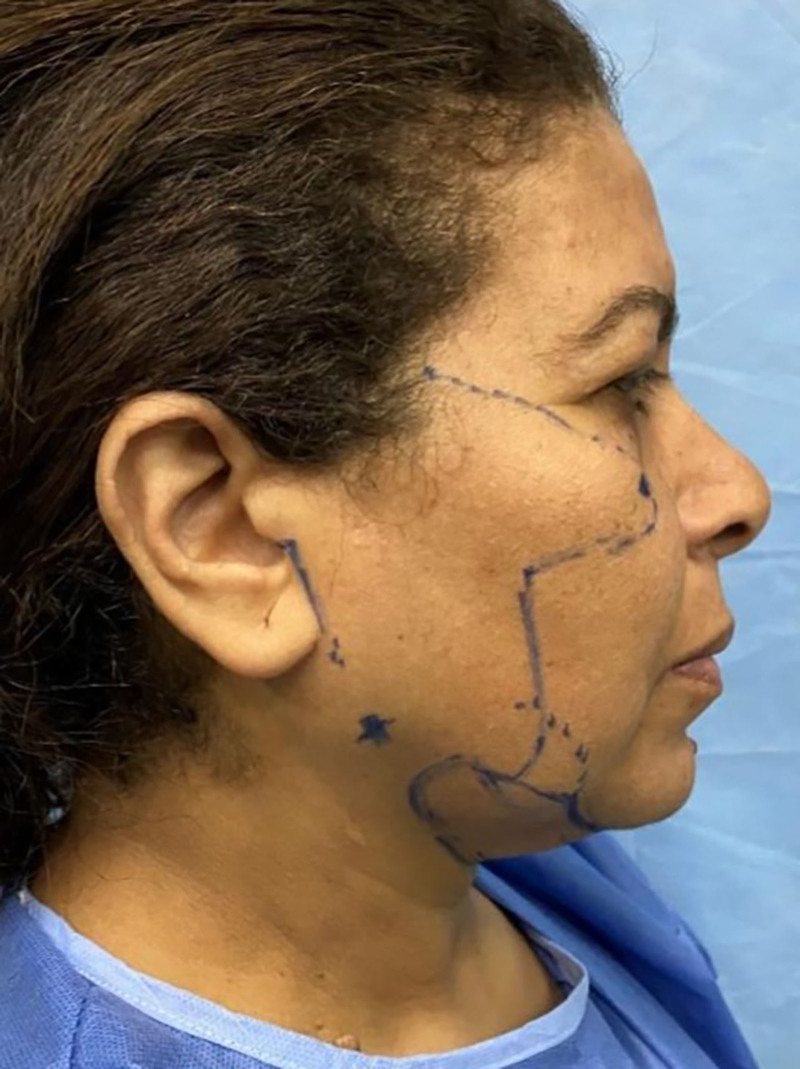
Preoperative markings were done with the patients in a sitting position. The double chin and the lower parts of the jowls were marked while the neck was flexed. The mandibular angle and the anterior border of the parotid were marked. Incision markings for the face lift began at the lower limit of the lobule, progressing upward in the preauricular crease up to a small hairless recess between the sideburn and the auricle. The incision line then followed the inferior edge of the sideburn and the hairline in a zigzag pattern, 2 mm within the sideburn. The postauricular incision extended to the middle of the helix, then perpendicular to it, reaching the scalp. The lower blepharoplasty incision line was drawn 2 mm below the eyelashes; medially, it stopped short of the inferior punctum, and laterally, the incision stopped slightly beyond the lateral canthus, directed along the crow’s feet. For the upper blepharoplasty, 3 points were marked: 1 above the midpupil, 8 mm from the eyelash line; the other 2 medial and lateral at 6 mm from the medial and lateral canthi. An ellipse was drawn as determined by the Pinch test (Fig. 1 ). Preoperative photographs were taken in the sitting position, in the anteroposterior and lateral views, and while gazing up and down.
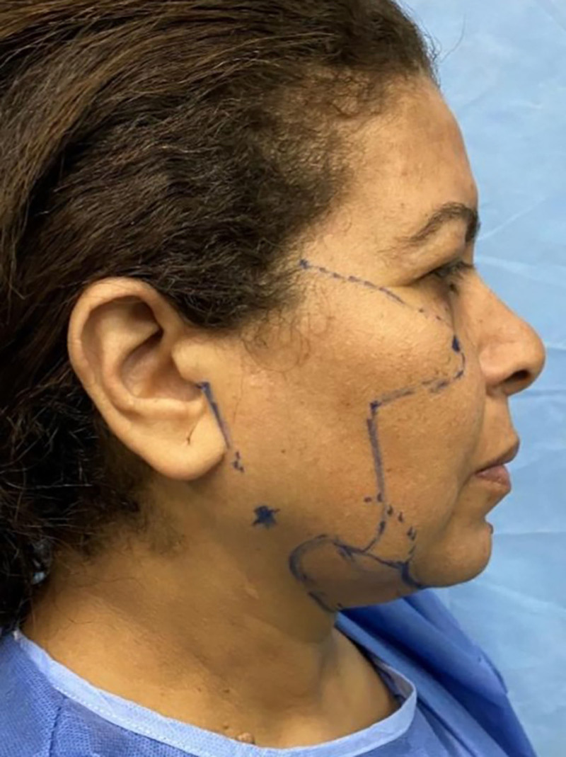
Fig. 1. Preoperative markings were performed with the patient seated, including the jawline, submental contour, and neck fold; incisions planned from the lower lobule through the preauricular crease to the temporal hairline, and from the postauricular region to the scalp and hairline.坐位术前标记(下颌线、颏下轮廓、颈),规划切口与悬吊路径。
Surgery was performed under local anesthesia with sedation or under general anesthesia. A small submental incision was used for neck liposuction. Tumescent infiltration, 30–50 mL, was followed by power-assisted liposuction with a 3-mm cannula in the preplatysmal plane.
Planned incision lines were infiltrated with a lidocaine–epinephrine solution. Tumescent fluid, 40 mL per side, was infiltrated into the subcutaneous plane in the dissection areas of the face.
Preauricular incision was followed by elevation of the supra-SMAS skin flap under direct vision. A no. 15 blade and Iris scissors were used for the initial dissection, maintaining a 10- to 15-mm flap thickness. Kaye face lift scissors were then used to elevate the flap. Meticulous hemostasis was achieved using bipolar diathermy (Fig. 2 ). The postauricular flap was then dissected inferiorly and anteriorly into the neck with diathermy until plasma fibers were visualized.
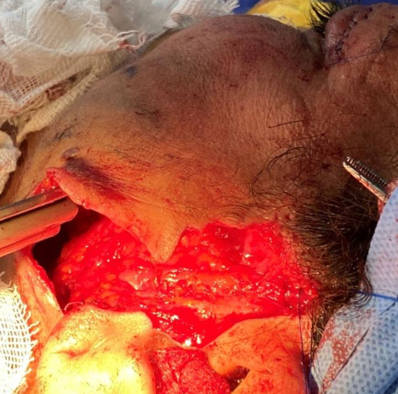
Fig. 2. The skin flap was elevated in the preauricular/SMAS plane under direct vision during face lift; tumescent infiltration was performed for hydrodissection; meticulous hemostasis was achieved with bipolar diathermy.面部提升中于耳前/SMAS 平面直视下提升皮瓣,肿胀麻醉下操作。At an anchor point marked 1 cm anterior to the helical rim, minimal dissection was done, exposing the temporal fascia above the zygomatic arch. Purse-string loop designs were marked. Polydioxanone 3-0 on a rounded needle was used.
The first bite is invertedly secured to the temporal fascia at the anchor point, then firm bites about 1 cm long and 0.3 mm deep were taken into the SMAS tissue, reaching a point just inferior to the angle of the mandible. The suturing direction then changed, creating a narrow 1-cm U-shaped loop, which continued back upward till the anchor point. The second purse-string suture originated at the same starting point. It was fashioned slightly wider and was directed toward the jowls. Both loop knots were tied under tension. A superficial inverted horizontal mattress suture using 4-0 Vicryl (polyglactin 910) was used to cover and bury the knots.
Skin flaps were placed on traction mainly cephalically. With the skin under no tension, the flap was incised and fixed with staples at 2 key points, 1 postauricular and 1 preauricular. A single key suture secured the earlobe to rest comfortably at the newly created junction. The excess skin was removed, followed by secure hemostasis and skin closure using Vicryl 4-0 inverted sutures and Monocryl (poliglecaprone 25) 4-0 intradermal sutures.
Before starting blepharoplasty, an eye shield was inserted for protection. Local anesthesia (lidocaine 1%, epinephrine 1:100,000) was injected into the subcutaneous plane, about 1 mL into each eyelid.
Excess upper lid skin was clamped using Allis forceps and then cut with sharp tenotomy scissors. A horizontal 5–8 mm strip of the orbicularis muscle was excised if redundant; otherwise, it was just split open. The orbital septum was cautiously opened to access the fat pads below. Excess fat was excised conservatively. Meticulous hemostasis was achieved, and the wound was then closed.
A subciliary incision was used for the lower blepharoplasty. A stab incision was used to access the submuscular plane at the lateral end, followed by dissection and completion of the skin–muscle incision using the tenotomy scissors. The attenuated orbital septum was exposed along with the underlying fat pads. Confluent fat from the central and nasal compartments was often redraped by advancing beyond the infraorbital rim, then holding in place beneath the orbicularis muscle using 5-0 Vicryl. Fat from the temporal fat pad was clamped, cauterized, and removed, followed by meticulous hemostasis.
The midface lift was approached from the same incision. The malar fat pad was dissected supraperiosteally starting caudally from the orbital rim. Dissection was kept lateral to the limbus to avoid the infraorbital neurovascular structures. The skin–muscle–malar pad complex was suspended using an inverted suture loop (Prolene 4-0) attached to the lateral orbital periosteum opposite the lateral canthus, then with 2 sutures to the temporal fascia. After tailoring the skin–muscle flap, overlapping skin and muscle were conservatively excised and closed with a few interrupted 5-0 Prolene sutures (Fig. 3 ). Steri-Strips and compression garments were then applied.
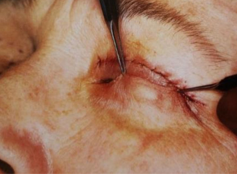
Fig. 3. A pinch test assessing skin laxity to determine the skin–orbicularis flap and the extent of midface lift needed from the subciliary incision.pinch test 评估皮肤松弛,决定皮瓣-眼轮匝肌瓣与中面部提升范围。
Cold compresses were used for the first 48 hours. Patients received antibiotic–steroid eye drops and a mild analgesic for 7 days. Follow-up started on postoperative day 3, then on day 7 when the sutures were removed; then continued weekly until the sixth week, when postoperative photographs were taken (Figs. 4 A, B).
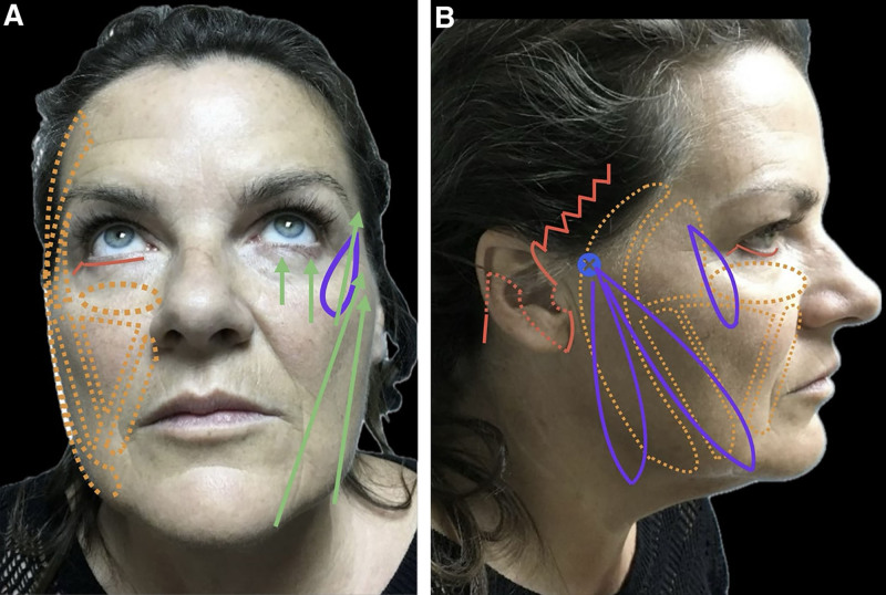
Fig. 4. Preoperative markings. A, Preoperative markings and planned incisions with the following color codes: orange dotted points indicate fat‑pad boundaries; red solid lines indicate incisions; red dotted points indicate posterior extensions; green arrows indicate elevation vectors and direction of pull; purple lines show suspension loops; and the blue X denotes the anchoring point. B, The schematic of the operative technique: same color conventions for incision lines, posterior extensions, fat‑pad boundaries, elevation vectors, suspension loops, and the blue X anchoring point.术前标记与计划切口(色码示意),含中面部提升范围。
The data were presented using both descriptive and inferential statistics. Quantitative variables, such as the AB distance measurements and patient satisfaction scores, were summarized with means and SDs to describe central tendency and dispersion, whereas medians and interquartile ranges (IQRs) were used to provide additional insight into data distribution, especially for skewed data. The normality of the data was assessed using the Shapiro–Wilk test, which confirmed that these variables were approximately normally distributed ( P > 0.05), justifying the use of parametric tests such as the paired t test for comparisons. The differences between pre- and postoperative measurements were analyzed with the paired t test, and the results were reported with P values and confidence intervals, indicating statistical significance. Patient satisfaction scores were also summarized with the median and IQR to reflect their distribution and high satisfaction levels.
This retrospective case series was conducted between January 2019 and June 2020, involving 25 patients who were candidates for various facial rejuvenation procedures such as face lifts, neck lifts, midface lifts, and blepharoplasty. The participants’ ages ranged from 40 to 68 years, with an average age of 54 (SD ± 7.5) years. Most of the sample were women, comprising 92% (23 women and 2 men), reflecting a female-to-male ratio of 23:2. All patients exhibited signs of facial aging, including sagging skin in the midface and jawline, nasolabial fold creases, marionette lines, facial fat loss or displacement, and jowls. Most of the patients reported experiencing at least 2 of these issues, with common concerns being sagging eyelids and excess skin (15 cases), eyelid puffiness with or without additional skin (19 cases), periorbital wrinkles and dark hollows (20 cases), and a drooping midface combined with a sad appearance and lid puffiness (23 cases). These complaints highlighted the diverse manifestations of facial aging among the participants.
All patients sought correction primarily for aging-related facial and cervical changes. The follow-up period varied from 2 to 8 months, with a median duration of 4 months. Throughout the follow-up visits, photographs were taken to document progress, and patient satisfaction levels were assessed to evaluate the outcomes of the interventions and their impact on the patients’ perceptions of facial rejuvenation. The duration of follow-up ranged from 10 to 15 months (median, 12 mo).
Palpebromalar groove position was measured as the linear distance from point A (the oral commissure) to point B (the perpendicular projection on the palpebromalar groove). All pre- and postoperative AB measurements underwent Shapiro–Wilk normality testing—a method favored in small sample analyses (<50)—and yielded nonsignificant results ( P > 0.05), indicating no statistical evidence of deviation from a normal distribution. No datapoints exceeded ±2 SDs from the mean, and thus no outliers were present. These results justified the use of a paired t test to evaluate differences. For full transparency, medians and IQRs were also reported: preoperative median AB distance was 54 mm (IQR, 52–56 mm), increasing to 62 mm (IQR, 60–64 mm) postoperatively. The mean absolute improvement was 8 mm (95% confidence interval, 6.4–9.6 mm), confirming a precise and statistically significant change in position.
Patient-reported satisfaction was self-assessed at 6 weeks and 12 months using a standardized 1–10 scale administered in writing during follow-up visits. The median satisfaction score was 9, suggesting that most patients rated their satisfaction quite favorably. The IQR spanned from 8 to 9, further reflecting that most scores clustered around the high end of the scale.
This study observed no major complications such as facial or orbital hematomas, infections, skin necrosis, or persistent ectropion among the 25 patients. However, 2 cases experienced delayed resolution of subcutaneous petechial hemorrhages. Additionally, 3 (12%) patients developed postoperative ecchymosis, which was effectively managed with local ointments and resolved within 2 weeks. Throughout the study, which involved 25 patients, the postoperative appearance of edema and ecchymosis improved notably, likely due to increased surgical experience and refined techniques. More careful tissue handling and shorter intraoperative durations contributed to less postoperative swelling and bruising. A few patients, specifically 3 (12%), also experienced mild conjunctivitis, which was successfully treated with topical antibiotic drops, indicating that the complications encountered were minor and easily managed (Figs. 5 , 6 ).
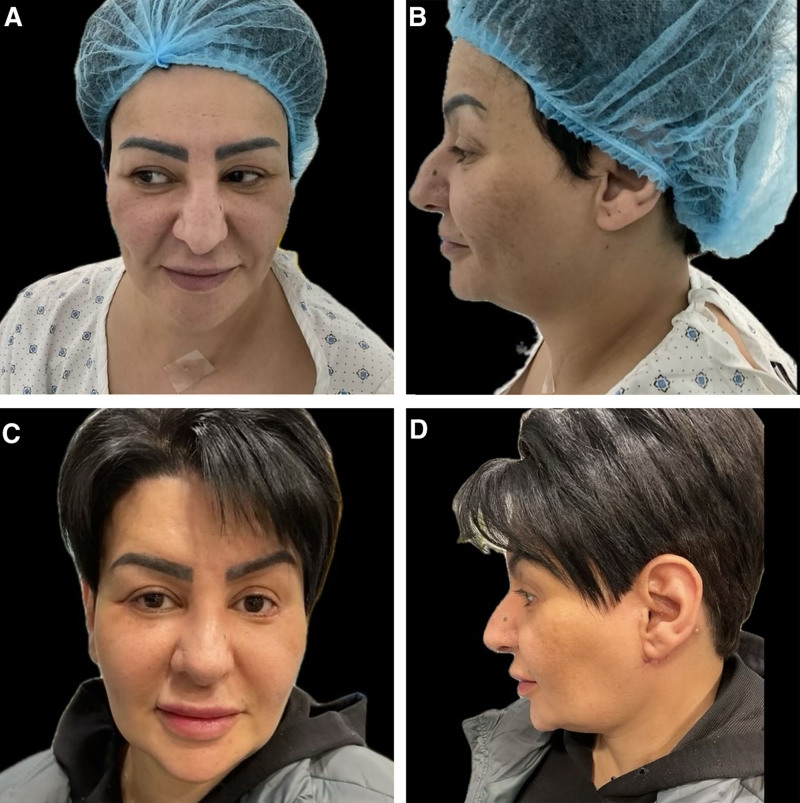
Fig. 5. Preoperative markings. Preoperative AP and lateral views (A, B) show baseline facial sagging and deep nasolabial folds; postoperative AP and lateral views (C, D) reveal improved contour, reduced jowls, and a lifted midface with the shallowing of the nasolabial fold. AP, anteroposterior view.术前 AP/侧位示基线面部下垂与深鼻唇沟。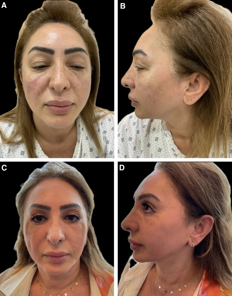
Fig. 6. Preoperative AP and lateral views (A, B) show midface ptosis; postoperative AP and lateral views (C, D) reveal enhanced cheek contour, reduced nasolabial depth, improved jawline, and overall rejuvenation and symmetry. AP, anteroposterior view.中面部下垂术前(A/B)与术后(C/D)AP/侧位对照,示中面部上提与轮廓改善。
Face lift has markedly evolved during the past several decades. After its start with skin-only excision, face lift has developed to address the SMAS by excision or lifting, which became the benchmark procedure during the 1970s and 1980s. During the 1990s, the deep-plane/composite face lift concept emerged, using a more extensive procedure, trying to achieve fuller and more long-lasting results. 9
The MACS lift was introduced in the early 2000s. It was presented as a safe and efficient alternative, and since then has been included as part of the aesthetic face surgeon’s armamentarium for surgical face lift. 4 , 10 , 11 , 12
MACS lift offers good aesthetic results with a high rate of patient satisfaction. 3 , 9 Its efficacy and longevity are comparable to more extensive techniques, with remarkable safety, shorter operative time, faster recovery, and a relatively steeper learning curve. 3 , 13
Management of the midface is an inseparable part of managing the aging face. The addition of a third loop to the original 2-loop MACS lift became standard for this procedure, as described by Verpaele et al, 7 Tonnard and Verpaele, 14 and Tonnard et al. 15 The use of facial fillers or autologous fat transfer is quite common in the malar region and helps to lift the midface and improve the nasolabial folds. 16 , 17 Fat grafting was included with the MACS lift, replacing the third loop. 18 Chopan et al 19 suggested using a broader second loop and omitting the third loop. Since its introduction in the 1990s, midface through a subciliary approach has become a well-recognized technique that offers a safe and effective lifting of the midface and treatment of deep nasolabial folds. 6
Our results demonstrate significant improvements in facial aging signs following midface and facial rejuvenation procedures, with a highly significant reduction in the palpebromalar groove distance ( P < 0.001). The average postoperative increase of 8 mm in the AB distance indicates effective lifting and contour restoration. The 8-mm elevation of the palpebromalar groove represents a clinically meaningful improvement, as a 5- to 10-mm correction is widely regarded as significant for restoring a youthful midface contour by counteracting age-related descent of the malar fat pad. Comparable midface elevation has been reported with endoscopic and trans conjunctival midface lifts (5–12 mm), aligning with the 8 mm improvement achieved in our series. 12 , 15 Unlike the traditional, 3-loop MACS lift, our modification replaces the third loop with a subciliary midface suspension, providing vertical repositioning of the malar fat pad while maintaining the benefits of a simplified MACS approach. Patient satisfaction scores were predominantly high, with a mean of 8.68 and most ratings between 8 and 10, reflecting positive perceptions and acceptance of the surgical outcomes.
Clinically, the procedures were safe, with no major complications such as hematomas or necrosis. Minor issues such as ecchymosis, edema, and mild conjunctivitis resolved quickly, and their occurrence was minimized through surgical refinement. Overall, the data suggest that the techniques used were effective, safe, and well received, supporting their use for facial rejuvenation with high patient satisfaction and low complication rates. In this study, a midface lift was performed using a transcutaneous subciliary incision in all cases to achieve midface rejuvenation, replacing the third purse-string suture used in the MACS lift. We adopted a superficial dissection technique for the midface lift, undermining only the skin–muscle–malar pad complex, which is flexible and safe. 20
This approach avoids dissection near facial nerve branches, thus eliminating the risk of injury; it resembles a subperiosteal midface lift. However, with superficial plane dissection, there is no need to manipulate the infraorbital nerve, reducing the risk of numbness from injury or traction and eliminating the necessity of a transverse caudal periosteal release incision. 21 , 22 A conservative strategy was applied for orbital fat management, involving minimal resection when necessary, with redraping of the central and nasal fat toward the tear trough region. 23 , 24 All patients underwent canthopexy to support the redundant orbicularis oculi muscle sling, and the skin–orbicularis–malar flap was suspended to the lateral orbital wall and temporal fascia. The orbicularis anchoring served as the handle of the cheek lift, aiding in cheek redraping and providing a sling-like support for the lower lid. The lift was performed along a vertical vector to counteract aging effects and the descent of the malar fat pad and skin. The subciliary approach for midface lifting has been well documented and validated in prior literature, notably supported by influential studies from Burnett et al 25 and Sullivan and Dailey 26 that demonstrate the validity and effectiveness of this technique. The excess skin in the upper eyelid was serially clamped and crushed with Allis forceps into a skin fold, then excised with scissors in a near bloodless manner. This maneuver helped approximate the skin edges, ensured hemostasis, and provided a safety check for the appropriate extent of skin removal. The improvement in the position of the palpebromalar groove was assessed to evaluate the effectiveness of the midface lift. In addition, the correction of the excess depth of the groove and tear trough, owing to the redraping of the sub–orbicularis oculi fat and the cranial bound anchoring of the skin–muscle–malar pad, was noted by the majority of the patients postoperatively. The overall result was a younger look with a smoother orbital midface transmission.
The limited follow-up duration constrains the ability to assess long-term durability. Larger, prospective studies with extended follow-up are warranted. One limitation of this study is the absence of simultaneous fat grafting, which may reduce the ability to fully address facial volume loss and skin quality rejuvenation. Fat transfer was intentionally deferred to isolate and evaluate the core surgical technique without adjunct procedures; however, it can be easily done postoperatively. Additionally, the MACS lift used in our approach does not involve SMAS undermining, a feature in other face lift techniques, but was intentionally excluded to maintain focus on a simplified, reproducible method. Although every effort was made to conceal the anterior hairline incision, some patients may still need to address excess skin and achieve balanced redraping adequately. Furthermore, the absence of central platysmaplasty, which was not part of our study scope, can contribute to less neck contouring than other techniques. This retrospective case series without a control group cannot provide direct comparisons to other face lift techniques. Findings should be interpreted as preliminary. Further research should include patients from different centers and may include comparative studies between different variations of the MACS lift and other face lift techniques to fine-tune the safest, most effective, and most satisfying techniques for patients from each age group and with all different rejuvenation demands and requirements. A limitation of the study is the absence of validated patient-reported outcome measures. Although we used a simple self-administered 1–10 satisfaction scale, which is widely applied in surgical outcomes research for its practicality, validated patient-reported outcome measures such as FACE-Q would provide a more rigorous assessment. We plan to incorporate such tools in future prospective studies. Future prospective trials with standardized measures, comparative cohorts, and extended observation periods are essential to validate these findings.
Combining a supraperiosteal midface lift through a lower blepharoplasty incision with a MACS lift helps respect vertical vectors associated with facial aging and reverses them, thus yielding a youthful yet natural appearance. This combined approach seems promising, with favorable safety and satisfaction outcomes in our series. However, further prospective studies with larger cohorts and control groups are required to validate and generalize these findings.
The authors have no financial interest to declare in relation to the content of this article.
Informed consent was obtained from all patients enrolled in the study.
This study was performed in accordance with the principles of the Declaration of Helsinki. Approval was granted by the ethics committee of the Cairo University Faculty of Medicine, approval number CMDRF132701.
声明:中文精读 · 仅供学术参考。内容来自公开文献检索,不代表本人观点,不构成诊疗建议。 医疗美容需在正规医疗机构由执业医师实施。
基于相同主题推荐 · 继续深挖
不同人群面部年轻化:专家共识与 AART 方法学临床应用经验/PMC13558784_fig1.webp)
这是一项基于改良 Delphi 法的专家共识(马来西亚6名具8–10年生物刺激剂经验的医师),并附4例 PLLA-SCA(Sculptra)临床应用个案,属专家意见与病例系列,并非
研究设计:回顾性病例系列,采用 MACS 提升(2 个悬吊 purse-string 环)联合上/下睑成形,并加经睑缘下切口(subciliary)骨膜上中面部提升。
研究背景:除皱术从单层皮瓣演进到针对 SMAS(表浅肌筋膜系统)等深层结构,效果更自然持久;但经耳前大切口仍有面神经损伤与皮瓣坏死风险。本研究评估两种内镜入路(含深部 SMAS 提