微针联合脂肪源性干细胞外泌体改善面部毛孔:AI 多模态客观量化研究
2026年10月2日28 min read
Plastic and Reconstructive Surgery Global Open
Plastic and Reconstructive Surgery Global Open
结论与实践要点
本研究的证据等级为 单中心回顾性病例系列 (n=30,20–60 岁),非随机、无空白或对照臂,亦未设单用微针或单用外泌体的对照分组,因果推断受限。
其价值在于 多模态客观量化 :① QuantifiCare 3D 毛孔评分(6 个区域,−10~+10 量纲);② Gwyddion 三维表面粗糙度 Rz(µm);③ AI 轮廓分割的毛孔计数与平均面积(像素²)。三套独立方法方向一致,为「微针+外泌体」改善毛孔与肤质提供了可重复的影像证据。
落地建议:可作为「客观评估+患者教育」的参考范式;若用于临床决策,建议结合前瞻随机对照与组织学证据,并关注外泌体产品的合规与质控要求。
Background:Enlarged facial pores are a common cosmetic concern, exacerbated by age-related collagen degradation and sebaceous hyperactivity. Despite clinical demand, objective quantification methods for pore improvement remain limited. This study used 3 independent analytical platforms—QuantifiCare 3-dimensional (3D) imaging, Gwyddion-based surface roughness analysis, and artificial intelligence (AI)–assisted image segmentation—to evaluate the efficacy of microneedling combined with adipose-derived stem cell exosome (ASCE+) therapy.
Methods:Thirty subjects underwent 3 sessions of microneedling with topical ASCE+ application. Standardized high-resolution facial images were obtained via the LifeViz Mini system under controlled lighting and positioning. QuantifiCare software generated regional pore scores across 6 facial zones. Gwyddion software quantified 3D surface roughness ( R z , µm) in 4 subunits using paired anatomical landmarks. AI-based contour segmentation assessed pore count and mean pore area (pixels 2 ) in forehead, nose, malar, and buccal zones. Paired t tests were used for statistical comparisons.
Results:QuantifiCare pore scores significantly increased across all regions ( P < 0.001), reflecting improved skin texture. Gwyddion R z values decreased by 43%–61%, indicating smoother surface topography. AI analysis showed marked pore size reduction (forehead, –58.2%; nose, –56.1%; malar region, –36.4%; P < 0.001), and modest reduction in the buccal zone (–10.7%, P = 0.0132). These multimodal findings consistently support enhanced dermal remodeling and reduced pilosebaceous prominence.
Conclusions:This is the first study to integrate 3D imaging, roughness mapping, and AI segmentation for pore analysis after microneedling and exosome therapy. The combined modality demonstrates significant improvement in pore architecture and skin texture, highlighting its utility in aesthetic dermatology.
Question: Can microneedling combined with adipose-derived stem cell exosome (ASCE+) therapy improve facial pores and skin roughness using objective, artificial intelligence–assisted imaging?
Findings: In this retrospective study of 30 patients, 3 sessions of microneedling with ASCE+ therapy resulted in significant improvements across multiple facial regions. QuantifiCare 3-dimensional analysis showed consistent increases in pore scores, Gwyddion mapping demonstrated up to 75.6% reduction in surface roughness, and artificial intelligence–based pore morphometry confirmed reductions of up to 58.2%. Patient satisfaction was high, with a mean Global Aesthetic Improvement Scale score of 4.83 out of 5.
Meaning: This combination offers a noninvasive, regenerative option for refining pores and enhancing skin texture.
Enlarged facial pores remain a common cosmetic concern that often worsens with age due to dermal matrix degradation, increased sebaceous activity, and structural skin laxity. Despite their high prevalence, the pathophysiology of visible facial pores is multifactorial and not yet fully understood, encompassing sebaceous gland hypertrophy, perifollicular elastosis, and structural changes in the dermoepidermal junction. 1
Microneedling has long been used for skin rejuvenation, showing efficacy in improving texture and fine lines by inducing dermal remodeling through controlled microinjury. 2 More recently, exosome-based biologics—particularly those derived from mesenchymal stem cells—have gained traction as potent adjuncts to skin rejuvenation therapies due to their regenerative, anti-inflammatory, and collagen-stimulating properties. 3 Preclinical and clinical evidence has suggested that the combination of microneedling with stem cell–derived exosomes can synergistically enhance neocollagenesis, dermal density, and surface texture improvement. 4
However, although studies have focused on parameters such as wrinkle depth or overall skin quality, few have systematically quantified pore-specific improvements using objective, reproducible imaging modalities. Traditional visual scales are limited by subjectivity, interobserver variability, and lack of anatomical precision. Furthermore, prior studies often lack robust 3-dimensional (3D) analysis of facial subunits or consistent facial landmark alignment.
To address these gaps, the present retrospective study used 3 advanced imaging platforms—(1) QuantifiCare 3D stereophotogrammetry, 5 , 6 (2) Gwyddion 2.62–based surface roughness mapping, 7 , 8 and (3) artificial intelligence (AI)–based analysis for pore number and pixel area—to assess changes in pore visibility and skin roughness across 6 anatomically distinct facial subunits. 9 , 10 The study aimed to objectively quantify treatment outcomes following 3 sessions of microneedling combined with topical adipose-derived stem cell exosome (ASCE+) application during a 12-week period. This research offered not only the first AI-assisted, multimodal analysis of pore reduction across the forehead, nose, malar, and buccal regions, but also provided histologically and functionally relevant evidence supporting the integration of exosome-based therapies in noninvasive pore management.
This single-center, retrospective clinical study was conducted at Kim Jongseo Plastic Surgery Clinic, Seoul, South Korea, in accordance with the Declaration of Helsinki. All participants provided written informed consent that included permission for facial photography, 3D imaging, roughness analysis, and anonymized AI-based image processing for research purposes. The consent form was reviewed and approved by the institutional review board (no. P01-202506-01-040). The study evaluated quantitative changes in facial pore size and skin surface texture following a 3-session microneedling protocol combined with topical application of a human adipose-derived stem cell exosome formulation.
Thirty patients (aged 20–60 y, both sexes) who underwent 3 consecutive microneedling and ASCE+ treatments between July 2023 and May 2025 were retrospectively enrolled. Inclusion criteria were (1) no aesthetic treatments, including injectables or energy-based devices, within 6 months prior; (2) availability of standardized 3D pre- and posttreatment imaging; and (3) no history of keloid formation or active dermatologic disease. Exclusion criteria included incomplete imaging data or posttreatment complications.
All subjects underwent a standardized skin preparation protocol before each treatment session. Cleansing was performed using a hydro-papaya enzyme cleanser (Reteenage, Seoul, South Korea), 11 followed by 30 minutes of ambient air drying to stabilize the skin surface before imaging and intervention. Topical anesthetic cream (9% lidocaine) was applied for 30 minutes under occlusion using plastic film to ensure adequate dermal penetration. After anesthesia removal, the face was irrigated using high-pressure sterile water from a Clear Jet device (Eunsung Global, Korea) to mechanically debride follicular openings and remove sebum plugs. 12 The entire face was then wiped with an alcohol sponge for aseptic preparation.
Microneedling was performed using a 2.0-mm depth, 540-needle dermaroller (manual, sterile, single-use) across 5 facial zones: forehead, malar region, buccal region, jawline, and nose. Each zone received 10 linear back-and-forth passes in horizontal and vertical directions until pinpoint bleeding and mild erythema were observed, indicating effective stratum corneum penetration. Following microneedling, manual comedone extraction was performed using a sterile extractor to remove impacted sebum and follicular debris.
Immediately after microneedling and pore cleansing, 2 full vials of ASCE+ SRV-S (20 mg of freeze-dried powder containing 970,000 ppm exosome and 5 mL of solution; ExoCoBio Inc., Korea), a good manufacturing practice–certified exosome formulation derived from human adipose stem cell culture media, were applied topically. 13 High-velocity dermal jet delivery was performed using the Clear Jet system to enhance transdermal penetration, with half of the mixed vial applied via jet spray and the remainder reserved for at-home application. To enhance skin barrier recovery, an ASCE+ Soothing Gel sheet mask was applied for 10 minutes posttreatment.
Patients were instructed to avoid facial washing or contact for the remainder of the treatment day. The remaining half of each ASCE+ vial was provided for patient-administered topical application in divided amounts over the subsequent 48 hours. On posttreatment day 1, patients were allowed to wash with water only. Patients were advised to avoid cosmetics and sunscreen for at least 5 days, as occlusive products may interfere with pore contraction and optimal treatment response. 14 , 15
All patients received 3 identical treatment sessions at 4-week intervals (week 0, week 4, and week 8). Final imaging and clinical assessments were conducted at week 12, 4 weeks after the third session.
Three-dimensional facial images were captured at baseline and week 12 under consistent lighting. The 3D surface roughness was quantified using online software, and pore characteristics were evaluated via AI.
Images were acquired using the LifeViz Mini system (QuantifiCare, France) under standardized and controlled imaging conditions. All photographs were taken in a dedicated photograph room with a black background and blackout curtains to eliminate ambient light. 16 , 17 Patients were instructed to stand with their shoulders aligned against predefined corner markers to ensure consistent positioning. Lighting was standardized using both ambient studio lights and the integrated QuantifiCare flash source, maintaining the same distance and using 2 red lights. Images were obtained from 4 fixed positions: (1) front view aligned with the glabellar midpoint, (2) left and (3) right oblique views captured using lateral wall markings, and (4) submental upward views from the patient’s beltline level to assess inferior facial planes. These methods enabled highly reproducible image acquisition. 18 , 19
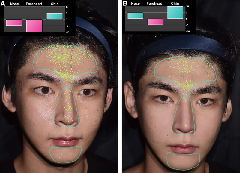
Quantitative analysis of pore severity was performed using QuantifiCare’s built-in Skincare software module, which assigns automated regional pore scores based on a proprietary algorithm. 20 , 21 Six predefined facial subunits were evaluated: (1) central forehead, (2) nasal dorsum (front view), (3) left lateral eye area, (4) left malar region and cheek, (5) right lateral eye area, and (6) right malar region and cheek zones (45-degree oblique views). The system generates unitless digital scores ranging from –10 (severe pore prominence) to +10 (optimal smoothness with minimal visible pores). Negative scores represent a high density and size of visible pores, whereas positive scores reflect refined skin texture. Both pre- and posttreatment values were recorded for each subunit and used for longitudinal comparison (Figs. 1 , 2 ).
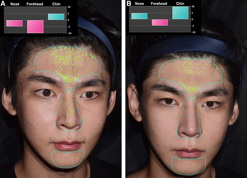
Fig. 1. Three-dimensional QuantifiCare skincare program–assisted pore mapping and regional score changes in the forehead, nose, and chin. A, Pretreatment image demonstrating high pore density in the forehead and nose regions, with a negative regional pore score on the forehead. B, Posttreatment image reveals a marked reduction in forehead pore count and average size, accompanied by an improved pore score.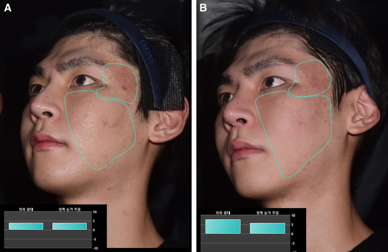
Fig. 2. Quantitative method to analyze the reduction in periorbital and malar pore density using a 3D program. Pretreatment image (A) revealing a high density of facial pores and visible fine wrinkles in the periorbital and malar subunits, indicated by green 3D program–detected pore markers. Posttreatment image (B) demonstrating a marked reduction in pore count and periorbital wrinkle depth, supported by QuantifiCare digital scores.
Three-dimensional surface roughness was quantified using Gwyddion software 22 , 23 based on high-resolution images obtained with the LifeViz Mini system. Roughness values ( R z , µm) were extracted after postacquisition alignment of pre- and posttreatment images using anatomical landmarks to ensure spatial correspondence. This standardized approach allowed for sensitive detection of topographical skin changes, enabling robust quantification of posttreatment reductions in surface irregularities. All images were precisely matched and maximally magnified to clearly visualize surface pores and microcontours. Region-specific protocols were used for consistent and reproducible measurements. Forehead region: 2 separate measurements were taken—1 horizontally along the glabellar crease (mid-brow line), and the other vertically along the midline of the forehead from hairline to supraorbital ridge. Nasal region: 3 longitudinal segments were measured from nasion to tip, capturing the upper, mid, and lower dorsum to comprehensively assess pilosebaceous irregularities. Malar region (cheekbone): 2 transverse axes extending from the lateral ala base (alar-facial groove) to the lateral canthus and from the mouth corner to the lateral canthus were selected to encompass pore-dense skin over the zygomatic prominence. Images were magnified to emphasize pore detail within the region of interest. Buccal region (cheek): 2 oblique measurement lines were defined—one following the marionette line toward the lateral canthus and the other oriented toward the midpoint between the lateral canthus and the hairline (Figs. 3 , 4 ).
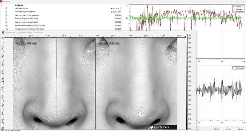
Fig. 3. Ultrasound-based analysis of fibroseptal network structure before and after treatment, demonstrating changes in septal orientation and density.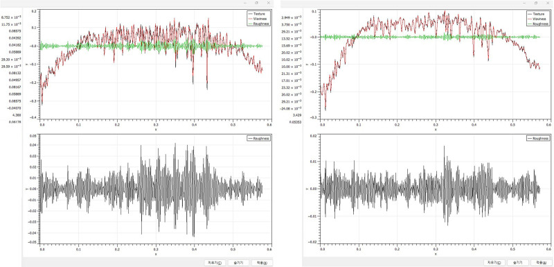
Fig. 4. Quantitative program output illustrating structural remodeling of the fibroseptal network following treatment, including changes in septal length and alignment.
To quantitatively evaluate changes in skin surface microstructure, particularly pore characteristics, high-resolution frontal images of the forehead region were analyzed before and after treatment using an AI-assisted image analysis pipeline. 24 , 25 To objectively quantify treatment-related improvements in skin texture, mean pore size (in pixels 2 ) was analyzed across 4 major facial subunits—forehead, nose, malar region (cheekbone), and buccal region (cheek)—in 30 patients using an AI-assisted image analysis algorithm. The results demonstrated consistent and statistically relevant reductions in pore area across all regions posttreatment. Pore regions were segmented using an AI (ChatGPT4o) algorithm that detected round depressions. Mean pore area (pixels 2 ) was calculated across 4 zones: forehead, nose, malar region, and cheek.
The analysis incorporated adaptive thresholding and contour-based segmentation to reliably detect and measure skin pores, which were defined as round to oval hypopigmented depressions on the skin surface. 26 , 27 For example, pretreatment analysis of the subject frontal view identified 1558 pores with an average pore area of 6.60 ± 5.20 pixels 2 . Posttreatment analysis demonstrated 1587 pores, with an average pore area reduced to 6.37 ± 4.86 pixels 2 , representing a 3.49% reduction in mean pore size (Fig. 5 ). This pattern implies an overall contraction of individual pores and a homogenization of surface topography, which may reflect histological improvements in the pilosebaceous unit and surrounding dermal matrix. In the same way, the number of pores and the average pixel area of pores in the nose, malar region, and cheek were analyzed using AI.
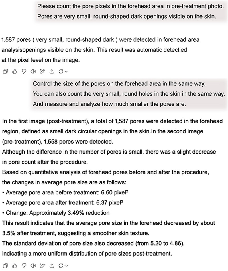
Fig. 5. AI-assisted pixel-level analysis detected 1558 pores in the pretreatment image and 1587 pores in the posttreatment image. The mean pore area decreased from 6.60 to 6.37 pixels2 (approximately 3.49% reduction), suggesting improved skin smoothness and a more homogeneous pore distribution after treatment.
At the week 12 visit, all patients completed the Global Aesthetic Improvement Scale (GAIS) to assess subjective perception of skin quality improvement.
All numeric data are presented as mean ± SD. Paired t tests were used to assess statistical differences between pre- and posttreatment measurements. A P value less than 0.05 was considered statistically significant. Statistical analysis was performed using SPSS version 27.0 (IBM Corp., Armonk, NY). 28 , 29
Quantitative evaluation of regional skin texture was performed using the QuantifiCare skin analysis system in 30 subjects. Six anatomically distinct facial zones were analyzed: central forehead, nasal dorsum, right infraorbital area, right malar region, left infraorbital area, and left malar region.
Before treatment, negative pore scores were observed in the central forehead (−7.11 ± 1.61) and nasal dorsum (−5.76 ± 1.92), indicating severe skin irregularity and dilated pores. Posttreatment, these values significantly improved to 0.42 ± 1.50 ( t = 21.92, P < 0.0001) and 2.62 ± 1.23 ( t = 19.47, P < 0.0001), respectively. Similarly, pore scores increased in the right lateral eye area (from 1.98 ± 1.01 to 5.16 ± 1.35) ( t = 10.90, P < 0.0001), right malar region (4.02 ± 1.20 to 7.26 ± 1.43) ( t = 8.51, P < 0.0001), left lateral eye area (2.36 ± 1.07 to 5.22 ± 1.36) ( t = 10.44, P < 0.0001), and left malar region (3.89 ± 1.21 to 6.09 ± 1.76) ( t = 5.56, P < 0.0001) following the intervention. QuantifiCare pore score analysis demonstrated a statistically significant improvement across all 6 facial zones following the intervention. Paired t tests revealed a substantial increase in scores. These findings collectively support the treatment’s efficacy in promoting dermal remodeling and smoothing the skin surface (Figs. 6 – 10 ) (Table 1 ).
Table 1. QuantifiCare Pore Score Improvement Across 6 Facial Subunits Before and After Treatment
| Region | Pretreatment Score (Mean ± SD) | Posttreatment Score (Mean ± SD) | Percentage Increase | t | P |
|---|---|---|---|---|---|
| Forehead | −7.11 ± 1.61 | 0.42 ± 1.50 | ↑ 105.9 | 21.92 | <0.0001 |
| Nose | −5.76 ± 1.92 | 2.62 ± 1.23 | ↑ 145.5 | 19.47 | <0.0001 |
| Right lateral eye | 1.98 ± 1.01 | 5.16 ± 1.35 | ↑ 160.6 | 10.9 | <0.0001 |
| Right malar region and cheek | 4.02 ± 1.20 | 7.26 ± 1.43 | ↑ 80.6 | 8.51 | <0.0001 |
| Left lateral eye | 2.36 ± 1.07 | 5.22 ± 1.36 | ↑ 121.2 | 10.44 | <0.0001 |
| Left malar region and cheek | 3.89 ± 1.21 | 6.09 ± 1.76 | ↑ 56.6 | 5.56 | <0.0001 |
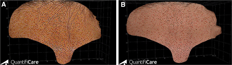
Fig. 6. Three-dimensional pore distribution mapping of the forehead before and after treatment using the 3D QuantifiCare skincare program. A, The pretreatment 3D analysis reveals a dense and irregular distribution of skin pores across the forehead. B, The posttreatment mapping shows a visibly reduced pore count and more homogenous spatial distribution, supporting the observed 3.49% reduction in average pore area and decreased SD (from 5.20 to 4.86), indicating a smoother and more even skin texture.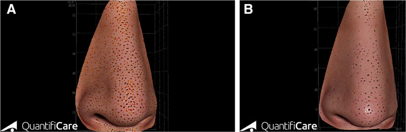
Fig. 7. Three-dimensional pore distribution mapping of the nose before and after treatment using the 3D QuantifiCare skincare program. A, The pretreatment image shows a high density of enlarged pores scattered across the nasal surface. B, The posttreatment image demonstrates a marked reduction in both pore count and size, supporting the efficacy of exosome-based microneedling therapy in refining sebaceous skin areas such as the nose.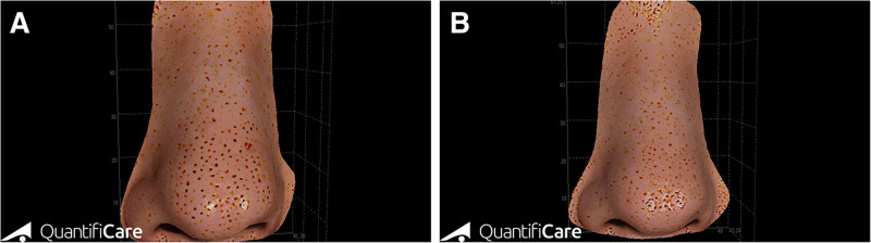
Fig. 8. Pore reduction on the nasal skin surface (39-y-old woman). High-resolution 3D pore mapping of the nose demonstrates a visible reduction in both pore number and distribution density after treatment. Quantitative visual analysis suggests improved skin surface smoothness and decreased pore prominence, particularly along the alar and tip regions. A, Before treatment. B, After treatment.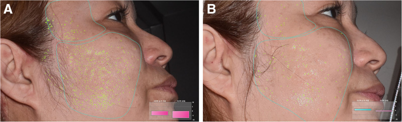
Fig. 9. AI-based analysis of pore reduction on the right cheek (55-y-old woman). Pretreatment imaging shows a high density of pores, particularly in the malar and suborbital zones. Posttreatment analysis demonstrates a marked reduction in pore count and density, indicating significant improvement in skin texture and tightening. A, Before treatment. B, After treatment.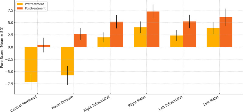
Fig. 10. QuantifiCare 3D pore score analysis before and after treatment across 6 facial regions. Quantitative analysis revealed significant posttreatment improvement in pore scores across all evaluated facial regions, with the most notable changes observed in the central forehead and nasal dorsum. These results demonstrate consistent enhancement in skin texture and pore refinement following treatment, as evidenced by increased mean scores and reduced SD.This consistent increase in pore scores across all evaluated regions suggests a marked enhancement in skin texture, reduced pore dilation, and improved dermal smoothness following treatment. These findings may reflect enhanced dermal remodeling, collagen neosynthesis, and sebum regulation induced by the applied modality.
To evaluate treatment-induced improvements in skin surface texture, roughness scores were analyzed using high-resolution 3D images processed through the Gwyddion program. Measurements were conducted at 4 key facial subunits: forehead, nose, malar region (cheekbone), and buccal region (cheek) in 30 patients.
The mean roughness score significantly decreased from 16.46 ± 1.93 (pretreatment) to 5.88 ± 1.14 (posttreatment), reflecting a 64.3% reduction ( t = 25.23, P = 2.79 × 10 −21 ) ( P < 0.001).
A reduction from 8.79 ± 0.99 to 3.05 ± 0.57 was observed (65.3% improvement), indicating marked smoothing in pilosebaceous-dense regions ( t = 24.48, P = 6.46 × 10 −21 ) ( P < 0.001).
Roughness scores dropped from 8.64 ± 0.56 to 3.39 ± 0.74 (60.7% decrease), suggesting substantial dermal remodeling in the midface ( t = 30.10, P = 2.00 × 10 −23 ) ( P < 0.001).
The buccal area showed a roughness reduction from 13.56 ± 1.42 to 3.31 ± 0.73 (75.6% reduction), representing the most prominent improvement among all regions ( t = 33.01, P = 1.49 × 10 −24 ) ( P < 0.001). These findings support the hypothesis that RF-based interventions promote significant dermal smoothing and topographical normalization, which were objectively validated through 3D roughness profiling (Fig. 11 ) (Table 2 ).
Table 2. Quantitative Reduction in 3D Skin Surface Roughness Following RF-based Microneedling Treatment
| Facial Region | Pretreatment Roughness (Mean ± SD) | Posttreatment Roughness (Mean ± SD) | Percentage Reduction | t | P |
|---|---|---|---|---|---|
| Forehead | 16.46 ± 1.93 | 5.88 ± 1.14 | 64.30 | 25.23 | 2.79 × 10 −21 |
| Nose | 8.79 ± 0.99 | 3.05 ± 0.57 | 65.30 | 24.48 | 6.46 × 10 −21 |
| Malar region | 8.64 ± 0.56 | 3.39 ± 0.74 | 60.70 | 30.1 | 2.00 × 10 −23 |
| Cheek | 13.56 ± 1.42 | 3.31 ± 0.73 | 75.60 | 33.01 | 1.49 × 10 −24 |
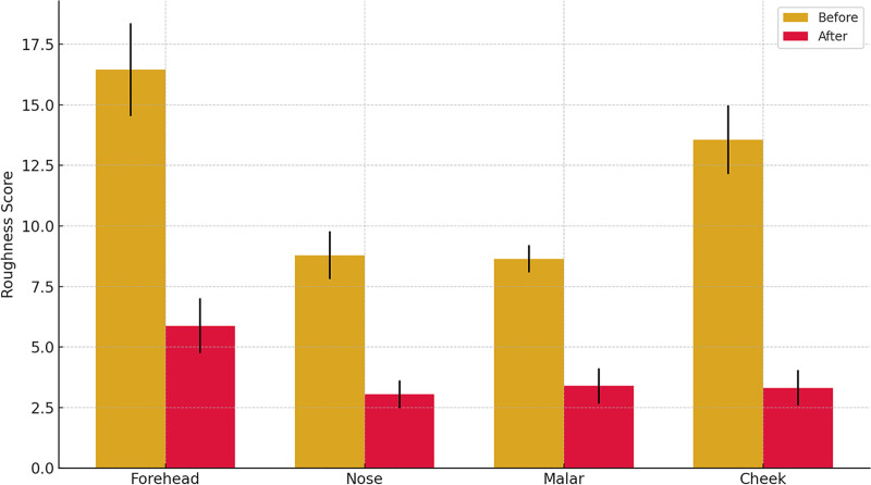
Fig. 11. Quantitative roughness reduction across facial subunits following the Gadion protocol (Gwyddion analysis). Surface roughness scores significantly decreased across all facial regions, with the forehead showing the largest improvement from 16.4 ± 1.9 to 5.8 ± 1.1. This demonstrates the efficacy of the Gadion program in reducing microtopographical skin irregularities, particularly in high-mobility areas.
The mean pore area decreased from 8.82 ± 1.37 pixels 2 (pretreatment) to 3.69 ± 0.89 pixels 2 (posttreatment), representing a 58.2% reduction in pore size ( t = 16.68, P = 2.12 × 10 −16 ) ( P < 0.001). This substantial improvement suggests remodeling of the superficial dermis and restoration of skin elasticity in the glabellar and frontal subunits.
The nasal unit exhibited the greatest absolute reduction, with mean pore size declining from 8.96 ± 1.08 to 3.93 ± 1.19 pixels 2 , reflecting a 56.1% reduction ( t = 18.75, P = 9.35 × 10 −18 ) ( P < 0.001). This significant change may indicate contraction of dilated pilosebaceous structures and enhanced dermal matrix density induced by RF-mediated thermal coagulation.
The average pore size was reduced from 8.00 ± 0.89 to 5.09 ± 1.14 pixels 2 , a 36.4% decrease, suggesting improvement in skin surface topography and collagen reorganization in the midface area ( t = 11.15, P = 5.29 × 10 −12 ) ( P < 0.001).
In the buccal zone, the mean pore area reduced from 7.87 ± 1.16 to 7.03 ± 1.25 pixels 2 , indicating a 10.7% reduction ( t = 2.64, P = 0.0132) ( P > 0.001). Although less pronounced, this may reflect enhanced dermal tightening or sebum regulation in the lower facial region. Overall, these data support the hypothesis that monopolar RF treatment promotes histological dermal remodeling and functional normalization of pilosebaceous units, contributing to smoother and more uniform skin surface architecture (Fig. 12 ) (Table 3 ).
Table 3. Quantitative Changes in Mean Facial Pore Size (Pixels2) Following RF-based Microneedling Treatment
| Facial Region | Pretreatment (Mean ± SD) | Posttreatment (Mean ± SD) | % Reduction | t | P | Statistical Significance |
|---|---|---|---|---|---|---|
| Forehead | 8.82 ± 1.37 | 3.69 ± 0.89 | 58.2 | 16.68 | 2.12 × 10 −16 | P < 0.001 |
| Nose | 8.96 ± 1.08 | 3.93 ± 1.19 | 56.1 | 18.75 | 9.35 × 10 −18 | P < 0.001 |
| Malar region | 8.00 ± 0.89 | 5.09 ± 1.14 | 36.4 | 11.15 | 5.29 × 10 −12 | P < 0.001 |
| Cheek | 7.87 ± 1.16 | 7.03 ± 1.25 | 10.7 | 2.64 | 0.0132 | P > 0.001 |
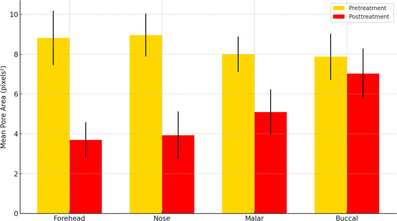
Fig. 12. Quantitative reduction in pore size across facial subunits following treatment. Mean pore area (pixels2) significantly decreased across all facial subunits after treatment, with the most pronounced reduction observed in the forehead and nasal regions. These results demonstrate the efficacy of the intervention in minimizing pore size, as confirmed by AI-based image analysis.
Subjective aesthetic improvement in pore appearance was evaluated using the GAIS. The mean GAIS score was 4.83 ± 0.38 out of 5, indicating a very much improved outcome in the majority of participants. Notably, 80% of subjects reported the highest score (GAIS 5), reflecting high satisfaction and visible clinical improvement in skin smoothness and pore appearance following the full treatment regimen.
Recent advances have highlighted the expanding role of exosomes as a biologically active, cell-free therapeutic modality in the context of regenerative medicine and aesthetic biostimulation, driven by their emerging ability to modulate tissue repair, stimulate fibroblast activity, and enhance extracellular matrix (ECM) remodeling through paracrine molecular signaling pathways. 30 , 31
This retrospective study presented compelling multimodal evidence supporting the efficacy of microneedling combined with ASCE+ therapy in reducing facial pore size and improving skin texture across 6 anatomically distinct facial zones. Using advanced imaging modalities—QuantifiCare 3D stereophotogrammetry, Gwyddion-based roughness analysis, and AI-assisted pore morphometry—we objectively documented statistically significant improvements in both structural and perceptual parameters of skin quality.
Recent advancements in plastic and aesthetic surgery have emphasized the increasing integration of digital technologies and AI-based methods for prognostic and morphological assessment across both reconstructive and aesthetic domains. In line with this evolving paradigm, the present study applied multimodal digital technologies—including AI-based pore detection, Gwyddion-based skin surface roughness measurement, and 3D photogrammetry—to provide a comprehensive, quantitative analysis of facial rejuvenation outcomes following exosome-based therapy.
Pore scores on the QuantifiCare system improved markedly across all regions, with particularly pronounced effects in the forehead and nasal dorsum, areas typically characterized by high sebaceous density and enlarged follicular openings. These improvements suggest a functional normalization of pilosebaceous units, likely mediated by a combination of controlled dermal injury from microneedling and regenerative signaling from exosome therapy. The AI-based pore area analysis reinforced these findings, demonstrating up to 58.2% reduction in mean pore size in the forehead and 56.1% in the nasal unit. These changes support the hypothesis that exosome-mediated pathways enhance dermal remodeling and matrix tightening.
Furthermore, the significant reduction in 3D surface roughness—up to 75.6% in the buccal region—suggests that the combined therapy not only reduces pore diameter but also promotes global topographic smoothness. Prior studies have demonstrated that microneedling enhances dermal remodeling via fibroblast activation and neocollagenesis. Exosomes are known to further amplify this process by delivering growth factors, anti-inflammatory cytokines, and microRNAs that modulate fibroblast behavior, increase elastin synthesis, and improve ECM organization. These findings align with recent literature demonstrating that exosomes derived from adipose-derived stem cells can stimulate fibroblast proliferation, type I and III collagen production, and vascular endothelial growth factor-mediated neovascularization, collectively contributing to enhanced dermal remodeling and matrix reorganization. 32 , 33
Patient satisfaction was notably high, with an average GAIS score of 4.83 ± 0.38, reflecting visible and meaningful clinical outcomes. The strong alignment between objective imaging data and subjective assessments underscores the translational relevance of combining microneedling with exosome-based biologics in real-world aesthetic settings.
This study, although comprehensive, has inherent limitations. The retrospective design limits control over confounding variables. Additionally, histological confirmation of ECM remodeling and inflammatory modulation such as immunohistochemical staining for collagen I/III or transforming growth factor–beta is absent. AI pore detection, although innovative, may be influenced by lighting or image resolution variability despite standardization efforts. Future prospective, randomized controlled trials with histopathological validation, longer follow-up periods, and comparison to monotherapy arms (microneedling alone versus exosome alone) are warranted to delineate the independent contributions of each component. Moreover, incorporation of biomolecular markers (eg, matrix metalloproteinases, vascular endothelial growth factor) may provide mechanistic insight into the regenerative cascade induced by this combination therapy. This combined microneedling and exosome protocol, although evaluated in an aesthetic dermatology setting, may have broader applicability across general aesthetic medicine, including plastic surgery and integrative rejuvenation practices.
This study provided compelling, multimodal evidence that the combination of microneedling and topically applied adipose-derived stem cell exosomes results in significant improvements in facial pore morphology and skin surface texture. Using 3 complementary and objective analytical platforms such as QuantifiCare 3D imaging, Gwyddion-based surface roughness analysis, and AI-assisted pore morphometry, statistically significant reductions in pore size, enhanced dermal smoothness, and improved regional pore scores across 6 anatomically distinct facial subunits were demonstrated.
The central forehead and nasal dorsum, which typically exhibit high sebaceous activity and enlarged pilosebaceous units, showed the greatest improvements in both pore scores and roughness reduction. AI-based analysis confirmed consistent contraction of individual pore areas across all evaluated regions, whereas Gwyddion profiling revealed up to 75.6% reductions in 3D roughness—indicative of substantial dermal remodeling. These objective changes were strongly supported by patient-reported outcomes, with the majority of participants rating their aesthetic improvement at the highest level on the GAIS scale.
Clinically, the findings support the use of this dual-modality approach as a noninvasive, repeatable treatment option for patients presenting with enlarged pores and textural irregularities. Unlike traditional topical or energy-based therapies, this protocol offers a biologically active alternative that may exert longer term regenerative effects with minimal downtime.
The author has no financial interest to declare in relation to the content of this article.
The author wishes to express sincere gratitude to Byungseong Cho for his interest and encouragement in this research.
声明:中文精读 · 仅供学术参考。内容来自公开文献检索,不代表本人观点,不构成诊疗建议。 医疗美容需在正规医疗机构由执业医师实施。
基于相同主题推荐 · 继续深挖
研究类型:韩国单中心回顾性病例系列(n=30,20–60 岁),接受 3 次微针(2.0 mm、540 针)联合 ASCE+ 脂肪源性干细胞外泌体治疗,间隔 4 周。
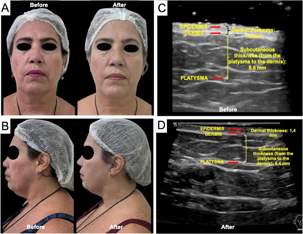
一项巴西前瞻性单中心研究(n=16,PASMFRS 1–4 级颔下脂肪)采用 Megaderme Duo 多层微针射频(40 针镀金微针,深度 2/4/6 mm,15–30 W,1
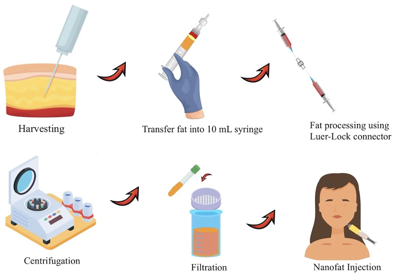
PRISMA 系统综述(2024-12),纳入 18 项研究、768 例患者;纳米脂肪为机械乳化/过滤的自体脂肪,富集 SVF 与脂肪来源干细胞,侧重再生而非单纯容积充填。