无针皮下分离联合透明质酸注射改善水平颈纹的影像学评估
2026年10月4日38 min read
Plastic and Reconstructive Surgery Global Open
结论与实践要点
证据等级: 这是一项回顾性、单臂、单中心观察性研究(病例系列),由单一作者完成,纳入 20 例中重度水平颈纹患者,采用治疗前后自身对照,无随机分组、无平行对照组。证据等级较低,结果可视为「探索性、提示性」而非确证性结论。
适用人群: 25–60 岁、中至重度水平颈纹、近 6 个月未接受颈部填充治疗者;排除了瘢痕体质、既往颈部手术、影响皮肤愈合的系统性疾病者。研究人群为韩国人,Fitzpatrick II–V 型,平均年龄 35.4 岁,以轻中度皮肤松弛为主。
关键结果:
机制要点: CureJet 利用电磁力驱动活塞,将透明质酸以高压微射流(本研究 3–10 Hz、约 1000 发)射入组织,通过剪切效应机械性松解颈纹下方的瘢痕样条索与粘连,起到类似传统皮下分离的作用,并为后续 30G 针注射的透明质酸创造更均匀的分布空间。超声可见注射后真皮深层填充物横向弥散(深度约 0.44 mm、分离宽度约 2.17 mm)。
证据边界与局限:
临床提示: 对于以脂肪与真皮容量缺失为主的水平颈纹,「先松解、后填充」的两步策略在短期影像学评估中显示了可行性与可量化的改善,且不良反应轻微自限。但在更大样本、对照、长期随访研究出现之前,宜将其视为一种有前景的探索性方案,而非成熟标准疗法;实际应用中需严格把握适应证并充分告知瘀青等预期反应。
Background:Horizontal neck folds (HNF) result from fibrous connections between the platysma and skin, often becoming more prominent with aging and skin laxity. This study aimed to assess the effectiveness of combining needle-free subcision and hyaluronic acid (HA) filler injections in improving the dermal and subcutaneous architecture of HNF using sonographic and 3-dimensional (3D) imaging tools.
Methods:Twenty patients underwent 3 sessions of HA (Cleviel Wrinkle) injections for HNF. A needle-free injector (CureJet) was used in the first session to perform subcision via high-pressure microjets of HA, mechanically disrupting adhesions in the subcutaneous tissue. The second and third sessions involved 29G needle injections for volumization. Ultrasound and 3D imaging measured changes in dermal thickness, subcutaneous fat, and skin roughness.
Results:Ultrasound showed significantly thinner subcutaneous fat in wrinkled areas (0.67 ± 0.26 mm) compared with normal regions (1.73 ± 0.32 mm), confirming fat loss as a key contributor to HNF. Posttreatment, subcutaneous fat thickness increased to 2.05 ± 0.41 mm, whereas dermal thickness improved from 1.02 ± 0.17 to 1.58 ± 0.26 mm. 3D analysis revealed a reduction in skin roughness, with the roughness average decreasing from 3.43 ± 0.73 to 1.79 ± 0.49. Mild bruising from subcision resolved within 7 days.
Conclusions:HA injections effectively increased dermal and subcutaneous fat thickness, reducing the appearance of HNF without complications such as nodules or lumps. The combination of needle-free subcision for adhesion release and subsequent HA volumization provided both structural and aesthetic benefits, making it a promising approach for noninvasive neck rejuvenation.
Question: Can needle-free subcision combined with hyaluronic acid (HA) filler effectively improve both the subcutaneous architecture and surface roughness of horizontal neck folds?
Findings: In 20 patients, needle-free injector subcision followed by HA filler significantly increased dermal and subcutaneous thickness and reduced skin roughness, as confirmed by ultrasound and 3-dimensional imaging, with only mild, self-limited bruising and no major complications.
Meaning: Needle-free subcision combined with HA filler provides a safe, minimally invasive treatment option that improves both structural and surface features of horizontal neck folds.
Horizontal neck folds (HNF) are characterized by fibrous connections between the platysma and the skin. HNFs are a common aesthetic concern with diverse etiologies, including congenital predisposition, weight gain, and aging-related structural changes such as dermal thinning and subcutaneous fat loss. Unlike dynamic wrinkles, these folds act as scar-like structures that resist yielding to weight gain, often deepening with increased skin tension. Many patients only seek treatment when aging exacerbates these lines with additional issues such as skin laxity. These factors lead to deepening horizontal lines, affecting overall neck and facial aesthetics. 1 Current treatments include minimally invasive techniques such as botulinum toxin, radiofrequency, laser treatments, and dermal fillers, with hyaluronic acid (HA) fillers widely used for volume restoration and skin smoothing. 2
However, the effectiveness of HA fillers for HNF is not well studied, especially with innovative delivery methods such as needle-free injectors. 3 Previous studies have attempted to treat HNF using a range of approaches. Lee and Kim 4 reported improvement in horizontal necklines following HA filler injections, but noted the challenges in treating congenital or deeply fibrotic folds. Tseng and Yu 1 retrospectively analyzed HA treatment for HNF and emphasized variability in response depending on the depth and elasticity of the skin. Although these studies demonstrated efficacy in superficial cases, they lacked imaging-based evaluation or mechanistic insights into filler–tissue interactions.
This study evaluated HNF improvement using a needle-free injector designed to perform subcision by delivering high-velocity microjets of HA. Unlike conventional needle-based techniques, this mechanism creates a controlled mechanical disruption of scar-like bands and adhesions, enabling improved tissue pliability and effective volume restoration with subsequent injections.
Advanced 3-dimensional (3D) imaging and high-frequency ultrasound were used to measure changes in skin roughness, dermal thickness, and subcutaneous fat, providing a detailed assessment of structural and surface skin improvements posttreatment. 5 This study aimed to fill a gap in the literature by systematically analyzing the impact of needle-free injectors in HNF treatment and comparing their outcomes with conventional methods. This retrospective study aimed to shed light on the use of HA fillers for HNF, highlighting their potential in enhancing skin integrity and appearance without complications such as nodules, lumps, or unintended filler migration.
This retrospective observational study was designed to evaluate the efficacy and structural effects of HA filler for HNFs using a needle-free injection technique, and absence of prior neck treatments in the author’s clinic for treating neck folds from January 2023 to October 2024. The inclusion criteria were (1) patients aged 25–60 years with moderate to severe HNF and (2) no prior neck filler treatments in the past 6 months. The exclusion criteria were (1) a history of keloid formation, (2) prior neck surgery, (3) systemic conditions affecting skin healing, and (4) any cosmetic procedures involving the abdominal skin—including liposuction, fillers, or botulinum toxin—performed within the past 6 months. This study was approved by the institutional review board (no. P01-202412-10-042), and written informed consent was obtained from all patients before participation. This retrospective study analyzed the improvement of HNF after HA injection using the 3D scanner and ultrasound evaluation. Cleviel Wrinkle (Pharma Research, Korea), a cross-linked HA filler, has shown success in facial rejuvenation, yet its use for HNF is underexplored. Patients received HA filler material using needle-free injection during the first session for subcision of adhesion and scar-like band along the HNFs and 30G needle injection during the second and third sessions.
High-frequency ultrasound has been widely used to evaluate dermal and subcutaneous structural changes after injectable treatments. 6 In this study, pre- and posttreatment 33-MHz ultrasound images (Aplio i-700, Cannon Medical System, Tokyo, Japan) were used to measure and analyze injection depths and improvements for all patients. Pre-procedure ultrasound imaging was performed to assess the soft tissue layers at the author’s clinic. Images were retrospectively collected to ensure consistency in injection depth. The thickness of the dermis and subcutaneous fat was measured before and after the procedure.
The needle-free injector (CureJet: C-Jet, Baz Biomedic, Seoul, Korea) uses electromagnetic force to drive a piston forward, creating high-pressure microjets of HA. Upon impact, these jets penetrate the tissue at high speed, generating a controlled mechanical effect. This disrupts adhesions and scar-like bands within the subcutaneous layer, mimicking the traditional subcision technique. The high-velocity injection produces a Venturi effect, enhancing distribution and enabling simultaneous subcision and filler delivery. The needle-free injector achieves subcision by delivering high-velocity microjets of HA. These jets generate a shearing effect within the subcutaneous tissue, disrupting adhesions and scar-like bands, as evidenced by ultrasound imaging showing loosening of tissue planes. 7 – 10
For each patient, 1000 shots were administered using the needle-free injector. 7 – 10 The C-Jet operates through an electromagnetic force that pushes a piston hammer forward very fast. When the hammer strikes the membrane of the needle free injector, its momentum drives the drug chamber to expel the filler through the nozzle into the tissue. Following the Venturi effect, the injecting fluid exits through the narrow nozzle at a very high speed. 11 Using the C-Jet, microjet injections can be repeated at a frequency of up to 20 Hz. In this study, the author used a rate of 3–10 Hz to maintain strong injection speed and penetration force. The C-Jet produces a stronger and deeper jet when applied in full contact with the skin. All shots were performed using the contact mode. A 1:2 diluted HA solution was injected using the contact method. The injectable used was HA, composed of 15 mg of HA. To avoid sticking of the injector nozzle, the HA was diluted at a 1:2 ratio. The nozzle tip was maintained in contact with the skin at a 45-degree angle to ensure deep and effective injections. When injections, the skin was stretched with the nondominant hand to allow complete coverage, whereas patients with smaller areas required approximately 1000 shots. ( See Video 1 [online] , which displays a needle-free injector for subcision of HNF.)
补充视频 / 附件(原文未随文提供原图,可于出版方页面查看)gox-13-e7340-s001.mp4Video 1. This video demonstrates the use of a needle-free injector to perform subcision, a minimally invasive technique designed to release fibrous adhesions and improve the appearance of horizontal neck folds.
Treatments were administered every 2 weeks, with an additional 2 sessions being the routine. During the second and third sessions, prefilled HA 1 mL was injected along the HNF into the subcutaneous fat layer using a 30G needle. ( See Video 2 [online] , which displays the 30G needle injection for volumization in a 33-year-old woman.)
补充视频 / 附件(原文未随文提供原图,可于出版方页面查看)gox-13-e7340-s002.mp4Video 2. This video demonstrates the use of a 30G needle to perform volumization in the treatment of horizontal neck folds for a 33-year-old female patient. The procedure involves injecting hyaluronic acid (HA) filler using a fine 29-gauge needle, which allows for precise placement of the filler in the subcutaneous fat layer. The purpose of this injection is to restore lost volume and improve the overall contour and smoothness of the skin in the neck area, which has been affected by wrinkles and folds. During the procedure, the practitioner inserts the needle into the subcutaneous plane, targeting the areas with visible volume loss, particularly along the horizontal neck folds. Small amounts of HA filler are deposited in a series of injections along the crease, gradually filling the tissue to lift the skin and reduce the depth of the folds. The use of a 30G needle ensures minimal trauma to the tissue and reduces the risk of bruising or discomfort for the patient. The prefilled HA filler is carefully massaged and molded into the treated area to ensure even distribution and natural results. The goal of the volumization process is not only to smooth out the visible folds but also to improve the hydration and elasticity of the skin in the treated area, providing a more youthful and rejuvenated appearance.
To evaluate treatment efficacy for HNF, a 3D scanner (3D LifeViz Mini; QuantifiCare, Sophia Antipolis, France) was used to measure skin changes immediately after the procedures. 12 The scanner captured standardized images with consistent lighting, distance, and angles, ensuring reliable and quantifiable data collection.
Open-source computer analysis software (Gwyddion data analysis software; Czech Metrology Institute, Jihlava, Czechia) was used to measure various skin roughness parameters and evaluate the degree of depression along the HNF. 13 These images were then analyzed using an online computer program for measuring skin roughness, roughness average, root mean square roughness, maximum height of roughness, maximum roughness valley depth, and waviness average. The figure shows the roughness average, root mean square roughness, maximum height of roughness, maximum roughness valley depth, and waviness average before and after the treatment on the same areas using the Gwyddion computer program. 14 After 3 sessions, patients visited the clinic without applying makeup or any other products to their faces. Three-dimensional photographs and sonographic images were then obtained. Paired t tests were conducted to compare pre- and posttreatment values for roughness, dermal thickness, and subcutaneous fat thickness. Statistical significance was set at a P value of less than 0.05.
After the treatment and taking the 3D images, 0.5 mL of a cosmetic product (ExtraGoldStem ample; Reteenage, Seoul, South Korea) was sprayed under high pressure to allow penetration of the ampoule into the needle holes and epidermis. The ampoule, containing growth factors and peptides, was applied to promote epidermal repair and hydration posttreatment, enhancing the overall healing process. After the first session, the treated area was covered with a Duoderm hydrogel bandage, and this occlusive dressing was maintained for 2 days.
The Global Aesthetic Improvement Scale was used for a comprehensive evaluation of the effects of the HA injection on the improvement of HNF (very much improved, 5; much improved, 4; improved, 3; no change, 2; worse, 1). Data were analyzed using paired t tests to compare pre- and posttreatment values for dermal thickness, subcutaneous fat thickness, and skin roughness parameters. A P value of less than 0.05 was considered statistically significant. Statistical analysis was performed using SPSS version 27.0 (IBM Corp., Armonk, NY).
This study included 20 patients (14 women, 6 men) with a mean age of 35.4 years (range 27–59 y). All participants were healthy without underlying systemic disease and had Fitzpatrick skin types II–IV. None had a history of previous neck filler treatments or neck surgery.
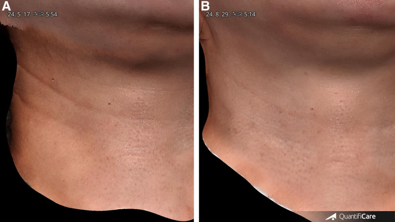
The 3D images showed the results of neck wrinkle reduction after a series of injections, captured using QuantifiCare 3D analysis technology.
In the pretreatment images, there are noticeable deep folds and wrinkles, particularly in the lower neck area. These creases suggest loss of specific soft tissue, which is typically unrelated to aging or skin sagging.
After 3 sessions of injections, there was a marked reduction in the depth and prominence of the neck wrinkles. The folds that were visible in the lower neck had diminished significantly. The skin appeared smoother and more even, with reduced furrows in the treated area.
The skin texture appeared much more uniform posttreatment, as indicated by the lack of deep, obvious ridges or valleys. This suggests that the soft tissue loss was treated in 2 layers (the dermis and the subcutaneous fat layer), which may have contributed to the improved appearance. The results provide clear evidence that the treatment has significantly reduced the visibility of neck wrinkles. The improvements are not just in the reduction of wrinkle depth but also in the overall smoothness, texture, and skin tightness. The 3D imaging highlighted the profound differences between the pre- and posttreatment stages, making it a valuable visual tool to support the efficacy of the procedure (Figs. 1 – 3 ). ( See Video 3 [online] , which displays before and after treatment of HNFs [40-year-old woman.]) ( See Video 4 [online] , which displays before and after treatment of HNFs [34-year-old man.])
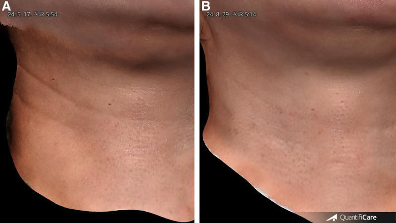
Fig. 1. Before and after treatment of horizontal neck folds (40-year-old woman). A, Pretreatment image shows noticeable, deep, and prominent horizontal folds and wrinkles on the lower neck. B, The posttreatment image shows that the depth and visibility of the horizontal folds have been greatly reduced, and the skin appears smoother and more even.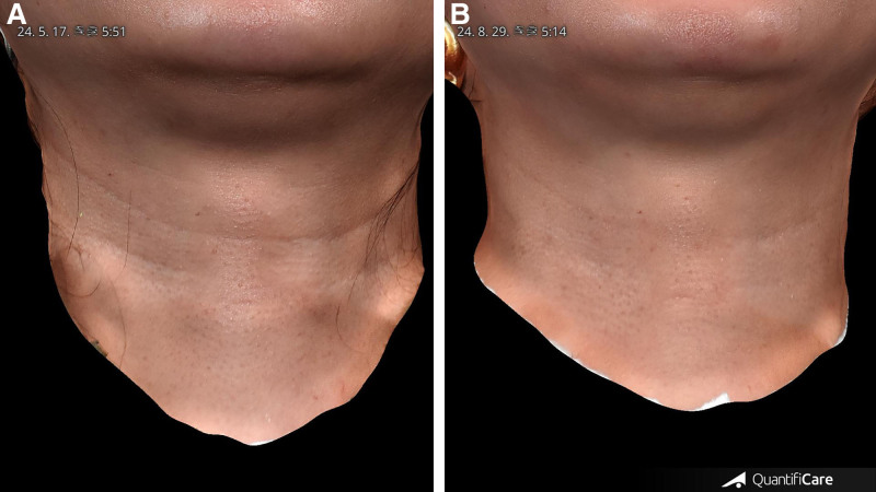
Fig. 2. Frontal view, before and after treatment of horizontal neck folds (40-year-old woman). A, The pretreatment image reveals deep lines and creases across the lower neck, highlighting the presence of significant skin laxity and subcutaneous volume loss, contributing to the overall aged appearance. B, The posttreatment image shows a marked improvement in the overall smoothness of the neck skin.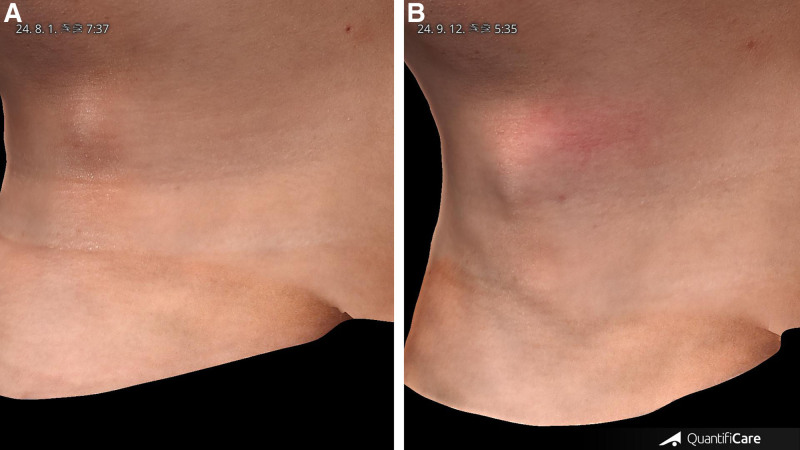
Fig. 3. Before and after treatment of HNFs (34-year-old man). A, The pretreatment image shows noticeable horizontal folds and creases in the lower neck region. B, The posttreatment image demonstrates a visible reduction in the depth and prominence of the HNFs.补充视频 / 附件(原文未随文提供原图,可于出版方页面查看)gox-13-e7340-s003.mp4Video 3. This video presents a side-by-side comparison of the results of treatment for horizontal neck folds in a 32-year-old female patient. The video highlights the improvements in skin texture and wrinkle depth following a series of treatments with Cleviel Wrinkle® hyaluronic acid (HA) filler. (left) In the before-treatment footage, deep horizontal neck folds are clearly visible. The skin in the lower neck region appears loose, with pronounced creases and irregularities in texture. This reflects a typical case of skin laxity and volume loss, which contributes to the formation of visible neck folds. (right) In the after-treatment footage, the results demonstrate a significant reduction in the depth of the horizontal neck folds. The skin appears smoother and firmer, with the previously deep creases now diminished or almost invisible. This improvement suggests successful volumization and structural support provided by the HA filler, resulting in a more youthful and even neck contour. The skin’s surface is more even, with fewer irregularities and improved texture overall. The side-by-side comparison provides a clear visual representation of the effectiveness of the treatment, emphasizing the reduction in wrinkle depth and the enhanced overall appearance of the neck skin.补充视频 / 附件(原文未随文提供原图,可于出版方页面查看)gox-13-e7340-s004.mp4Video 4. This video presents a comparison of the neck skin appearance in a 34-year-old male patient before and after treatment targeting horizontal neck folds. (left) Before treatment. In the initial footage, the horizontal neck folds are clearly visible, with noticeable creases and texture irregularities along the neck. The skin shows typical signs of volume loss and laxity, with prominent lines forming deep creases, particularly in the lower neck area. The wrinkles indicate both structural changes in the subcutaneous tissue and surface irregularities. (right) After treatment. The post-treatment footage demonstrates a significant improvement in the appearance of the neck folds. The depth of the horizontal lines has been reduced, and the overall skin texture appears smoother and more even. There is a visible restoration of volume, which has helped to lift the skin and minimize the appearance of the folds. The treatment has successfully improved the contour and surface of the neck, leaving it looking more youthful and firm.
The graphs compare the pre- and posttreatment values for key surface parameters, including roughness average, root mean square roughness, maximum height of roughness, maximum roughness valley depth, and waviness average. Before treatment, the roughness average was measured at 3.43 ± 0.73, indicating a relatively uneven surface texture. Following the procedure, this value significantly decreased to 1.79 ± 0.49, demonstrating a much smoother skin surface. Similarly, the root mean square roughness, which reflects the variability in surface texture, showed a substantial reduction from 4.49 ± 1.22 before treatment to 2.21 ± 0.60 afterward. This decline in variability further supports the effectiveness of the treatment in achieving a more uniform skin surface.
The maximum height of roughness, which represents the presence of high peaks on the skin surface, was initially 17.22 ± 6.01. After treatment, this value was markedly reduced to 7.83 ± 2.66, indicating that the intervention effectively diminished the surface irregularities. Additionally, the maximum roughness valley depth, which quantifies the depth of depressions in the skin, showed a reduction from 8.49 ± 3.15 to 4.15 ± 1.51 posttreatment. This suggests that the valleys on the skin surface became significantly shallower, contributing to a smoother appearance.
The waviness average, which measures the overall undulation of the surface, also demonstrated improvement. Pretreatment values were 15.84 ± 4.69, reflecting significant variations in the skin’s surface profile. After treatment, the value decreased to 10.09 ± 3.30, showing that the treatment effectively reduced overall waviness, although some variability remained.
These trends collectively indicate that the treatment led to a noticeable improvement in skin surface smoothness by reducing both roughness and waviness, as evidenced by the significant reductions in each parameter (Table 1 ) (Figs. 4 , 5 ). ( See figure, Supplemental Digital Content 1 , which displays the comparison of pretreatment and posttreatment parameters for HNF surface roughness. The graphs demonstrate pre- and posttreatment changes in 5 surface roughness metrics, showing overall improvement in skin texture following HNF correction, https://links.lww.com/PRSGO/E530.)
Table 1. Pre- and Posttreatment Changes in Neck Fold
| Pretreatment | Average | SD | Posttreatment | Average | SD |
|---|---|---|---|---|---|
| Roughness average | 3.43266 | 0.72987 | Roughness average | 1.79375 | 0.491135 |
| Root mean square roughness | 4.48652 | 1.21532 | Root mean square roughness | 2.20975 | 0.603355 |
| Maximum height of roughness | 17.2162 | 6.00914 | Maximum height of roughness | 7.82991 | 2.659237 |
| Maximum roughness valley depth | 8.49130 | 3.15350 | Maximum roughness valley depth | 4.14865 | 1.513757 |
| Waviness average | 15.83915 | 4.69457 | Waviness average | 10.08842 | 3.303343 |
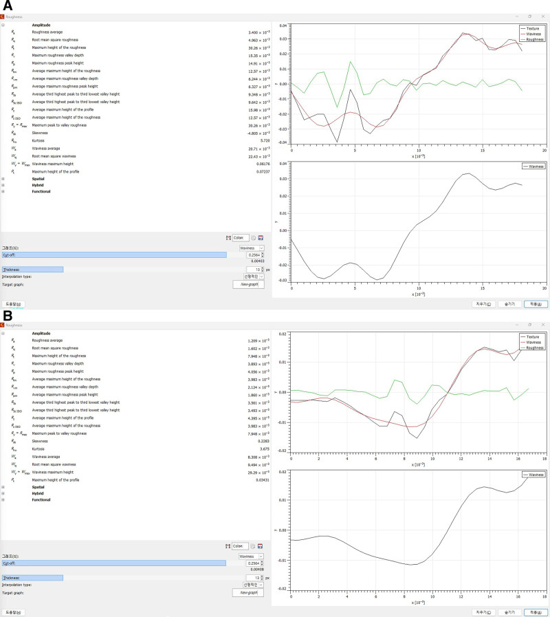
Fig. 4. Roughness average, root mean square roughness, maximum height of roughness, maximum roughness valley depth, and waviness average pre- and posttreatment. A, Roughness analysis of the HNF before treatment. B, The treatment shows significant improvements in roughness, root mean square roughness, maximum height of roughness, and valley depth.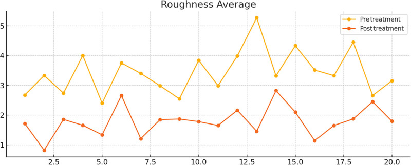
Fig. 5. Comparison of pretreatment and posttreatment parameters for HNF surface roughness. The pretreatment graph shows significantly higher values, with considerable variability across the surface, and the posttreatment values indicate a notable reduction in roughness, demonstrating smoother skin texture.To assess the statistical significance of these improvements, a paired t test was conducted, confirming that the reductions in roughness, valley depth, and waviness between pre- and posttreatment were statistically significant ( P < 0.001). This suggests that the observed smoothing effect was highly unlikely to be due to random variation. The paired t test was chosen as the appropriate statistical method because it compares related pre- and posttreatment measurements within the same individuals.
The statistical analysis was conducted under the framework of hypothesis testing, where the null hypothesis (H₀) assumed no difference between the pretreatment and posttreatment means, whereas the alternative hypothesis (H₁) proposed that a significant difference existed. The t statistic was calculated as the ratio of the mean of the differences to the SD of differences, adjusted for sample size.
The resulting P value provided a probability measure of whether the observed differences could have occurred by chance. Given that the P value was well under 0.05, the null hypothesis was rejected, confirming that the treatment had a statistically significant impact on improving skin texture. The consistent and significant reductions across all measured parameters underscore the effectiveness of the intervention in enhancing skin smoothness and reducing the prominence of HNFs.
The t statistic measures the size of the difference relative to the variation within the sample data. The P value helps determine whether that difference is statistically significant. In this case, the very low P values suggest that the treatment led to a real improvement in the skin surface properties, not just random fluctuations. In the results calculation, the P values for all metrics (roughness average, root mean square roughness, maximum height of roughness, etc.) were very small (<0.001). This means the likelihood of the differences between pre- and posttreatment measurements being due to chance is extremely low. Therefore, we can confidently say that the treatment had a significant impact on these skin parameters (Table 2 ).
Table 2. Statistical Significance of Pre- and Posttreatment Improvements in Neck Fold
| Neck Fold | t Statistic | P |
|---|---|---|
| Roughness average | 7.14843 | 4.95 × 10− 6 |
| Root mean square roughness | 7.791364 | 1.86 × 10− 6 |
| Maximum height of roughness | 5.79126 | 4.67 × 10− 5 |
| Maximum roughness valley depth | 6.04673 | 3.00 × 10− 5 |
| Waviness average | 4.474196 | 0.000524 |
Subcision achieved with the needle-free injector disrupted adhesions and scar-like bands, as evidenced by ultrasound imaging showing loosening of subcutaneous tissue planes. This effect allowed for uniform filler distribution and contributed to improved structural support in the treated areas. The observed increase in subcutaneous fat thickness posttreatment was attributed to the volumizing effect of HA filler, which effectively restored lost volume and improved support for the overlying skin.
This retrospective study found a significant difference in subcutaneous fat thickness between normal neck areas and regions with HNF. Normal areas showed an average subcutaneous thickness of 1.73 ± 0.32 mm, whereas HNF regions had only 0.67 ± 0.26 mm, highlighting the role of reduced subcutaneous fat in wrinkle formation. Posttreatment, subcutaneous thickness in HNF regions increased to 2.05 ± 0.41 mm. A paired t test showed this increase to be statistically significant ( t = −13.90, P = 2.25 × 10 −12 ), confirming the treatment’s effectiveness in enhancing subcutaneous thickness (Tables 2 and 3 ) (Figs. 6 – 8 ).
Table 3. Subcutaneous Fat Thickness in Neck Fold Areas, Pre- Versus Posttreatment Effect Following Injection of 1 mL HA
| Pretreatment Fat Thickness (Normal) | Pretreatment Fat Thickness (Fold) | Posttreatment Fat Thickness (Fold) | |
|---|---|---|---|
| Mean thickness, mm | 1.732173 | 0.675217 | 2.053478 |
| SD | 0.313479 | 0.259311 | 0.401731 |
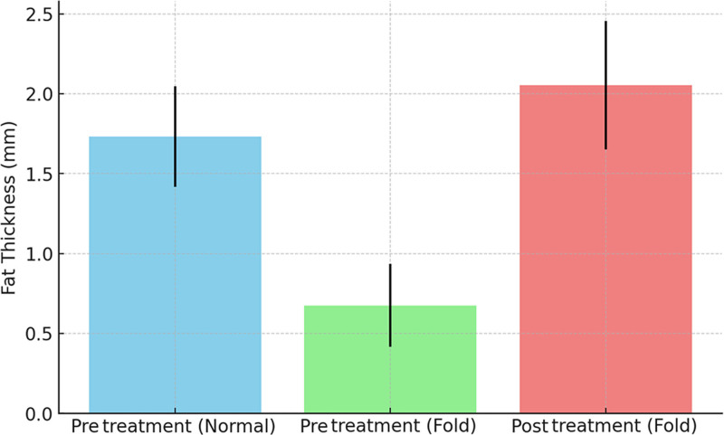
Fig. 6. Subcutaneous fat thickness in neck fold areas, pre- vs posttreatment. In wrinkled regions of the neck, subcutaneous fat thickness significantly decreased from 1.73 mm (normal areas) to 0.67 mm, and following treatment, it increased to 2.05 mm, indicating substantial volumization.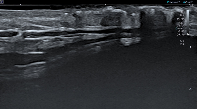
Fig. 7. Sonography imaging of a 34-year-old man immediately after needle-free injection. On August 1, 2024, a longitudinal ultrasound scan along the neck fold showed a consistent dermal-depth distribution of HA immediately after needle-free injection.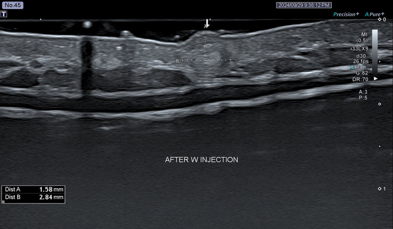
Fig. 8. Vertical cross-section imaging of a 34-year-old man (Fig. 7) immediately after 30G manual injection. On August 14, 2024, HA was injected using a 30G needle at a depth of 1.58 mm with a diameter of 2.84 mm to volumize the dermis and subcutaneous fat layer.
In this study, dermal thickness was measured in both the normal skin areas and the areas with neck wrinkles before and after treatment using a needle-free injector (Figs 6 , 7 , and 9 ).
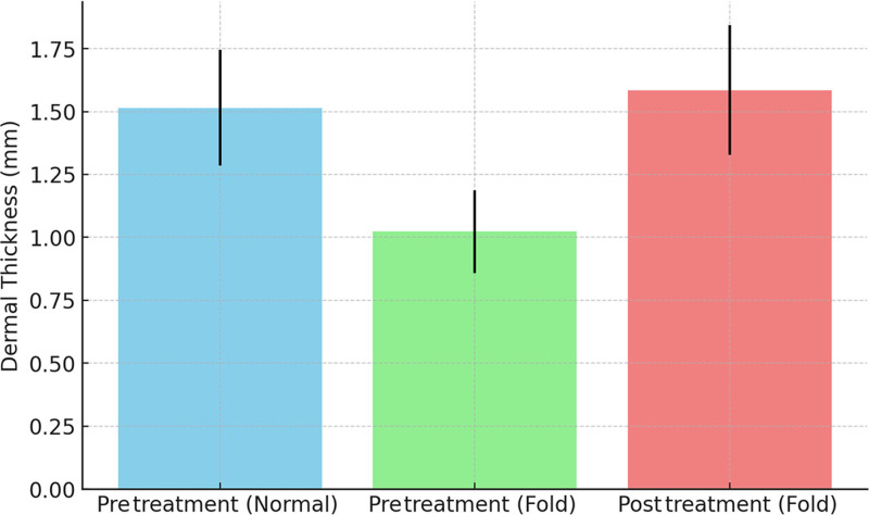
Fig. 9. Dermal thickness in normal skin (pretreatment), wrinkled skin (pretreatment), and wrinkled skin (posttreatment). In wrinkled neck areas, dermal thickness increased from 1.02 to 1.58 mm after treatment, approaching normal thickness (1.51 mm) and suggesting effective dermal regeneration and collagen restoration.
Normal AreasThe average dermal thickness in normal skin areas was 1.51 ± 0.23 mm, representing healthy, intact skin structure.
Wrinkled Areas (Before Treatment)In contrast, the dermal thickness in the wrinkle folds was significantly thinner, averaging 1.02 ± 0.17 mm. This reduction in dermal thickness likely contributes to the formation and deepening of wrinkles, as thinner skin is more prone to folding and sagging.
Wrinkled Areas (After Treatment)After the treatment, the dermal thickness in the wrinkle areas increased to an average of 1.58 ± 0.26 mm. This represents a substantial improvement, bringing the thickness of the dermal layer in the treated areas closer to that of normal, nonwrinkled skin.
The increase in dermal thickness suggests a successful therapeutic outcome, likely due to enhanced collagen production and dermal regeneration stimulated by the treatment. This thicker dermis may provide better structural support to the overlying skin, reducing the appearance of wrinkles.
The study demonstrates that the use of a needle-free injector led to a statistically significant increase in dermal thickness in the wrinkle-prone areas of the neck. This increase can be interpreted as a positive skin remodeling effect, as thicker dermal layers are associated with better skin elasticity, resilience, and a smoother surface appearance. The treatment’s ability to restore dermal thickness to near-normal levels indicates its potential in noninvasive rejuvenation therapies. By increasing dermal thickness, the skin gains more structural integrity, reducing the likelihood of deep wrinkle formation and improving overall skin appearance. This finding suggests that the needle-free injection method may offer a valuable alternative to traditional invasive techniques for patients seeking to reduce neck wrinkles and enhance skin quality.
The paired t test comparing dermal thickness before and after treatment resulted in a t statistic of −9.24 and a P value of 4.99 × 10 −9 . This extremely low P value indicates that the increase in dermal thickness posttreatment is statistically significant, meaning that the observed improvement in dermal thickness is highly unlikely to be due to chance. Therefore, we can confidently conclude that the needle-free injection treatment effectively increased dermal thickness in the wrinkle areas (Tables 4 , 5 ) (Figs. 10 – 12 ).
Table 4. Impact of Needle-free Injection With Booster Needle Injection Treatment on Dermal Thickness in Neck Wrinkle Areas: Pretreatment Versus Posttreatment
| Pretreatment Dermal Thickness (Normal) | Pretreatment Dermal Thickness (Fold) | Posttreatment Dermal Thickness (Fold) | |
|---|---|---|---|
| Mean | 1.514783 | 1.022609 | 1.585217 |
| SD | 0.229859 | 0.164851 | 0.258270 |
Table 5. Patient Demographics and Baseline Characteristics
| Characteristic | Value (N = 20) |
|---|---|
| Mean age, y | 35.4 ± 6.2 |
| Skin type (Fitzpatrick scale) | Type II: 2 (10%); type III: 8 (40%); type IV: 7 (35%); type V: 3 (15%) |
| Presence of neck wrinkles (mild/moderate/severe) | Mild: 6 (30%); moderate: 10 (50%); severe: 4 (20%) |
| Baseline neck skin laxity: mild: 5 (25%); moderate: 12 (60%); severe: 3 (15%) | Mild: 6 (30%); moderate: 10 (50%); severe: 4 (20%) |
| Mean baseline neck fold depth, mm | 2.35 ± 0.42 |
| Mean baseline neck skin thickness, mm | 1.51 ± 0.23 |
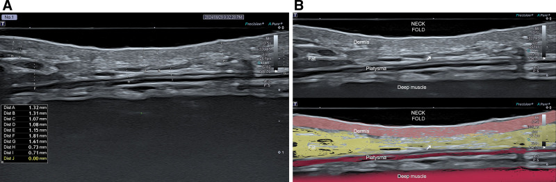
Fig. 10. A, Pretreatment vertical cross-section sonographic image of the neck fold in a 33-year-old woman. Dermal layer: the dermal thickness is measured at 1.31 mm at point B and 1.32 mm at point A, similar to the surrounding areas where the skin is smoother and without folds. Fat layer: the measurements indicate that the fat thickness is 0.71 mm at points I to 0.73 mm at points H, showing a substantial thinning of the fat layer in the folded area. B, Ultrasound sonographic image of the neck highlighting the anatomical layers in a patient with a neck fold. A clear sonographic distinction among the dermis (pink), subcutaneous fat (yellow), platysma (thin red line), and deep muscle layer (dark red) provides anatomical insight into neck fold formation and guides precise treatment planning.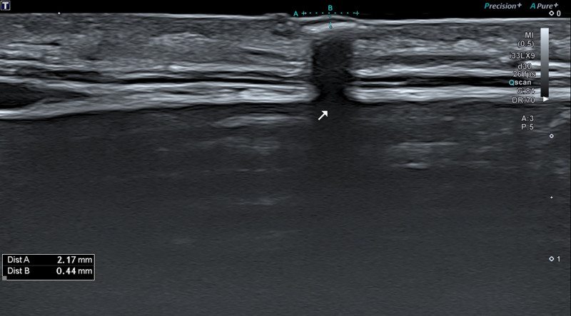
Fig. 11. Vertical cross-section sonographic image of the neck fold in a 33-year-old woman immediately after the treatment using a needle-free injector (August 8, 2024). Posttreatment sonography showed dermal thickening and lateral spread of HA at a depth of 0.44 mm, with a subcision width of 2.17 mm, indicating effective adhesion release under the neck fold.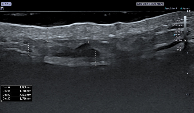
Fig. 12. Ultrasound imaging of the same female patient in Fig. 11, 1 week after 30G needle manual injection (September 13, 2024). Posttreatment ultrasound revealed substantial increases in dermal and subcutaneous fat thickness (1.83 and 2.63 mm, respectively), indicating restored volume and improved neck contour.
The Global Aesthetic Improvement Scale score after treatment was 4.52 ± 0.15.
This study provided valuable insights into the role of subcutaneous fat in the development of neck wrinkles and demonstrated the efficacy of HA fillers in both subcision and volumization. 1 – 3 Cleviel Wrinkle features a high HA concentration of 15 mg/mL, providing enhanced volumizing effects with minimal migration (<1 mm observed during follow-up). This compares favorably with standard fillers, which report migration up to 2.5 cm. Long-term studies are ongoing to evaluate its safety and efficacy. Although preliminary results demonstrate the safety of the HA for neck rejuvenation, longer term follow-up is required to confirm the durability of outcomes and the absence of delayed adverse effects.
One of the most important findings from this study is the significant reduction in subcutaneous fat thickness in the wrinkled areas of the neck compared with normal skin, as confirmed by high-frequency ultrasound imaging. This finding highlights a key underlying cause of neck wrinkles: loss of subcutaneous fat, which diminishes the support for the overlying skin, leading to the formation of deeper creases and folds. By restoring this lost fat volume through HA injections, the skin can regain a more youthful and smooth appearance. Moreover, the use of a needle-free injector in the first session provided a crucial advantage by performing subcision to release scar-like bands and adhesions along the HNF. 7 – 10 This technique helped loosen the tight attachments in the subcutaneous tissue, allowing the injected HA filler to distribute more evenly and effectively within the fat layer in subsequent sessions. Subcision performed with the needle-free injector is especially advantageous because it is extremely fast (20 shots/s), enabling the operator to perform the procedure efficiently. However, 1 notable downside of this method is the occurrence of bruising at the treated sites, which is expected as part of the subcision process. Bruising was observed in all patients and typically resolved within 7–14 days, consistent with superficial trauma from subcision. Despite this temporary side effect, subcision remains a crucial first step in the treatment protocol, ensuring optimal outcomes for volumization in the subsequent sessions. 15 , 16
Our findings suggest that a multimodal approach, combining needle-free injectors for subcision and traditional needle injections for volumization, offers a robust solution for patients seeking nonsurgical neck rejuvenation. This approach ensures both structural release of adhesions and restoration of lost volume, leading to more durable and aesthetically pleasing results.
This study adds to the growing body of evidence supporting the use of HA fillers in neck rejuvenation and highlights the innovative application of needle-free injection technology in combination with traditional techniques. Future studies may explore the long-term effects of these treatments and their potential applications for other anatomical areas where adhesion and volume loss contribute to wrinkle formation. This combined approach offers a promising pathway for achieving noninvasive yet highly effective aesthetic improvements in the neck.
Limitations of this study include a small sample size and short-term follow-up. Future studies should explore long-term outcomes and compare needle-free versus traditional injection methods in a larger cohort.
This study demonstrated the efficacy of combining needle-free subcision with 30G needle-based HA injections for treating HNFs. Three-dimensional imaging and ultrasound confirmed improvements in skin texture and dermal and subcutaneous thickness. Subcision using high-velocity microjets disrupted scar-like adhesions, enhancing filler integration and skin smoothness. Although short-term outcomes are promising, further studies with long-term follow-up and histological analysis are needed to determine whether tissue thickening reflects true remodeling or filler deposition. This dual-action, minimally invasive technique offers a safe and effective option for neck rejuvenation.
The author has no financial interest to declare in relation to the content of this article.
The author gratefully acknowledges the Botox Filler Thread Lifting Association (BoFilTh) of the Korean Society of Plastic and Reconstructive Surgeons (KPRS) for their academic encouragement and intellectual support during the preparation of this study.
声明:中文精读 · 仅供学术参考。内容来自公开文献检索,不代表本人观点,不构成诊疗建议。 医疗美容需在正规医疗机构由执业医师实施。
基于相同主题推荐 · 继续深挖
研究类型:回顾性单臂观察性研究(单作者、单中心),共纳入 20 例水平颈纹患者(女 14 例、男 6 例),平均年龄 35.4 岁(27–59 岁),Fitzpatrick II–
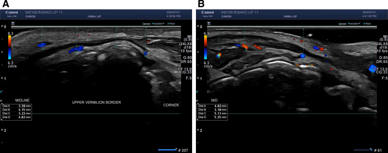
本研究为一项前瞻性研究,纳入25例韩国女性,采用透明质酸(Cleviel Volume)行唇部填充,结合注射前高频超声对上下唇动脉深度进行6点测绘。
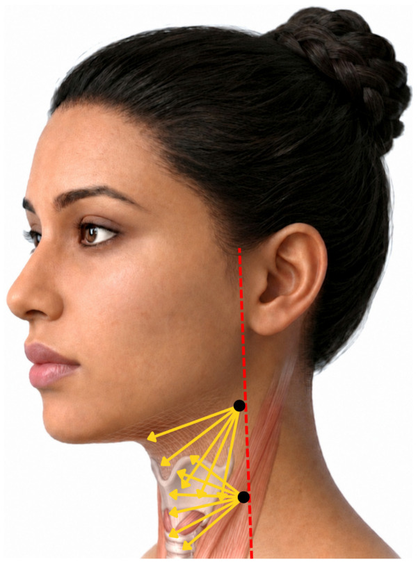
单次联合注射 CaHA、HA 与肉毒毒素 A 改善颈部松弛