东北亚女性面部轮廓化的解剖学导向美学设计
2026年10月6日28 min read
Clinical, Cosmetic and Investigational Dermatology
结论与实践要点
可将面部填充视为「解剖导向的设计过程」而非分区补凹:先以多视角评估「轮廓流中断」,定位中线/颧/下颌轴,沿轴规划连贯矢量(入针点→深部平面→相对分布),用钝针小容积、低压、广布连接相邻区域,优先恢复整体轮廓连续性而非制造局灶隆起。
东北亚案例中,Stage 1 以稀释 PCL 混合物在深部平面(骨膜上/深筋膜浅层)打底建立支撑,Stage 2 稳定后用 HA 精修(额颞 2 mL、前颊/颧下 4 mL、下颌 0.6 mL)。但须明确:稀释 PCL 为标签外用法,其流变与生物行为未经验证,4 月随访不足以排除迟发并发症,且原文明确该混合物不推荐为标准方案。
边界说明:本文为概念验证/个案报告,证据等级低、无对照、无标准化客观测量,不能据以断言疗效或长期安全;参考面部形态为定性模型、未经信效度检验。若借鉴其「矢量 + 轮廓连续」思路,应以合规材料、充分知情同意与标准化影像随访为前提,并警惕标签外 PCL 混合物的不确定风险。
Facial filler contouring is commonly planned within a volume-centric framework emphasizing product selection and localized augmentation. Although widely used, such approaches may not always clearly separate material selection from aesthetic design intent or fully incorporate population-specific anatomical characteristics, particularly in Northeast Asian faces. This proof-of-concept case report proposes an anatomy-informed, design-oriented conceptual framework that prioritizes whole-face contour continuity and anatomically appropriate pathways over isolated volumetric correction. A population-contextualized Reference Facial Form is introduced as a qualitative design reference from which aesthetic intent is translated into geometric principles and subsequently into predefined vectors. In this framework, vectors are structured cannula-based trajectories defined by specific entry points, advancement directions, withdrawal pathways, and deep anatomical planes. Procedural planning is guided by predefined procedural conditions—including anatomical entry points, vector orientation, and relative filler distribution within deep planes—rather than primarily by fixed injection volumes. The framework is illustrated through a single two-stage clinical case. The initial stage used an off-label diluted polycaprolactone-based admixture to establish broad deep-plane structural support, followed by selective contour refinement with hyaluronic acid fillers after initial stabilization. Early clinical images illustrated the correspondence between the planned vectors and early contour formation but were not suitable for establishing treatment efficacy or durability. The available follow-up was also insufficient to establish long-term safety. Fifteen de-identified supplementary cases constituted a criterion-defined consecutive illustrative series demonstrating recurrent vector and entry-point patterns across similar anatomical contexts; they were not analyzed as a validation or outcome cohort. By framing facial filler contouring as a design-first process, this framework may provide a conceptual language for clinical planning and education. Its clinical efficacy, inter-operator reproducibility, generalizability, and long-term safety require evaluation in larger, systematically designed studies.
Keywords: facial contouring、dermal fillers、anatomy-driven aesthetic design、vector-based planning、facial anatomy、northeast asian women
Facial filler treatment planning commonly integrates anatomical assessment with product selection, injection technique, and volume determination. These elements are essential for safe and precise procedural execution. 1 , 2 However, when planning is organized primarily around individual sites of deficiency, the relationship between each correction and adjacent facial regions may remain insufficiently defined. Consequently, technically appropriate augmentation of individual areas does not necessarily ensure coherent transitions across the overall facial contour.
Facial shape is determined by the layered relationships among skeletal support, retaining ligaments, soft-tissue spaces, and deep and superficial fat compartments. 3–7 These structures influence tissue mobility, projection, and transitions between neighboring facial regions and provide the anatomical basis for selecting injection planes and trajectories. Population-associated differences in craniofacial morphology, soft-tissue distribution, and aesthetic treatment goals may also influence full-face planning. Studies and consensus recommendations addressing Asian and Korean faces therefore support adapting treatment planning to the patient’s individual anatomy and intended facial form rather than directly applying a single population-based template. 8–13
Although facial anatomy and region-specific injection techniques have been extensively described, 1–7 , 14 a practical gap remains between identifying anatomical structures and translating a whole-face aesthetic intention into a coordinated procedural plan. To address this gap, we propose “anatomy-driven aesthetic design”, a design-first approach that begins with a patient-specific Reference Facial Form and reverse-translates the discrepancy from baseline into coordinated intervention vectors. The framework introduces contour continuity as a qualitative planning construct for organizing individual interventions according to their relationships across the whole face.
The purpose of this proof-of-concept case report is to define this conceptual framework and illustrate its clinical translation through a representative case. Supplementary cases are included to demonstrate recurrent vector and entry-point patterns in comparable anatomical contexts rather than to serve as an efficacy or validation cohort.
The proposed framework integrates aesthetic judgment and clinical anatomy under a single principle:
“Aesthetic intent defines the target; anatomy determines the pathway.”
Facial contour planning proceeds through two stages. First, the intended contour relationships are established using the qualitative Reference Facial Form described below. These relationships are then translated into patient-specific entry points, trajectories, tissue planes, and relative volume distribution according to skeletal support, retaining ligaments, soft-tissue spaces, fat compartments, and regional anatomy. 3–6 , 14 This conversion of aesthetic intention into an anatomically appropriate procedural plan is defined here as “anatomical translation.”
In the present Northeast Asian context, this planning process is informed by reported morphological tendencies, including relatively broad transverse midfacial proportions and limited anterior projection in certain facial regions. 10–13 These population-level observations provide anatomical context for planning, while the intended form remains individualized according to each patient’s baseline anatomy, proportions, and treatment goals.
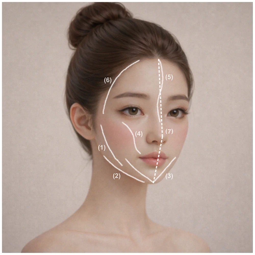
Figure 1 represents the Reference Facial Form through seven qualitative contour relationships: 1)A continuous curve extending from the zygomatic region to the mandibular angle2)A predominantly linear contour along the inferior mandibular border, with limited curvature to maintain structural definition3)A gently converging V-shaped contour toward the chin4)A curvilinear flow extending from the anterior cheek, modulated across the nasolabial region, and re-establishing continuity at the mid-cheek5)A continuous frontal–nasal axis connecting the glabella and nasal bridge in proportional relation to the nasal profile6)A smooth lateral curve extending from the lateral forehead through the temple to the zygomatic region, without marked protrusion or depression7)A stable facial midline axis supporting bilateral contour balance
These directional relationships provide a qualitative reference for identifying contour discontinuities and translating them into vector-based procedural planning. Their anatomical and population-specific context is informed by established literature, 3–6 , 8–13 whereas their organization into this seven-element structure represents the author’s conceptual synthesis. They are intended for individualized planning rather than as numerical morphometric norms or prescriptive treatment targets.
The framework specifically addresses whole-face contour continuity and is not intended as a comprehensive model of facial attractiveness. 15 Fillers serve as the medium through which the design is implemented, whereas product selection, rheological properties, and volume are procedural variables determined after the intended contour and anatomical pathways have been established. The framework therefore prioritizes coordinated structural support over isolated superficial volumization and provides the conceptual basis for the vector-based strategy described in the following section.
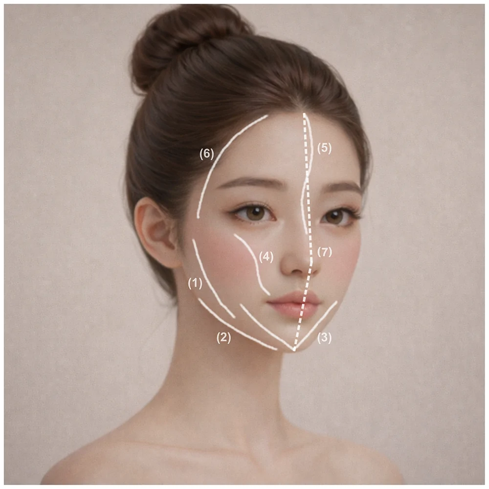
Figure 1 AI-generated conceptual illustration of the Reference Facial Form and contour continuity in anatomy-driven aesthetic design. This conceptual illustration was created using Google Gemini’s Nano Banana image-generation tool (Gemini 3 Pro Image). It does not depict an actual patient or clinical outcome and is presented solely to illustrate the design principles proposed in this study. The illustrated lines represent seven author-proposed qualitative contour relationships used as references in aesthetic planning: 1. A continuous curve extending from the zygomatic region to the mandibular angle. 2. A predominantly linear contour along the inferior mandibular border with limited curvature. 3. A gently converging V-shaped contour toward the chin. 4. A curvilinear flow extending from the anterior cheek, modulated across the nasolabial region, and re-establishing continuity at the mid-cheek. 5. A continuous frontal–nasal axis connecting the glabella and nasal bridge in proportional relation to the nasal profile. 6. A smooth lateral curve extending from the lateral forehead through the temple to the zygomatic region without marked protrusion or depression. 7. A stable facial midline axis supporting bilateral contour balance. These elements constitute a structured qualitative design reference intended to identify perceived discontinuities between adjacent facial subunits and guide vector-based contour planning, whereas patient-specific anatomy determines procedural execution. They do not represent quantitative morphometric criteria, mandatory treatment targets, or validated aesthetic standards.
Rather than prescribing a fixed set of absolute filler volumes, the framework provides a structured sequence for translating aesthetic intent into procedural planning:
Consistency within this framework is pursued through a common planning logic rather than the replication of identical technical parameters. Vector definition thereby serves as the link between aesthetic intent and anatomically appropriate execution.
In this framework, a “vector” is defined as a predefined procedural trajectory comprising the cannula entry point, advancement direction, withdrawal pathway, injection plane, and relative filler distribution. It therefore functions as a practical planning unit through which design intent is translated into structured execution. Vector planning is guided by three core considerations:
This sequence provides a structured method for converting an intended whole-face form into patient-specific procedural planning.
Within the Northeast Asian clinical context addressed by this framework, several recurrent patterns of contour discontinuity may lead to similar procedural considerations. These patterns are not assumed to occur uniformly across individuals and should be interpreted in relation to each patient’s skeletal support, retaining ligaments, facial spaces, and fat-compartment distribution. 3–6 Commonly encountered patterns include: 1)Midfacial volume deficiency associated with the palpebromalar groove and limited anterior malar projection 3–7 2)Nasolabial contour discontinuity associated with underlying skeletal and deep soft-tissue support rather than solely with superficial skin folding 4–6 3)Developmental or age-associated deficiency in the deep submalar region 3–7 4)Interruption of the mandibular contour associated with marionette lines and pre-jowl sulcus formation 1 , 3 , 6 , 9 5)Limited chin projection or chin retrusion, which may be particularly relevant when planning lower-face proportions in Northeast Asian patients 8–11 , 13 6)Temporal hollowing disrupting the lateral forehead–temple–zygomatic transition 3 , 6 , 7 , 9 7)Forehead flattening or localized contour depression in the superior or lateral supraorbital region and lateral forehead 3–7 , 9–11 , 13 8)Contour discontinuity before overt surface aging, potentially associated with limited deep structural support, individual skeletal morphology, or early changes in ligamentous and soft-tissue relationships 3–11
These patterns are not treated as independent diagnostic categories or mandatory injection targets. Rather, they represent recurrent anatomical considerations for identifying where contour continuity is interrupted in an individual patient. Within the framework, these sites are assessed as interconnected components of the whole-face contour and translated into coordinated entry points, trajectories, tissue planes, and relative filler distribution.
These recurrent patterns provide the anatomical basis for the subsequent clinical illustrations. Their inclusion is intended to show how commonly treated regions can be reinterpreted within an integrated, anatomy-driven strategy that prioritizes whole-face contour continuity over isolated volume replacement.
A Northeast Asian woman in her early 30s presented with persistent concerns regarding imbalance in her overall facial contour. Despite previous consultations at multiple institutions, she reported that she had not received a clear explanation of how the individual contour irregularities contributed to the perceived whole-face disharmony or how they might be addressed through a coordinated treatment plan.
Multi-view clinical assessment identified contour discontinuity as the predominant finding, most apparent in the oblique and lateral views. The principal contour-disrupting features included relative volume deficiency in the submalar and anterior cheek regions, a visible palpebromalar groove, and nasolabial shadowing associated with limited malar and deep midfacial support. Together, these findings interrupted the transition from the zygomatic region through the midface to the mandibular contour. Additional findings included hollowing of the temple and lateral forehead. The chin showed mild anterior projection but lacked a clearly defined apex. Mild soft-tissue laxity and limited deep structural support were also observed clinically.
Based on these findings, a comprehensive treatment plan was formulated to restore whole-face contour continuity rather than correct each region in isolation.
Written informed consent was obtained for the procedures and for the publication of the patient’s clinical data and images.
Treatment was performed in two stages.
Stage 1 focused on establishing whole-face contour continuity and deep structural support through coordinated vectors across adjacent anatomical regions. A diluted admixture was selected with the clinical intention of facilitating broad distribution within the planned deep-plane framework rather than producing focal volumization.
The admixture consisted of 3 mL of PCL-based filler, 3 mL of normal saline, 3 mL of lidocaine, and 3 mL of non-cross-linked HA. Using a 23G cannula, the admixture was distributed broadly within the deep planes of the submalar, anterior cheek, and jawline regions to establish the foundational framework.
HA filler was subsequently used in regions requiring localized support or precise contour adjustment. Specifically, 1 mL of high-viscoelasticity HA filler was injected into the chin to reinforce the apex, and 0.5 mL was injected into each paranasal (piriform) region to enhance structural support. Lip contour balance was achieved using 1 mL of HA filler delivered with a 30G needle.
The diluted PCL-based admixture represented an off-label application and was administered after specific informed consent had been obtained.
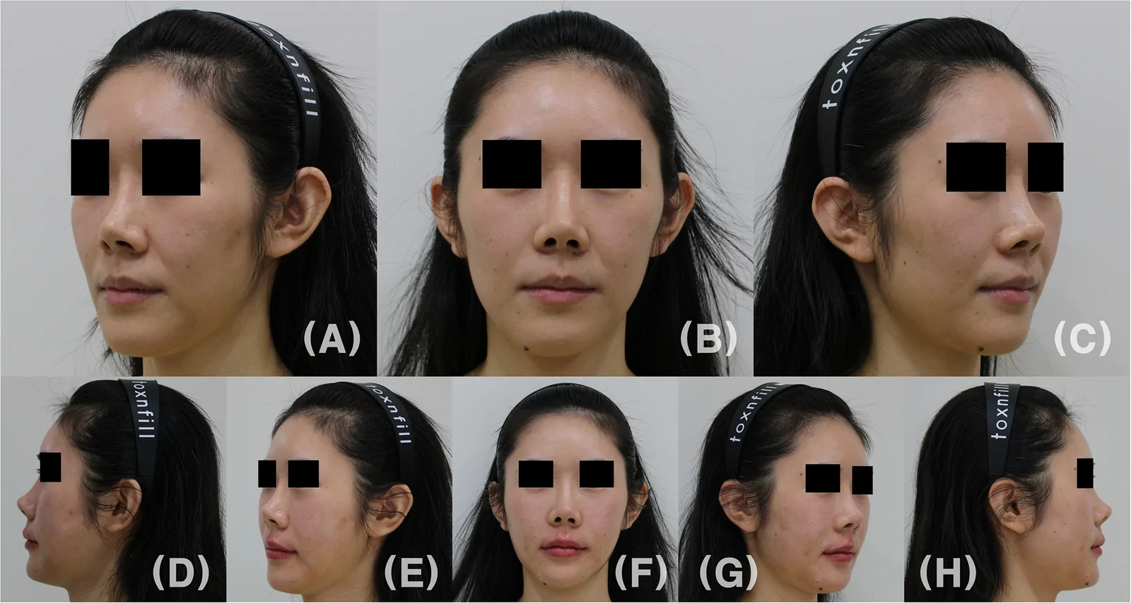
Figure 2 Pre- and post-treatment comparison after Stage 1 structural intervention. Patient: 30/F. Pre-treatment photographs (A–C) and immediate post-treatment photographs (D–H) obtained after Stage 1 structural contouring. Pre-treatment images include left oblique (A), frontal (B), and right oblique (C) views acquired using the standard three-view protocol available at baseline. Post-treatment images include frontal (F), left and right oblique (E and G), and left and right lateral (D and H) views obtained immediately after injection, reflecting an expanded imaging protocol adopted to better visualize global contour continuity. Stage 1 focused on establishing foundational structural support and restoring overall contour continuity using vector-guided deep-plane injections rather than definitive volumization. All images are shown at identical magnification. The post-treatment images were intentionally captured immediately after the procedure to accurately depict injection entry points, vector trajectories, and the resulting immediate structural contour changes prior to tissue settling or secondary adaptation. Only views with corresponding pre- and post-treatment orientations are presented for visual comparison. Minor differences in lighting and head positioning remain; therefore, the images should be interpreted as procedural illustrations rather than standardized objective outcome assessments. No contour-altering image manipulation was performed. Lateral post-treatment views are provided only as additional descriptive views because corresponding pre-treatment lateral photographs were unavailable; they should not be interpreted as direct before-and-after comparisons.
Four months later, after stabilization of the framework established during Stage 1, residual contour discontinuities were selectively refined using HA filler. A total of 2 mL was injected into the forehead and temporal regions to create a smooth lateral curvature, and an additional 4 mL was administered to the anterior cheek and submalar regions to reinforce contour continuity. A further 0.6 mL was used for fine adjustment of the mandibular line.
At the patient’s request, limited filler correction of post-surgical nasal contour asymmetry was also performed after confirming sufficient anatomical space for treatment. All injections followed the predefined entry points and vectors illustrated in Figure 4 .
In this case, material selection and treatment staging reflected case-specific clinical judgment rather than a required sequence within the proposed framework. The diluted PCL-based admixture was selected to provide broadly distributed deep-plane support in the setting of diffuse contour insufficiency, whereas HA was used for localized support, projection, and subsequent refinement. The framework could also have been implemented using HA alone. Material selection therefore remains adaptable, whereas the underlying design logic—based on intended contour relationships, anatomically appropriate pathways, and coordinated vectors—remains consistent.
In the main case, a 23G cannula was used to restore disrupted contours through continuous anatomical pathways rather than to augment isolated defects. Entry points were selected according to the intended vector direction, regional anatomy, accessible deep planes, cannula reach, and planned injectate distribution. They were generally positioned near the lateral boundaries of the contour-discontinuity sites described in Recurrent Contour-Discontinuity Patterns in Northeast Asian Faces .
The injectate was distributed along predefined deep-plane vectors to reconnect adjacent regions while limiting focal overprojection. Relative volume distribution was adjusted according to the extent and anatomical characteristics of each contour discontinuity rather than determined by predetermined absolute replacement volumes.
Forehead and Temporal Region1) Central Forehead : A paramedian entry accessed the supraperiosteal plane. Small-to-moderate volumes, adjusted according to the degree of depression, were distributed broadly to create a curved vertical support axis.2) Lateral Forehead and Temple : Given the lack of immediate bony contact, the cannula targeted deep planes, either supraperiosteal or superficial to the deep temporal fascia, through hairline entry points. Small-to-moderate volumes were distributed in the deep layer to connect the central forehead axis with the lateral forehead and temporal contour without creating focal protrusion.3) Zygomatic Connection : Using an inferior or existing lateral entry with inferior advancement, moderate volumes were distributed along deep vectors to reinforce curvilinear connectivity from the forehead to the zygomatic arch.
Midface (Anterior and Lateral Cheek)1) Anterior Cheek : From the palpebromalar origin, the cannula advanced into the sub-SMAS areolar plane. Small volumes were distributed at low pressure along the groove-oriented vector to address contour discontinuity rather than create focal projection.2) Mid-Cheek Transition : A lateral zygomatic entry with anteroinferior advancement in the sub-SMAS plane allowed broad distribution of moderate volumes to create a smooth transition between the anterior zygoma and cheek.3) Submalar Region : From the submalar groove origin, the cannula advanced posteriorly into the pre-masseteric space within the deep areolar plane. Moderate-or-greater volumes were distributed along the planned vector across the deep compartment to address the broader area of contour discontinuity.
Lower Face (Nasolabial, Chin, and Jawline)1) Nasolabial Fold and Alar Base : Entry at the apex of the nasolabial–alar triangle accessed the supraperiosteal pyriform plane. Small structural volumes were used to reinforce the alar base and support continuity with the midface.2) Chin : Moderate volumes were distributed across the supraperiosteal plane rather than placed as a single focal bolus, with the intention of producing an integrated contour without excessive localized projection.3) Mandibular Border : Bilateral entry points adjacent to the chin accessed the deep supraperiosteal plane. Sequential small increments were distributed along the mandibular border to create a continuous linear contour rather than a segmented profile.
These predefined vectors, anatomical planes, and relative volume distributions represent the clinical translation of the design principles described in Design Logic and Vector Definition . Although their regional application was adjusted according to the patient’s anatomy and the extent of each contour discontinuity, the underlying planning sequence remained consistent: identification of the intended contour relationship, selection of an anatomically appropriate entry point and plane, and distribution along the corresponding vector.
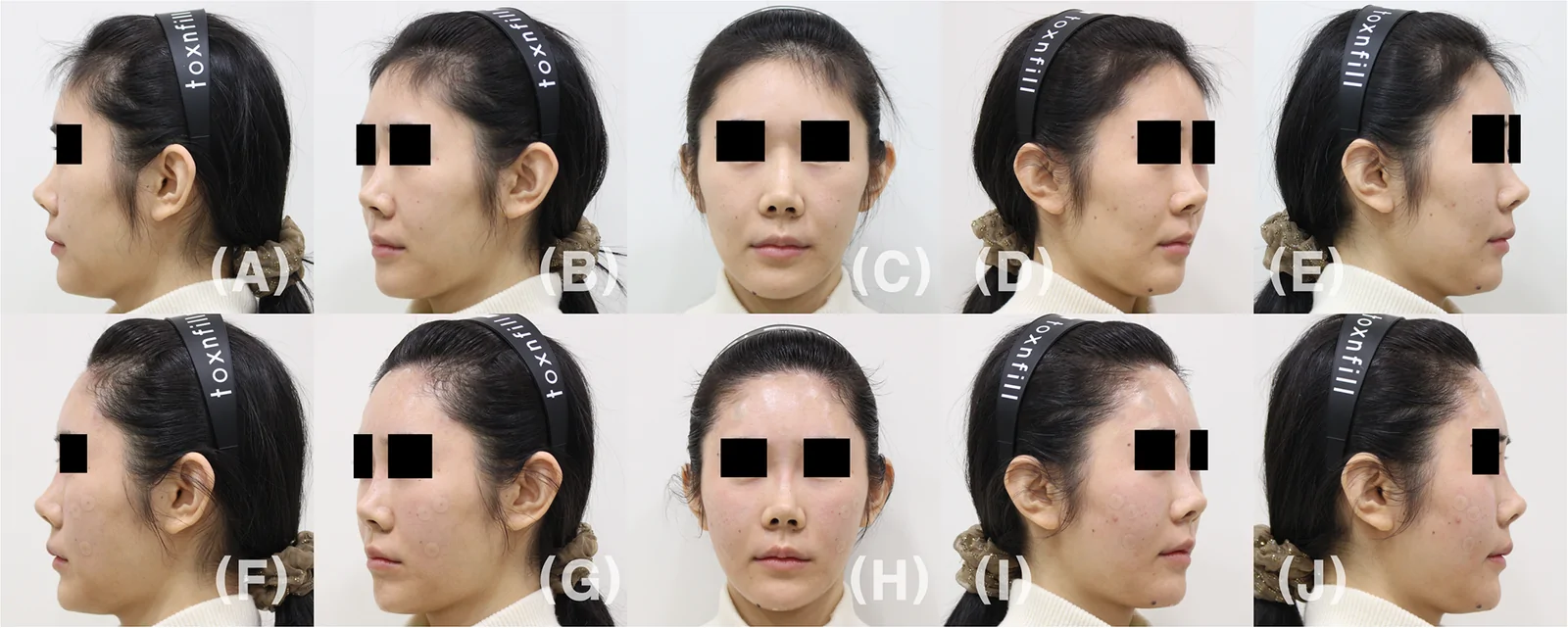
Figure 3 Pre- and post-treatment comparison after Stage 2 refinement. Pre–Stage 2 photographs (A–E) and immediate post–Stage 2 photographs (F–J) shown in frontal (C and H), left and right oblique pre-treatment (B, D) and post-treatment (G, I), and left and right lateral pre-treatment (A and E) and post-treatment (F and J) views at identical magnification. Stage 2 focused on refinement and reinforcement of the pre-defined contour lines following stabilization of the structural framework established during Stage 1. Post-treatment images were obtained immediately after the procedure to illustrate the early contour formation corresponding to the planned vectors before tissue settling. Only views with corresponding pre- and post-treatment orientations are presented for visual comparison. Minor differences in lighting and head positioning remain; therefore, the images should be interpreted as procedural illustrations rather than standardized objective outcome assessments. No contour-altering image manipulation was performed.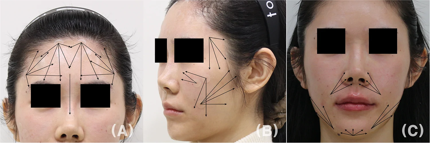
Figure 4 Vector-based injection design and anatomical planes. All procedures were performed using a 23G cannula following predefined entry points and vector directions derived from the anatomy-driven aesthetic design framework. (A) Forehead, temporal, and zygomatic vector design. Forehead and temporal regions were treated in deep planes to establish global structural support: supraperiosteal placement centrally, and supraperiosteal or deep temporal plane placement laterally to restore smooth lateral convexity and continuity. (B) Midfacial vector design involving the anterior cheek, submalar region, and zygomatic connection. In the anterior cheek (palpebromalar region), filler was delivered along predefined vectors within the sub-SMAS areolar plane to restore anterior cheek support while minimizing superficial contour irregularities. Submalar augmentation was achieved by advancing the cannula into the deep areolar plane of the pre-masseteric space, allowing broad, uniform elevation of the submalar contour. (C) Lower-face vector design involving the paranasal region, chin, and mandibular border. For nasolabial and alar base support, vectors were directed to the supraperiosteal pyriform plane via the nasolabial–alar triangle to reinforce deep structural support beyond superficial fold correction. Chin and mandibular contouring were performed in the supraperiosteal plane using distributed deposition rather than focal bolus injection to preserve linearity and avoid surface distortion. This vector- and plane-based strategy illustrates how anatomical depth and directional planning are integrated to achieve contour continuity within the proposed design framework.
The 15 supplementary cases illustrate recurrent regional vector and entry-point patterns applied in comparable anatomical contexts (Supplementary Figures 1–5). The series comprised all patients aged 20–50 years who underwent facial contouring filler treatment at the study clinic between April and November 2025. Cases were not selected according to treatment outcome.
The restricted age range was intended to illustrate structural contour enhancement with less potential confounding from advanced age-related volume loss or soft-tissue laxity. The series was included solely for procedural illustration and was not analyzed as an efficacy, outcome, or validation cohort.
Pre-procedural evaluation included a review of anticoagulant use and previous filler treatments. Procedures were performed under aseptic conditions with topical or local anesthesia. A 23G cannula was used for vector-based injections, whereas a 30G needle was used for lip contouring. Patients were positioned semi-upright or supine according to the treatment region, with repeated multi-view assessment performed during the procedure.
Visual review of the available frontal and oblique photographs showed smoother transitions between adjacent facial regions without obvious focal overprojection ( Figures 2 and 3 ). However, differences in lighting and head position, together with the absence of matched lateral pre-treatment photographs, precluded objective comparison. The images therefore illustrate the correspondence between the planned vectors and the observed contour formation rather than establish treatment efficacy.
No adverse events were observed during the 4-month interval between Stage 1 and Stage 2. Following Stage 2, mild bruising was noted at the local anesthesia sites, but follow-up communication 4 hours after the procedure identified no concerning symptoms. No delayed adverse events following Stage 2 had been reported by the time of manuscript preparation. Nevertheless, the available follow-up was insufficient to assess late-onset complications associated with PCL-based filler.
This proof-of-concept report presents facial filler contouring as a design-first process in which a patient-specific whole-face form is envisioned before the injection plan is determined. The discrepancy between the existing and intended forms is then translated into coordinated regional vectors, anatomically appropriate pathways, and relative filler distribution. The conceptual contribution of the framework lies in formalizing this planning sequence rather than proposing a new filler product, injection technique, or anatomical plane.
The framework shares with the MD Codes™ and broader consensus approaches an emphasis on structured, anatomy-based, full-face assessment. 1 , 2 , 16 The principal distinction lies in the organizational starting point. Established approaches, including the MD Codes™, structure treatment through standardized anatomical units and treatment codes selected according to individual facial attributes. 1 The present framework instead begins with an intended patient-specific whole-face form and translates the relationships within that form into a coordinated procedural plan. It is therefore proposed as a complementary planning perspective rather than as a replacement for established methodologies.
Regional anatomy—including tissue layers, vascular territories, and established injection planes—determines the anatomically appropriate pathways through which the intended contour may be created. 3–6 , 14 The framework therefore operates within established anatomical safety principles while allowing entry points, trajectories, planes, and relative filler distribution to be adapted to the individual patient. 6 In this context, continuity refers to the external three-dimensional contour across adjacent facial regions, rather than to filler placement within a single uninterrupted anatomical plane.
The Reference Facial Form serves as a qualitative working reference that is adapted to the patient’s baseline anatomy, facial proportions, and treatment goals. In the present Northeast Asian context, the zygomatic, mandibular, and chin regions were considered interconnected components of whole-face contour continuity, informed by relevant anatomical, anthropometric, and Asian aesthetic literature. 3–6 , 8–13 This population-specific context supports individualized anatomical translation rather than prescribing a uniform ethnic aesthetic ideal. 8 , 9 The framework may be particularly applicable when contour imbalance extends across multiple adjacent regions and may not be fully represented by a single localized correction.
In the main case, the diluted PCL-based admixture was used off-label during Stage 1 to provide broadly distributed structural support across anatomically appropriate deep planes, followed by selective refinement with HA. Dilution was intended to reduce injection resistance and facilitate broader distribution. However, the rheological and biological properties of the resulting admixture were not experimentally characterized. Its elastic modulus, viscosity, cohesivity, injectability, tissue interactions, and longer-term material behavior therefore remain uncertain. 17–19
Distributed deep-plane deposition was used with the intention of limiting focal material accumulation. However, whether this distribution pattern reduces the risk of nodule formation or other PCL-related complications has not been established. 20 The absence of adverse events in this single case cannot establish the safety, efficacy, or reproducibility of the admixture, and the available 4-month follow-up was insufficient to exclude late-onset complications. 20 , 21
This material sequence represents a case-specific implementation of the proposed design logic rather than a standardized formulation or a required component of the framework. The same planning principles could be implemented using HA fillers alone or other appropriately selected materials. Accordingly, this case should not be interpreted as a general recommendation for the described admixture. Any off-label application requires careful patient selection, material-specific clinical judgment, explicit informed consent, and appropriate long-term follow-up.
The primary clinical evidence in this proof-of-concept report was derived from a single case treated by a single operator, without a comparator or standardized objective outcome measures. The 15 supplementary cases were included solely to illustrate recurrent vector and entry-point patterns and were not analyzed as an efficacy or validation cohort. These features limit conclusions regarding clinical effectiveness, generalizability, reproducibility, and comparative safety.
The Reference Facial Form was not operationalized using numerical landmarks, ratios, or treatment thresholds, and its construct validity and interobserver reliability were not evaluated. The framework should therefore be understood as a qualitative conceptual model rather than a validated or prescriptive treatment protocol.
Clinical photography was not fully standardized, matched lateral pretreatment views were unavailable, and neither morphometric assessment nor validated patient-reported outcomes were used. The available images therefore illustrate early contour formation but do not establish effectiveness, durability, or long-term stability.
Follow-up was also insufficient to assess late-onset complications associated with the off-label diluted PCL-based admixture, whose safety and long-term behavior require separate investigation. Future studies should evaluate the framework using standardized multi-view imaging, predefined morphometric measures, and validated instruments such as the FACE-Q. 22
Facial filler contouring may be approached as an anatomy-informed design process that translates an intended patient-specific whole-face form into coordinated vectors, anatomical planes, and relative filler distribution. This proof-of-concept case illustrates a qualitative framework centered on contour continuity rather than isolated volumetric correction. Further prospective studies are required to evaluate its clinical utility, reproducibility, and generalizability.
声明:中文精读 · 仅供学术参考。内容来自公开文献检索,不代表本人观点,不构成诊疗建议。 医疗美容需在正规医疗机构由执业医师实施。
基于相同主题推荐 · 继续深挖
上睑下垂手术的报道成功率很大程度上取决于“成功”如何定义,而非单纯反映手术本身的效果。
不同人群面部年轻化:专家共识与 AART 方法学临床应用经验/PMC13558784_fig1.webp)
这是一项基于改良 Delphi 法的专家共识(马来西亚6名具8–10年生物刺激剂经验的医师),并附4例 PLLA-SCA(Sculptra)临床应用个案,属专家意见与病例系列,并非
单中心、单臂前瞻性病例系列(证据等级 III),纳入 2024 年河北医科大学第一医院 786 例男性乳腺发育内镜腺体切除术患者(773 例双侧、13 例单侧),平均随访 96.5