多模式腹部塑形:注射溶脂联合超声空化与神经肌肉电刺激的回顾性病例系列
2026年10月9日31 min read
Clinical, Cosmetic and Investigational Dermatology
Clinical, Cosmetic and Investigational Dermatology
结论与实践要点
本文为回顾性病例系列(非随机、无对照),纳入5名规律抗阻训练、低碳饮食但仍存在局限性腹部脂肪堆积的体能活跃受试者,采用标准化6个月多模式方案。方案前期为脱氧胆酸钠+己酮可可碱+L-肉碱注射溶脂联合超声空化(Mesolipo);方案后期(Abdomen Definition)加入皮下注射塑造腹沟、腹直肌内代谢支持及俄式电流神经肌肉电刺激。描述性前后对比显示:中位腹围由94cm降至88cm、体脂率13.7%降至10.3%、脂肪量11.3降至8.7kg、腹部皮褶22降至16mm;多数受试者肌肉量维持或增加,标准照片示腹直肌、白线与半月线轮廓更明显,部分病例在体重变化很小的情况下呈现身体成分重塑。
局限与边界:研究为回顾性、非对照、单中心、小样本(n=5),且多种干预与受试者自身训练/营养习惯同时叠加,故无法确立疗效,也不能区分方案各组分、运动或饮食各自的贡献,属描述性与假设生成性观察,外推须谨慎。本方案含处方注射与有创操作,应在合规医疗机构由有资质医师评估适应证与风险后实施;结论尚待前瞻性、大样本、对照研究验证。
Abdominal contouring has progressively evolved from isolated fat reduction techniques toward multimodal approaches that simultaneously address adipose tissue, muscular definition, and body composition. This retrospective case series describes preliminary clinical observations in five physically active patients with persistent localized abdominal adiposity despite regular resistance training, adherence to a low-carbohydrate diet, and protein supplementation. All patients underwent a standardized six-month multimodal protocol consisting of two sequential phases. The first phase combined injectable lipolysis using sodium deoxycholate, pentoxifylline, and L-carnitine with ultrasonic cavitation (Mesolipo). The second phase (Abdomen Definition) comprised targeted subcutaneous injections intended to enhance abdominal grooves, intramuscular metabolic support of the rectus abdominis, and neuromuscular electrical stimulation using Russian current. Treatment outcomes were assessed through standardized clinical photography, anthropometric measurements, and multifrequency bioimpedance analysis before and after completion of the protocol. Descriptive pre- and post-treatment comparisons showed reductions in median abdominal circumference from 94 to 88 cm, body fat percentage from 13.7% to 10.3%, fat mass from 11.3 to 8.7 kg, and abdominal skinfold thickness from 22 to 16 mm. Skeletal muscle mass was preserved or increased in most patients, while standardized photographs showed visually apparent changes in abdominal contour and definition, including greater visibility of the rectus abdominis, linea alba, and semilunar lines. In several cases, changes in body composition were observed despite minimal variation or slight increases in total body weight, a pattern compatible with body recomposition rather than weight loss alone. Given the retrospective, uncontrolled design, the small sample size, and the simultaneous use of multiple interventions, these observations cannot establish treatment efficacy or determine the relative contribution of individual protocol components, resistance training, or nutritional habits. Nevertheless, the findings provide preliminary, descriptive, and hypothesis-generating observations regarding the feasibility of a multimodal approach to abdominal contouring. Prospective controlled studies with larger samples are needed to evaluate efficacy and determine the contribution of individual therapeutic components.
Keywords: abdominal contouring、case series、mesolipo、abdomen definition、muscle definition、multimodal therapy、injectable lipolysis、sodium deoxycholate、neuromuscular electrical stimulation
Abdominal contouring has evolved from interventions focused exclusively on reducing localized adiposity toward more comprehensive approaches addressing fat distribution, muscular definition, and overall body composition. Beyond simple volume reduction, contemporary aesthetic medicine increasingly considers the anatomical relationship between subcutaneous adipose tissue and the underlying musculature as an important determinant of abdominal appearance. Accordingly, minimally invasive procedures have been explored as adjuncts to regular physical activity and nutritional management in individuals seeking greater abdominal definition. 1
Despite adherence to resistance training and dietary interventions, localized abdominal adiposity may persist in some individuals and remain an important concern in body contouring. Regional adipose tissue characteristics, including differences in adrenergic responsiveness, tissue perfusion, and lipolytic activity, may influence the mobilization of stored lipids and contribute to heterogeneous responses to lifestyle interventions. 1 This has supported interest in minimally invasive procedures designed to target localized adipose deposits.
Injectable lipolysis is among the nonsurgical approaches investigated for localized fat reduction. Sodium deoxycholate acts primarily through a detergent-mediated disruption of adipocyte cell membranes, resulting in adipocytolysis followed by an inflammatory response, macrophage-mediated clearance, and subsequent tissue remodeling. 2–5 Clinical evidence, particularly from studies of localized adiposity, indicates that treatment outcomes and adverse-event profiles are influenced by patient selection, dose, anatomical site, and injection technique. 3–5 Blunt-tip microcannulas may facilitate broader subcutaneous distribution of injected solutions and potentially reduce the likelihood of inadvertent vascular injury; however, procedural safety remains dependent on anatomical knowledge, appropriate dosing, and careful technique. 4 , 5
Energy-based modalities have also been investigated as adjunctive approaches to localized adiposity. Ultrasound-based treatments deliver acoustic energy to subcutaneous tissues and have been proposed to induce mechanical effects that may contribute to adipocyte disruption and lipid mobilization. 5 , 6 However, the extent to which ultrasonic cavitation enhances the effects of injectable lipolysis, particularly when both modalities are used sequentially within the same protocol, remains insufficiently established. Therefore, any complementary or synergistic interaction between these interventions should currently be regarded as a therapeutic rationale rather than a demonstrated biological or clinical effect.
Reduction of subcutaneous adiposity alone may not fully account for the visual perception of abdominal definition, which also depends on the visibility of anatomical landmarks such as the linea alba, semilunar lines, tendinous intersections, and the underlying rectus abdominis. 7 Neuromuscular electrical stimulation (NMES) provides a more established physiological basis for addressing the muscular component, as electrically induced contractions can recruit motor units and, depending on stimulation parameters and repeated exposure, promote neuromuscular adaptations. 8 , 9 Nevertheless, the magnitude and clinical relevance of these adaptations vary according to stimulation characteristics, treatment duration, and the population studied.
Nutritional and metabolic factors may also influence preservation and adaptation of skeletal muscle during changes in body composition. 10 , 11 Compounds such as beta-hydroxy-beta-methylbutyrate (HMB), creatine, glutamine, L-carnitine, ornithine alpha-ketoglutarate, and vanadium-containing compounds have been investigated individually in nutritional, metabolic, or exercise-related contexts because of their proposed roles in protein metabolism, cellular energetics, substrate utilization, or muscle physiology. Importantly, however, most available evidence relates to systemic or oral supplementation and established physiological mechanisms rather than localized intramuscular administration for aesthetic abdominal contouring. Evidence specifically supporting the local intramuscular injection of these compounds into the rectus abdominis, either individually or in combination, remains limited. Their inclusion in the present protocol should therefore be regarded as a biologically motivated and exploratory component rather than an established strategy for inducing localized muscle hypertrophy or definition.
From a safety and regulatory perspective, multimodal injectable protocols require particular caution because compounded formulations, anatomical injection sites, concentrations, and routes of administration may differ from those evaluated for individual ingredients in conventional therapeutic or nutritional settings. Appropriate aseptic technique, anatomical knowledge, conservative dosing, patient selection, and monitoring are therefore essential. Moreover, the use of multiple interventions within the same treatment course limits the ability to determine the safety and clinical contribution of each individual component.
The protocol evaluated in the present series was structured sequentially to first address the subcutaneous adipose compartment and subsequently focus on anatomical definition and muscular stimulation. The initial phase combined injectable lipolysis with ultrasonic cavitation to target persistent localized adiposity. This was followed by a second phase incorporating targeted subcutaneous injections along abdominal anatomical landmarks, intramuscular administration of metabolically oriented compounds into the rectus abdominis, and NMES. This sequence was based on the clinical rationale that reducing the overlying adipose compartment before emphasizing anatomical landmarks and muscular recruitment could facilitate visualization of abdominal definition. However, this sequence has not been independently validated, and potential additive or synergistic effects among its components remain hypothetical.
Although multimodal strategies combining injectable, energy-based, and muscle-focused interventions have been proposed for body contouring, the available literature predominantly evaluates individual modalities, and evidence supporting integrated protocols remains limited. In particular, there is insufficient evidence to determine whether combining these interventions provides benefits beyond those attributable to individual procedures, resistance training, nutritional management, or changes in body composition over time.
The present retrospective case series therefore describes preliminary clinical observations in five consecutive patients with localized abdominal adiposity who underwent a standardized six-month multimodal protocol combining injectable lipolysis, ultrasonic cavitation, targeted subcutaneous and intramuscular injections, and neuromuscular electrical stimulation. Anthropometric measurements, multifrequency bioimpedance analysis, standardized clinical photography, and treatment-related adverse events were descriptively evaluated before and after the protocol. Given the uncontrolled design and simultaneous use of multiple interventions, the objective was not to establish efficacy or causal relationships, but rather to document the clinical course, feasibility, safety observations, and body-composition changes associated with this exploratory multimodal approach and to generate hypotheses for future controlled investigation.
This study was designed as a retrospective case series conducted between 2022 and 2023, including five patients presenting localized abdominal adiposity. All participants were engaged in regular resistance training programs and followed structured dietary strategies characterized by reduced carbohydrate intake and protein supplementation. Exercise and dietary habits were maintained throughout the treatment period and were not experimentally controlled as part of the protocol; therefore, they were considered potential confounding factors when interpreting changes in body composition. None of the patients reported the use of anabolic agents, glucagon-like peptide-1 (GLP-1) receptor agonists, glucose-dependent insulinotropic polypeptide (GIP) agonists, or other pharmacological interventions that could significantly influence body composition. Given the retrospective case-series design and the sample size of five patients, no formal sample-size or statistical power calculation was performed, and outcomes were analyzed descriptively without inferential efficacy testing.
The study protocol was reviewed and approved by the Research Ethics Committee of Hospital Mater Dei, Minas Gerais, Brazil, through Plataforma Brasil (CEP Protocol No. 82947324.1.0000.5134). Prior to participation, all patients provided written informed consent to participate in the study. In addition, all patients signed a separate written consent authorizing the publication of their clinical information, photographs, and the use of their image and voice for scientific publication. Institutional ethical approval covered the conduct of the study and the publication of the case details. The study was conducted in accordance with the principles of the Declaration of Helsinki.
The treatment protocol was performed over a six-month period and was structured into two sequential phases, each targeting specific physiological components involved in abdominal contouring. The first phase was directed primarily toward localized subcutaneous adiposity, whereas the second phase focused on anatomical contouring and muscular stimulation. This sequence was selected based on the clinical rationale that initially addressing the overlying adipose compartment could facilitate subsequent visualization of abdominal anatomical landmarks; however, this sequence has not been independently validated. All injectable formulations used in both phases were compounded by Biometil Laboratory (Santa Catarina, Brazil) as sterile galenic preparations.
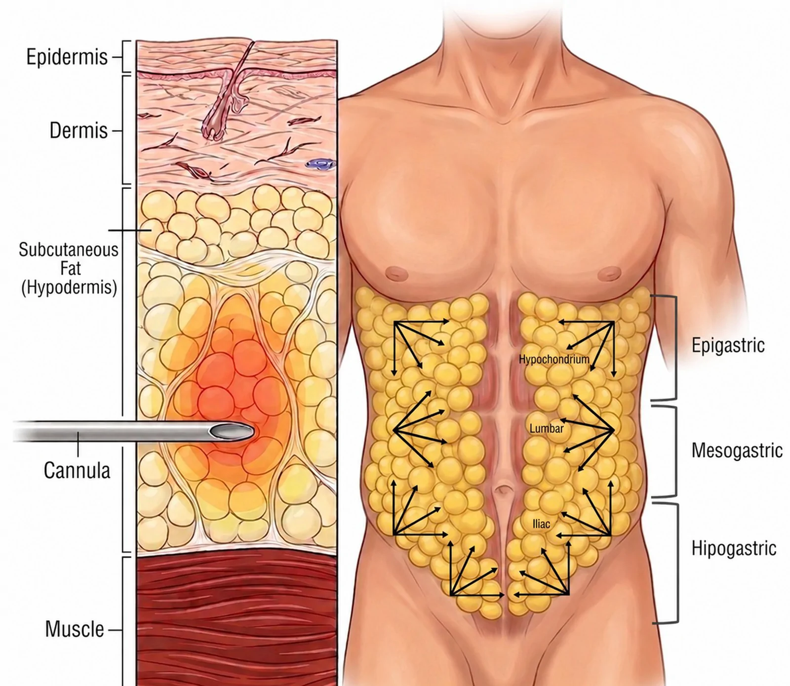
The first phase consisted of a modified injectable lipolysis technique combined with energy-based therapy, referred to as the Mesolipo Technique, as illustrated in Figure 1 . The injectable formulation included sodium deoxycholate at a concentration of 4.75%, pentoxifylline 40 mg/2mL, L-carnitine 600 mg/2 mL, and lidocaine 2%/2 mL. The total formulation volume administered per session ranged from 10 to 14 mL. The solution was administered into the subcutaneous tissue using a blunt-tipped microcannula 22 G x 70 mm through a retrograde fan technique, allowing for homogeneous distribution across the treated area.
Sodium deoxycholate acts as a detergent agent capable of disrupting adipocyte membranes, leading to adipocytolysis and subsequent inflammatory clearance. 1 Pentoxifylline was included due to its phosphodiesterase inhibitory effect, which increases intracellular cyclic adenosine monophosphate levels and enhances lipolytic activity, in addition to improving local microcirculation. 2 L-carnitine was incorporated based on its role in facilitating mitochondrial transport of fatty acids, supporting beta-oxidation processes. 2
Six Mesolipo sessions were performed at two-week intervals. The total dose of sodium deoxycholate administered per session was individualized according to patient body weight and abdominal skinfold thickness, following a predefined dosing algorithm presented in Table 1 . The dosing algorithm was established based on available toxicological and preclinical data for sodium deoxycholate, which demonstrate a dose-dependent cytotoxic profile. Experimental studies report a no-observed-adverse-effect level of approximately 5 mg/kg and systemic tolerability up to 50 mg/kg in animal models. Considering the interspecies correction factor and the characteristics of subcutaneous administration, more conservative dosing thresholds, typically below 5 mg/kg, were adopted to enhance safety. 4 , 5 , 12 Within this context, the proposed dosing strategy was designed to allow proportional adjustment of the administered volume according to adipose tissue distribution while minimizing the risk of adverse effects. The toxic response of sodium deoxycholate is predominantly local, related to its detergent action on cell membranes, which induces adipocyte lysis and a controlled inflammatory process. Systemic toxicity remains uncommon and is generally associated with excessive dosing or inadvertent intravascular administration.
Table 1 Dosing Protocol for Sodium Deoxycholate (4.75% | 95 mg/ 2 mL) Based on Body Weight and Abdominal Skinfold Thickness. The Dosing Strategy Was Designed to Ensure Safe and Proportional Administration According to Adipose Tissue Volume, Following a Stepwise Escalation Model Grounded in Toxicological Safety Considerations
| Body Weight (kg) | Skinfold Thickness (cm) | Maximum Dose (mg) |
|---|---|---|
| ≤70 | 2 < Y ≤ 3 | 100 |
| 3 < Y ≤ 4 | 200 | |
| 70 < X ≤ 90 | 2 < Y ≤ 3 | 100 |
| 3 < Y ≤ 4 | 200 | |
| 4 < Y ≤ 5 | 300 | |
| 90 < X ≤ 120 | 2 < Y ≤ 3 | 100 |
| 3 < Y ≤ 4 | 200 | |
| 4 < Y ≤ 5 | 300 | |
| 5 < Y ≤ 6 | 400 | |
| ≥130 | 2 < Y ≤ 3 | 100 |
| 3 < Y ≤ 4 | 200 | |
| 4 < Y ≤ 5 | 300 | |
| 5 < Y ≤ 6 | 400 | |
| Y > 6 | 500 |
For greater reproducibility, the relationship between body weight, abdominal skinfold thickness, administered volume, and sodium deoxycholate dose is presented in Table 1 . The total formulation volume administered per session ranged from 10 to 14 mL and was individualized according to body weight and abdominal skinfold thickness following the predefined dosing algorithm. The administered volumes were 12, 10, 14, 12, and 10 mL for Cases 1 through 5, respectively. Sodium deoxycholate was supplied at a concentration of 4.75% (47.5 mg/mL; 95 mg/2 mL). The highest sodium deoxycholate dose was administered to Case 3, who had a baseline body weight of 92.1 kg and an initial abdominal skinfold thickness within the >5 to ≤6 cm range. The administered dose corresponded to 380 mg (four 2-mL ampoules), equivalent to approximately 4.13 mg/kg. Thus, even the highest dose administered in this series remained below the predefined conservative threshold of 5 mg/kg.
Immediately after each injection session, ultrasonic cavitation was applied to the treated area using the Vibria Maxx device (HTM, Brazil), with standardized parameters including an intensity of 1.2 W/cm 2 , power output of 12 W, emission frequency of 100 Hz, a modulation cycle of 20%, and a total application time of 5 minutes and 11 seconds. The same device settings and treatment duration were used across treatment sessions. This step was included based on the proposed mechanical effects of ultrasonic energy on adipose tissue and its potential role in facilitating lipid mobilization within the interstitial environment. 5 However, a synergistic effect between ultrasonic cavitation and injectable lipolysis was not independently evaluated in the present case series.
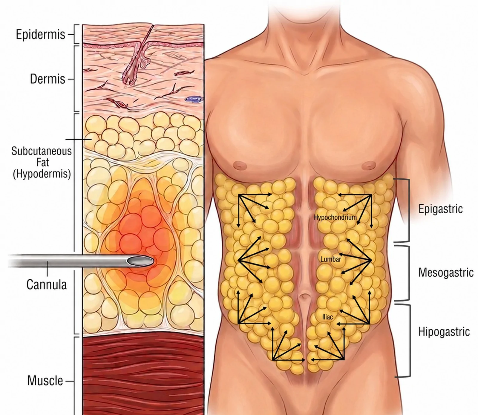
Figure 1 Mesolipo Technique (Phase 1) for localized abdominal adiposity. Schematic representation of the subcutaneous injection technique used during the first phase of the multimodal abdominal contouring protocol. The compounded formulation was administered into the abdominal subcutaneous tissue using a 22-G × 70-mm blunt-tip microcannula through a retrograde fan technique, followed immediately by ultrasonic cavitation using the Vibria Maxx device (HTM, Brazil). The injectable formulation contained sodium deoxycholate, pentoxifylline, L-carnitine, and lidocaine. This illustrative image was generated with the assistance of Gemini Pro.
The second phase consisted of a structured abdominal high-performance protocol, referred to as Abdomen Definition, performed over twelve weekly sessions and illustrated in Figure 2 . Each session was divided into three sequential steps.
In the first step, a subcutaneous injection technique was performed along predefined abdominal anatomical landmarks, including the linea alba, semilunar lines, and regions corresponding to the tendinous intersections. A formulation containing vitamin B12 2500 μg/ 2 mL, L-carnitine 600 mg/ 2 mL, caffeine 50 mg/ 2 mL, benzopyrone 10 mg/ 2 mL, sodium deoxycholate 120 mg/ 2 mL, and lidocaine 2%/2 mL was administered along the anatomical lines separating the rectus abdominis segments. 5 , 7 The injections were performed using a 30G needle inserted at approximately 45 degrees relative to the skin surface, with the bevel oriented upward. A manual skin fold was created to ensure proper positioning within the subcutaneous plane. The solution was delivered using a retroinjection technique, with volumes ranging from 0.2 mL to 0.4 mL per injection point, distributed along the predefined anatomical landmarks. A total of 25–50 injection points were used per session, corresponding to a total subcutaneous injection volume of 12 mL and a total sodium deoxycholate dose of 120 mg.
In the second step, intramuscular injections were performed directly into the rectus abdominis muscles using a formulation containing beta-hydroxy-beta-methylbutyrate (HMB) 200 mg/5 mL, ornithine alpha-ketoglutarate (OKG) 400 mg/ 5 mL, glutamine 600 mg/ 5 mL, vanadium 50 µg/ 5 mL, L-carnitine 800 mg/ 5 mL, and creatine 430 mg/ 5 mL. Applications were performed using a 26G × 13 mm needle inserted at a 90° angle into the rectus abdominis, with the bevel oriented parallel to the muscle fibers. Injection volumes ranged from 0.2 to 0.8 mL per point and were distributed across 6–25 intramuscular injection points, resulting in a total intramuscular volume of 5 mL per session. The compounds included in this formulation were selected based on their proposed roles in skeletal muscle metabolism and body-composition physiology. 7 , 11 However, because evidence supporting their localized intramuscular administration for aesthetic abdominal contouring is limited, this component should be regarded as exploratory rather than as an established method for inducing localized muscle hypertrophy or definition.
Immediately following the intramuscular injections, the third step consisted of neuromuscular electrical stimulation using Russian current delivered by the Vibria Maxx device (HTM, Brazil). Surface electrodes were positioned over the abdominal region corresponding to the rectus abdominis muscles, and electrical stimulation was applied to induce involuntary muscle contractions. According to the manufacturer’s specifications, the Russian current is a symmetrical, balanced biphasic current delivered through up to eight independent channels, with a fixed carrier frequency of 2500 Hz, an adjustable burst frequency ranging from 1 to 200 Hz, and a maximum output intensity of 150 mA. Stimulation intensity was progressively adjusted according to each patient’s tolerance threshold, aiming to produce visible and sustained muscle contractions without clinically relevant discomfort. Electrical stimulation has been associated with increased motor unit activation and may contribute to muscle adaptation processes through repeated contraction cycles. 8 , 9
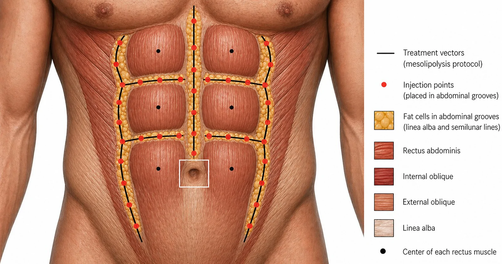
Figure 2 Abdomen Definition technique (Phase 2) illustrating the anatomical landmarks and injection strategy used for abdominal contouring. Treatment vectors follow the natural abdominal grooves, including the linea alba and semilunar lines, where subcutaneous fat accumulates, with red dots indicating injection points for Step 1, targeting selective reduction of intermuscular adipose tissue. In addition, the schematic highlights the central regions of the rectus abdominis, corresponding to Step 2, where intramuscular injections are performed. The diagram also demonstrates the spatial relationship between adipose tissue, rectus abdominis, and adjacent muscle groups. This illustrative image was generated with the assistance of Gemini Pro.
Treatment outcomes were assessed using anthropometric measurements, bioimpedance analysis, and standardized clinical photography. Anthropometric evaluation included body weight, height, body mass index (BMI), abdominal circumference, and abdominal skinfold thickness measured using a calibrated scientific adipometer (Avanutri Scientific Adipometer, Brazil). Abdominal circumference was measured at the level of the umbilicus with the patient in the standing position at the end of a normal expiration. Abdominal skinfold thickness was assessed at three standardized sites: above the umbilicus, at the level of the umbilicus, and below the umbilicus. Three measurements were obtained, and the mean value was used for analysis. All anthropometric assessments were performed at baseline and after completion of the treatment protocol using the same equipment and by the same evaluator. Individual baseline and post-treatment anthropometric measurements are presented in Table 2 .
Table 2 Integrated Bioimpedance and Anthropometric Parameters Before and After the Multimodal Abdominal Contouring Protocol
| Patient | Weight (kg) | BMI (kg/m 2 ) | AbdominalCircumference (cm) | BodyFat (%) | FatMass (kg) | LeanMass (%) | Muscle Mass (kg) | Skinfold (mm) | Phase Angle (°) |
|---|---|---|---|---|---|---|---|---|---|
| Case 1 | 83.1 → 82.8 | 25.1 → 25.0 | 94 → 88 | 13.7 → 10.3 | 11.4 → 8.5 | 86.3 → 89.7 | 40.9 → 41.4 | 22 → 16 | 8.5 → 7.9 |
| Case 2 | 68.9 → 66.3 | 24.7 → 23.8 | 89 → 83 | 20.0 → 18.2 | 13.8 → 12.1 | 80.0 → 81.8 | 27.1 → 27.4 | 26 → 22 | 9.0 → 9.1 |
| Case 3 | 92.1 → 95.3 | 26.3 → 26.7 | 101 → 96 | 11.2 → 9.2 | 10.3 → 8.7 | 88.8 → 90.8 | 44.0 → 45.4 | 18 → 14 | 7.5 → 8.2 |
| Case 4 | 93.8 → 88.9 | 27.1 → 25.7 | 105 → 98 | 7.7 → 7.3 | 7.3 → 6.5 | 92.3 → 92.7 | 47.0 → 45.0 | 14 → 12 | 8.9 → 8.6 |
| Case 5 | 58.6 → 60.0 | 21.8 → 22.3 | 82 → 77 | 19.3 → 17.1 | 11.3 → 10.3 | 80.7 → 82.9 | 25.8 → 27.7 | 24 → 20 | 7.6 → 8.6 |
Bioimpedance analysis was performed using the Tera Science device (Tera Science, Brazil) to assess body fat percentage, fat mass, lean mass, skeletal muscle mass, and phase angle. Baseline and final assessments were performed under standardized premeasurement conditions, including an 8-hour fasting period and avoidance of strenuous physical exercise for at least 8 hours before assessment. Participants were instructed to maintain their usual hydration status before the examination and to avoid excessive fluid intake immediately before measurement. The same premeasurement conditions were maintained as consistently as possible at baseline and final assessments to minimize the potential influence of hydration status, recent food and fluid intake, and physical activity on bioimpedance-derived body-composition estimates. Individual baseline and post-treatment bioimpedance-derived body-composition measurements are also presented in Table 2 .
Standardized two-dimensional clinical photographs were obtained at baseline and after completion of the six-month treatment protocol using the same digital single-lens reflex camera (Canon EOS Rebel T6i, Canon Inc., Japan), with uniform lighting, background, camera settings, and patient positioning. The camera was positioned at a fixed distance of 70 cm from the patient, with the lens aligned approximately at the level of the umbilicus. Photographs were obtained in frontal, lateral, and oblique views, with the abdomen photographed both relaxed and voluntarily contracted. The same positioning and contraction instructions were provided at baseline and final assessments. No digital modification of body contour was performed. The paired baseline and post-treatment clinical photographs for Cases 1–5 are presented in Figures 3–7 , respectively.
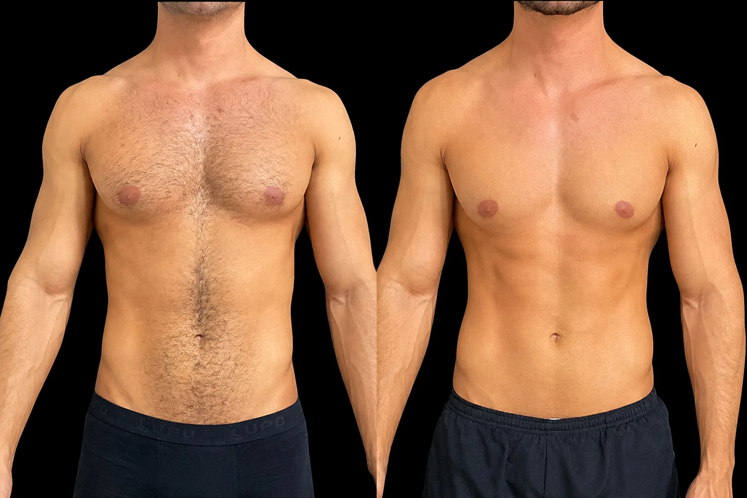
Figure 3 Case 1. Standardized clinical photographs obtained at baseline (left) and after completion of the six-month multimodal abdominal contouring protocol (right). The paired photographs show visually apparent differences in abdominal projection and contour, particularly in the infraumbilical region, together with greater visibility of the rectus abdominis, linea alba, and semilunar lines.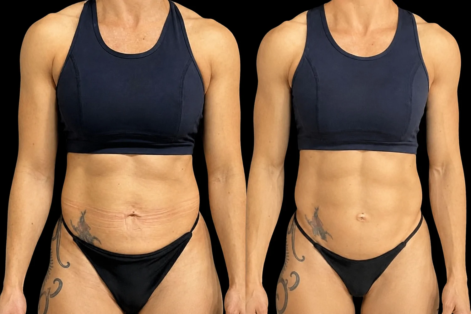
Figure 4 Case 2. Standardized clinical photographs obtained at baseline (left) and after completion of the six-month multimodal abdominal contouring protocol (right). The paired photographs show visually apparent differences in abdominal contour and infraumbilical fullness, together with greater visibility of the rectus abdominis and adjacent anatomical landmarks.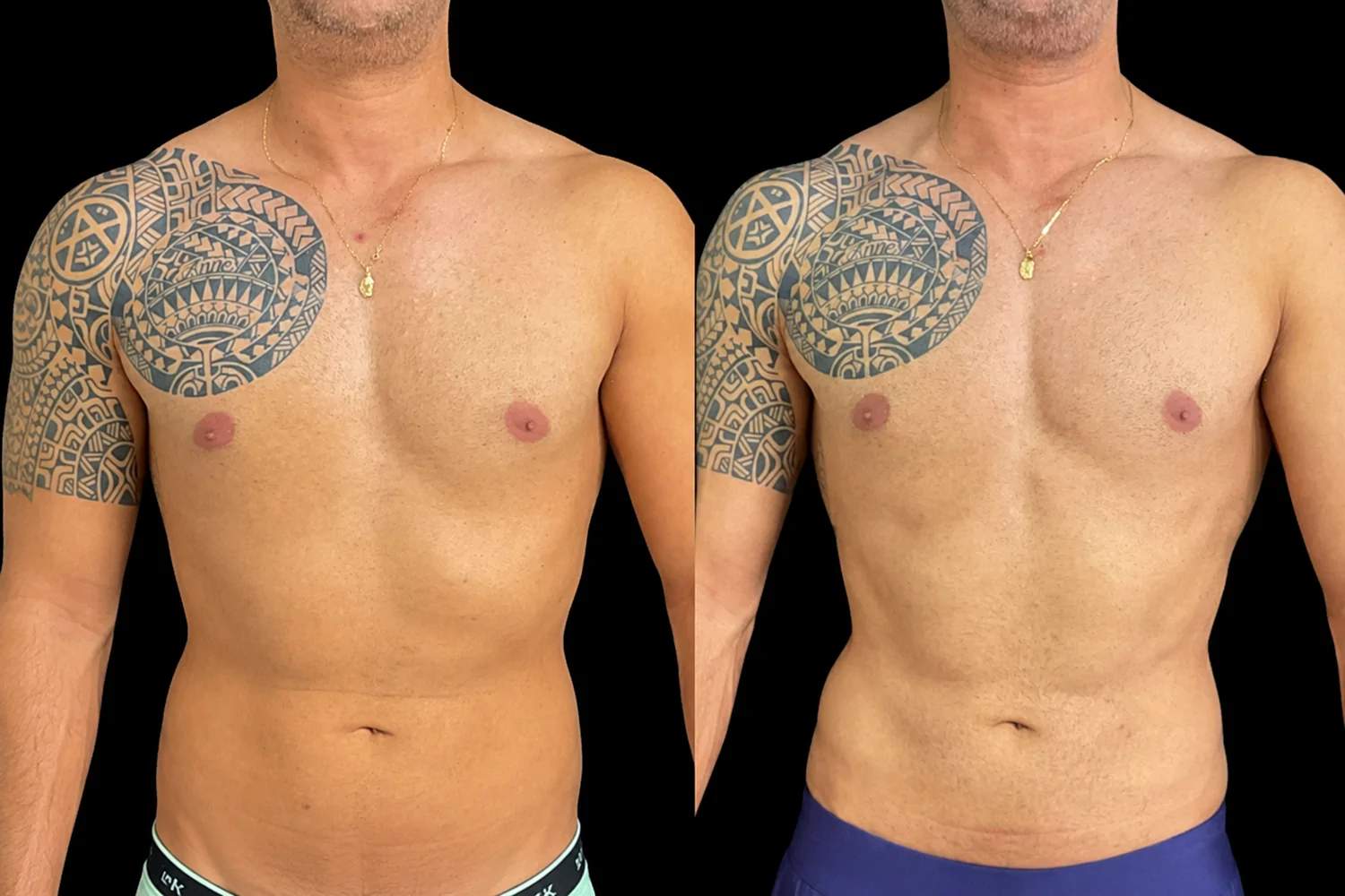
Figure 5 Case 3. Standardized clinical photographs obtained at baseline (left) and after completion of the six-month multimodal abdominal contouring protocol (right). The paired photographs show visually apparent differences in abdominal contour and definition, including greater visibility of the rectus abdominis and tendinous intersections. These photographic observations are presented descriptively and should be interpreted together with the corresponding anthropometric and body-composition measurements.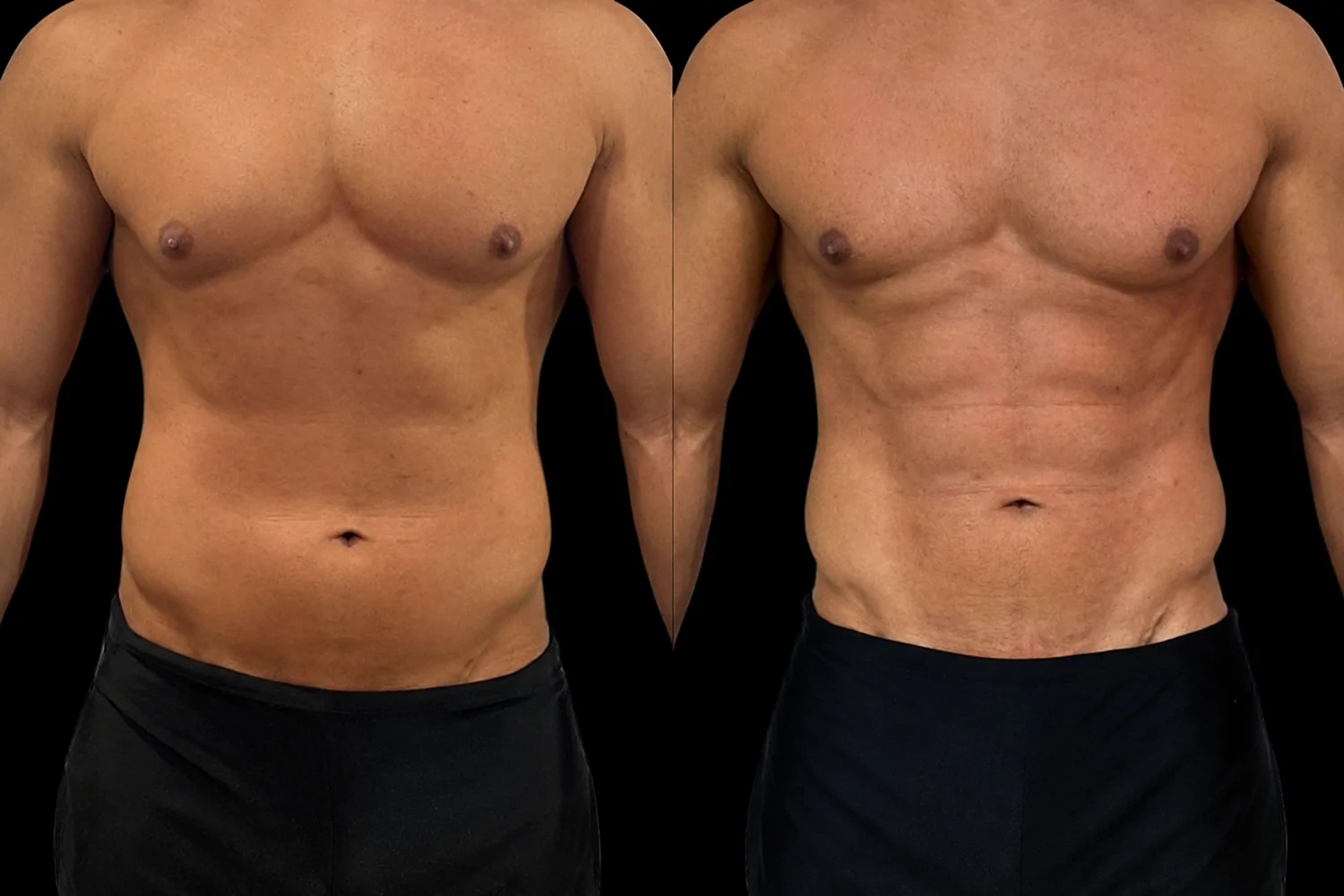
Figure 6 Case 4. Standardized clinical photographs obtained at baseline (left) and after completion of the six-month multimodal abdominal contouring protocol (right). The paired photographs show visually apparent differences in abdominal projection and contour, together with greater delineation of the rectus abdominis and adjacent anatomical landmarks.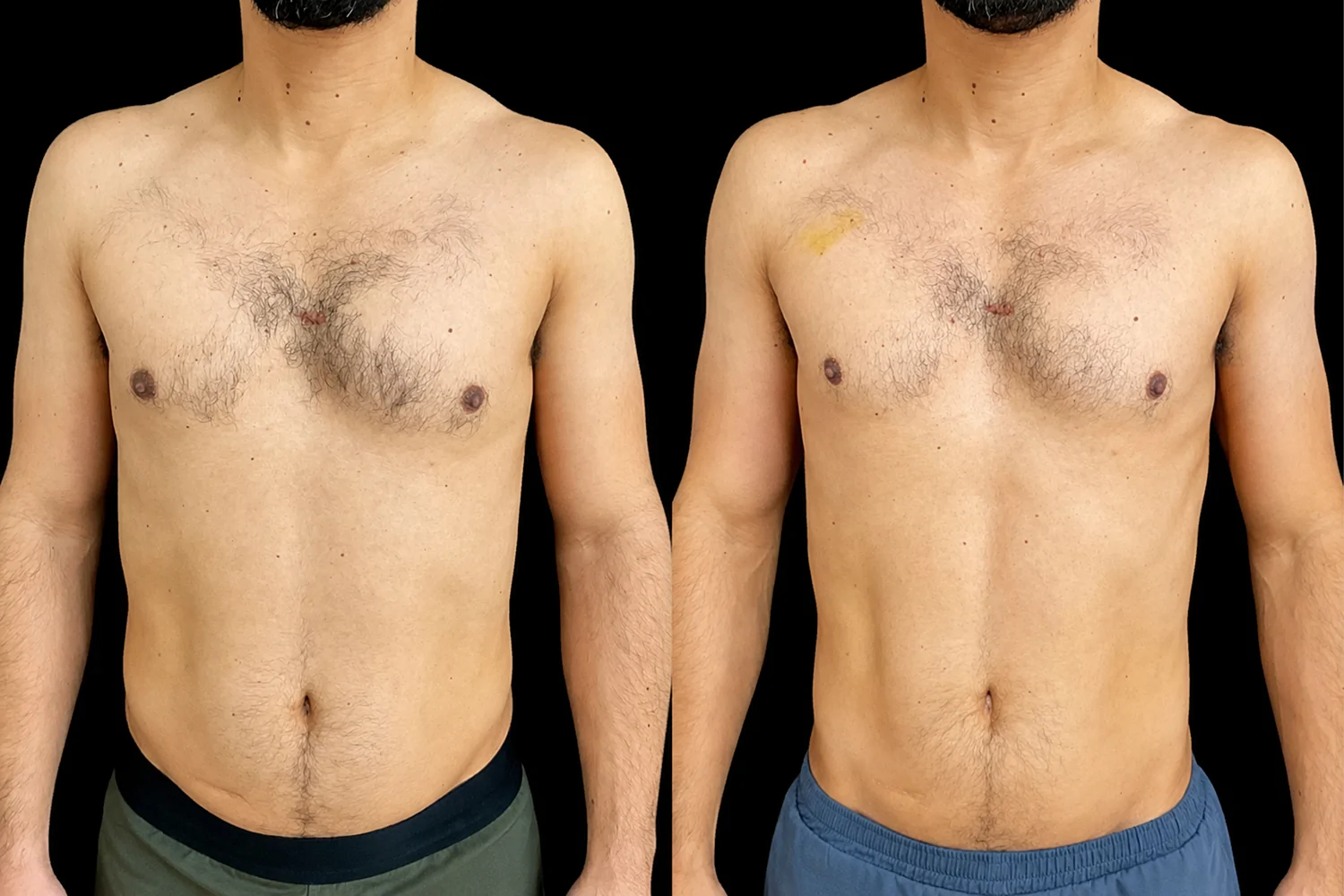
Figure 7 Case 5. Standardized clinical photographs obtained at baseline (left) and after completion of the six-month multimodal abdominal contouring protocol (right). The paired photographs show visually apparent differences in abdominal contour and localized abdominal fullness, together with greater visibility of the rectus abdominis, linea alba, and semilunar lines.
Safety was monitored throughout the six-month treatment period. At each treatment visit, patients were assessed for local and systemic adverse events, including pain, edema, erythema, ecchymosis, nodules, sensory disturbances, signs of infection, muscle-related symptoms, and systemic reactions. Mild and transient local reactions, when present, were recorded and managed conservatively. Treatment discontinuation or modification was considered in the event of clinically significant adverse reactions, newly identified contraindications, pregnancy or lactation, or any change in the patient’s clinical condition that could compromise treatment safety. No serious adverse events, clinically significant complications, or treatment discontinuations due to adverse events occurred during the study period.
The six-month period corresponded to completion of the sequential treatment protocol. The final anthropometric, bioimpedance, and photographic assessments were performed at the end of treatment rather than after a treatment-free follow-up period. Therefore, the present observations describe changes associated with completion of the six-month protocol and do not establish the persistence or durability of these changes after treatment cessation.
Because this was an uncontrolled retrospective case series involving five patients, no inferential statistical testing or formal efficacy hypothesis testing was performed. Results were reported descriptively using individual pre- and post-treatment anthropometric and bioimpedance measurements. The small sample size precluded statistical generalization or causal inference. Furthermore, concomitant resistance training and dietary practices represented important potential confounders, while the sequential multimodal design precluded determination of the independent contribution of injectable lipolysis, ultrasonic cavitation, targeted subcutaneous injections, intramuscular injections, or neuromuscular electrical stimulation to the observed changes.
This case series describes preliminary clinical observations in five physically active patients with persistent localized abdominal adiposity who underwent a standardized multimodal abdominal contouring protocol combining injectable lipolysis, ultrasonic cavitation, targeted subcutaneous and intramuscular injections, and neuromuscular electrical stimulation. Despite differences in baseline body composition, all patients showed reductions in abdominal circumference, body fat percentage, fat mass, and abdominal skinfold thickness ( Table 2 ), accompanied by visually apparent changes in abdominal contour and muscular definition ( Figures 3–7 ). These findings should be interpreted as descriptive associations observed after completion of the multimodal protocol rather than as evidence of treatment efficacy. Because all interventions were administered sequentially and patients continued resistance training and dietary management throughout the treatment period, the relative contribution of the protocol, its individual components, and concomitant lifestyle factors cannot be determined. Nevertheless, the observations provide a clinical basis for further investigation of approaches that simultaneously address adipose tissue and muscular definition.
The initial phase of the protocol focused on localized adipose tissue remodeling through the combination of sodium deoxycholate and ultrasonic cavitation. Sodium deoxycholate promotes selective adipocyte membrane disruption, resulting in adipocytolysis followed by macrophage-mediated clearance and progressive tissue remodeling. 2–5 The subsequent application of ultrasonic cavitation was based on its proposed mechanical effects on adipose tissue and potential to facilitate lipid mobilization. 5 , 6 However, the present study was not designed to evaluate whether ultrasonic cavitation enhanced the effects of sodium deoxycholate or whether a synergistic interaction occurred between these modalities. Although reductions in abdominal circumference, skinfold thickness, and fat mass were observed across all five cases, these changes cannot be attributed specifically to the combined lipolysis–ultrasound phase because of the absence of a comparator group and the subsequent use of additional interventions.
Unlike conventional lipolytic treatments, the second phase of the protocol was designed to address both superficial abdominal adiposity and the anatomical relationship with the underlying rectus abdominis musculature. Selective subcutaneous injections were performed along the linea alba, semilunar lines, and tendinous intersections, whereas intramuscular injections and neuromuscular electrical stimulation were incorporated as exploratory muscle-focused components. Preservation or increases in bioimpedance-derived skeletal muscle mass were observed in most patients and occurred concurrently with visually apparent changes in abdominal definition. However, these observations do not establish that the intramuscular formulation or neuromuscular electrical stimulation caused the changes in muscle mass or abdominal definition. Resistance training, nutritional practices, changes in adipose tissue, measurement variability, and the combined treatment components represent alternative or complementary explanations. Furthermore, evidence specifically supporting localized intramuscular administration of the compounded formulation for abdominal muscle definition remains limited. Accordingly, the findings should be regarded as hypothesis-generating observations supporting further evaluation of this integrated concept rather than evidence of a specific muscle-modulating effect.
An interesting descriptive observation emerging from the present series was the apparent variation in response patterns according to baseline body composition ( Table 2 and Figures 3–7 ). Patients presenting greater initial adiposity tended to show larger absolute changes in fat-related parameters, whereas leaner individuals appeared to show more visually apparent changes in muscular definition despite relatively modest quantitative changes. Given the sample size of only five patients, these patterns should not be interpreted as evidence of differential treatment response or as establishing baseline body composition as a predictor of outcome. Rather, they illustrate the potential relevance of considering both adipose distribution and underlying anatomical landmarks when evaluating abdominal contour. Abdominal aesthetics depend not only on the amount of adipose tissue but also on the interaction between fat distribution and visualization of the underlying musculature. 7 , 13
Changes compatible with body recomposition were observed in some patients ( Table 2 ). In two cases, body weight remained stable or increased slightly despite measurable reductions in body fat and abdominal circumference, accompanied by preservation or gains in bioimpedance-derived skeletal muscle mass. Similar patterns have been reported in resistance-trained individuals, in whom simultaneous fat reduction and maintenance or gain of lean tissue may occur in association with resistance training and appropriate nutritional support. 10 , 11 Importantly, the patients in the present series continued resistance training and dietary management throughout the study period. Therefore, the observed pattern cannot be attributed specifically to the multimodal treatment protocol, intramuscular injections, or neuromuscular electrical stimulation. These observations nevertheless illustrate why changes in total body weight alone may not adequately reflect concurrent changes in body-composition parameters in physically active individuals undergoing body contouring interventions.
Bioimpedance-derived body-composition parameters should also be interpreted cautiously. Although reductions in fat-related parameters and preservation or increases in lean or skeletal muscle mass were observed across the series, bioimpedance provides indirect estimates of body composition and is influenced by hydration status, recent exercise, food intake, and other physiological and technical factors. Phase angle is similarly sensitive to hydration and measurement conditions and should not be interpreted as a direct measure of treatment-related improvement in cellular integrity. Accordingly, the concordance between anthropometric measurements, bioimpedance-derived parameters, and standardized clinical photography strengthens the descriptive internal consistency of the observations but does not establish causal treatment effects. Future prospective studies should incorporate more objective imaging modalities, such as high-frequency ultrasound, dual-energy X-ray absorptiometry, or magnetic resonance imaging, where appropriate, to characterize changes in adipose and muscular compartments independently.
Several limitations should be acknowledged. The retrospective design, the small sample size (n = 5), the absence of a control group, and the inability to isolate the individual contribution of each therapeutic component preclude conclusions regarding efficacy, superiority, or causal mechanisms. No inferential statistical analysis or efficacy hypothesis testing was appropriate for this exploratory case series, and the findings cannot be generalized beyond the patients described. In addition, all participants maintained regular resistance training and nutritional interventions throughout follow-up, representing major potential confounding factors that may have independently contributed to changes in fat mass, lean mass, and muscular definition. The simultaneous and sequential use of multiple therapeutic modalities further prevents determination of whether any observed change was associated predominantly with injectable lipolysis, ultrasonic cavitation, targeted subcutaneous injections, intramuscular injections, neuromuscular electrical stimulation, lifestyle factors, or their combined effects.
Additional limitations relate to outcome assessment and follow-up. Bioimpedance-derived changes may be affected by hydration and other premeasurement conditions, while photographic evaluation of abdominal definition remains inherently susceptible to positioning, lighting, muscle contraction, and observer interpretation despite efforts toward standardization. Moreover, the final assessment coincided with completion of the six-month treatment protocol rather than a treatment-free follow-up period. Consequently, the present study provides no information regarding the durability or persistence of the observed changes after treatment cessation. Nevertheless, the use of multiple complementary assessment methods, including anthropometric measurements, bioimpedance-derived parameters, and clinical photographic documentation, provides descriptive triangulation of the clinical observations within this small series.
Taken together, this case series documents the feasibility and preliminary clinical observations associated with integrating injectable lipolysis, energy-based therapy, targeted subcutaneous and intramuscular interventions, and neuromuscular stimulation within a structured abdominal contouring protocol. The principal contribution of this report lies in describing the integrated and sequential multimodal concept rather than demonstrating the efficacy of the overall protocol or any individual component. The findings should be considered exploratory and hypothesis-generating. Prospective controlled studies with larger sample sizes, appropriate comparator groups, standardized control of exercise and nutritional variables, objective imaging-based assessment of adipose and muscular compartments, and post-treatment follow-up are required to determine reproducibility, evaluate clinical effectiveness, clarify the relative contribution of each therapeutic component, and establish the durability of the observed changes.
声明:中文精读 · 仅供学术参考。内容来自公开文献检索,不代表本人观点,不构成诊疗建议。 医疗美容需在正规医疗机构由执业医师实施。
基于相同主题推荐 · 继续深挖
回顾性病例系列,样本仅5例,证据等级偏低
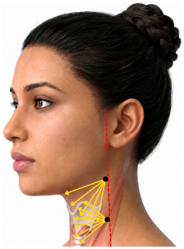
一项前瞻性、开放标签探索性临床研究纳入13名颈部皮肤松弛女性(平均48.5岁),单次采用STIIM UP方案(CaHA+HA+BoNT-A经22G钝针扇形注射至颈前皮下)。

这项前瞻性、开放标签、随机、多中心(德国11个中心)上市后研究纳入121名受试者,按1:1随机接受 Belotero Volume(CPM-V,n=58)或含利多卡因的 CPM-V