面神经麻痹后口-眼联带运动的肉毒毒素治疗策略:基于睑裂比(mPFR)的临床分型
2026年10月10日28 min read
Plastic and Reconstructive Surgery Global Open
Plastic and Reconstructive Surgery Global Open
结论与实践要点
本研究为回顾性病例系列,纳入2022年4月至2024年3月面神经麻痹后口-眼联带运动患者10例,依据口动时睑裂比(mPFR)分为四型(I型90%–近似正常、II型30%–89%、III型0%–29%、IV型波动型)。结果显示,I型与II型患者经肉毒毒素注射及镜像生物反馈后mPFR与主观不适改善,III型及部分难治II型从选择性神经/肌切除术获益,IV型对肉毒毒素反应不稳定甚至反常。肉毒毒素单睑用量约10–15 U,疗效多在3个月左右随药效消退而复现,联合镜像训练可延长获益。
研究局限性在于样本量小(n=10)、为单中心回顾性设计,缺乏对照与长期随访,mPFR分型阈值基于本队列初步拟定,尚待外部验证。肉毒毒素属对症、暂时性干预,不能改变神经误再生的根本,需结合功能训练或手术分层处理;文中影像与视频资料仅供示意,不替代个体化的神经电生理与影像评估,临床决策应综合病因、病程与瘫痪程度。
Background:Oral–ocular synkinesis is a disabling sequela of facial nerve palsy, characterized by involuntary orbicularis oculi contraction during oral movements. To better guide clinical management, we developed a novel classification system based on the palpebral fissure ratio with oral movement (mPFR).
Methods:This retrospective analysis considered 10 cases of oral–ocular synkinesis between April 2022 and March 2024. Patients were categorized into 4 types: type I (mild; mPFR, 90%–100%), type II (moderate; mPFR, 30%–89%), type III (severe; mPFR, 0%–29%), and type IV (fluctuating; unpredictable mPFR irrespective of intervention).
Results:Type I and II patients may respond well to botulinum toxin injections and/or mirror biofeedback, which can improve mPFR and reduce subjective discomfort. Type III patients or refractory type II cases may benefit from selective neurectomy or myomectomy. In patients with incomplete facial paralysis within 1 year of onset, nerve reconstruction using cross-facial nerve grafting, masseteric nerve transfer, or cross-link surgery to the hypoglossal can be considered, aiming to restore voluntary facial muscle movement while suppressing synkinetic activity. Notably, type IV patients showed minimal response to botulinum toxin, highlighting the importance of careful classification before intervention.
Conclusions:By integrating the objective metric of mPFR with individualized treatment strategies, this classification system may offer a practical framework to enhance both functional and aesthetic outcomes in patients with oral–ocular synkinesis. These findings highlight the importance of accurate evaluation and type-specific therapeutic approaches to improve the quality of life in individuals affected by synkinesis following facial nerve palsy.
Takeaways
Question: Can palpebral fissure ratio with oral movement (mPFR)–based classification guide optimal treatment selection for oral–ocular synkinesis after facial nerve palsy?
Findings: Patients were stratified by synkinesis severity using mPFR and treated with botulinum toxin, mirror biofeedback, or surgery accordingly. Mild and moderate cases responded well to conservative therapy, whereas severe or refractory cases required surgical intervention. Fluctuating cases showed limited response to invasive treatments.
Meaning: Objective mPFR-based classification enables rational, individualized treatment strategies, improving functional and aesthetic outcomes and supporting clinical decision-making in synkinesis management.
Facial nerve palsy affects approximately 32 individuals per 100,000 population annually. 1 The condition can significantly impair patients’ quality of life, leading to symptoms such as lagophthalmos with associated exposure keratopathy or excessive tearing, as well as drooping at the oral commissure, which can cause difficulty during meals. In clinical practice, about two-thirds of cases are classified as Bell palsy (idiopathic), with approximately 70% of patients achieving full recovery without residual complications.
In recent years, the emergence of novel pharmacological therapies beyond steroids, as well as decompression surgery, has improved outcomes in the acute phase of facial nerve palsy. 2 However, a certain proportion of patients continue to experience residual symptoms, manifesting as long-standing flaccid paralysis or incomplete paralysis.
Pathological synkinesis is one of the sequelae that can occur regardless of the degree of recovery from facial nerve paralysis. The most widely accepted theory attributes its cause to misdirected regeneration during the process of nerve recovery. Regenerating axons extend to muscles they were not originally intended to innervate, leading to aberrant reinnervation. 3 As a result, multiple muscles may contract involuntarily in response to neural signals from these aberrant nerve fibers, causing movements that occur against the patient’s intention.
The most frequent pattern of synkinesis involves ocular–oral synkinesis, 4 where unintended oral movements are accompanied by ocular muscle activity. For example, when the patient blinks, the corner of their mouth moves simultaneously. However, from a clinical standpoint, oral–ocular synkinesis often poses more significant challenges than ocular–oral synkinesis. This type of synkinesis is commonly characterized by involuntary eyelid movements occurring simultaneously with oral movements. Patients frequently find these unintended eyelid movements both irritating and embarrassing, particularly when they draw attention from others, such as when having a conversation or eating. In more severe cases, these involuntary eye closures can even result in visual field impairment. Given these significant impacts on patients’ quality of life, treatments targeting oral–ocular synkinesis have gained increasing attention in recent years.
Treatment options for pathological synkinesis include rehabilitation, botulinum toxin (BTX) therapy, and surgical intervention. Although it is widely recognized that treatment plans should be tailored to the severity and pattern of synkinesis, a definitive strategy has yet to be established. 5 , 6 This article discusses our classification system and treatment strategy for oral–ocular synkinesis.
This study included 10 consecutive cases of oral–ocular synkinesis following facial nerve palsy, treated between April 2022 and March 2024, and classified using our severity grading system, as detailed further. Based on medical records, we retrospectively analyzed patient demographics (age and sex), etiology of paralysis, electroneuronography at the onset of paralysis, duration from onset to initial consultation, and the Yanagihara score. 7
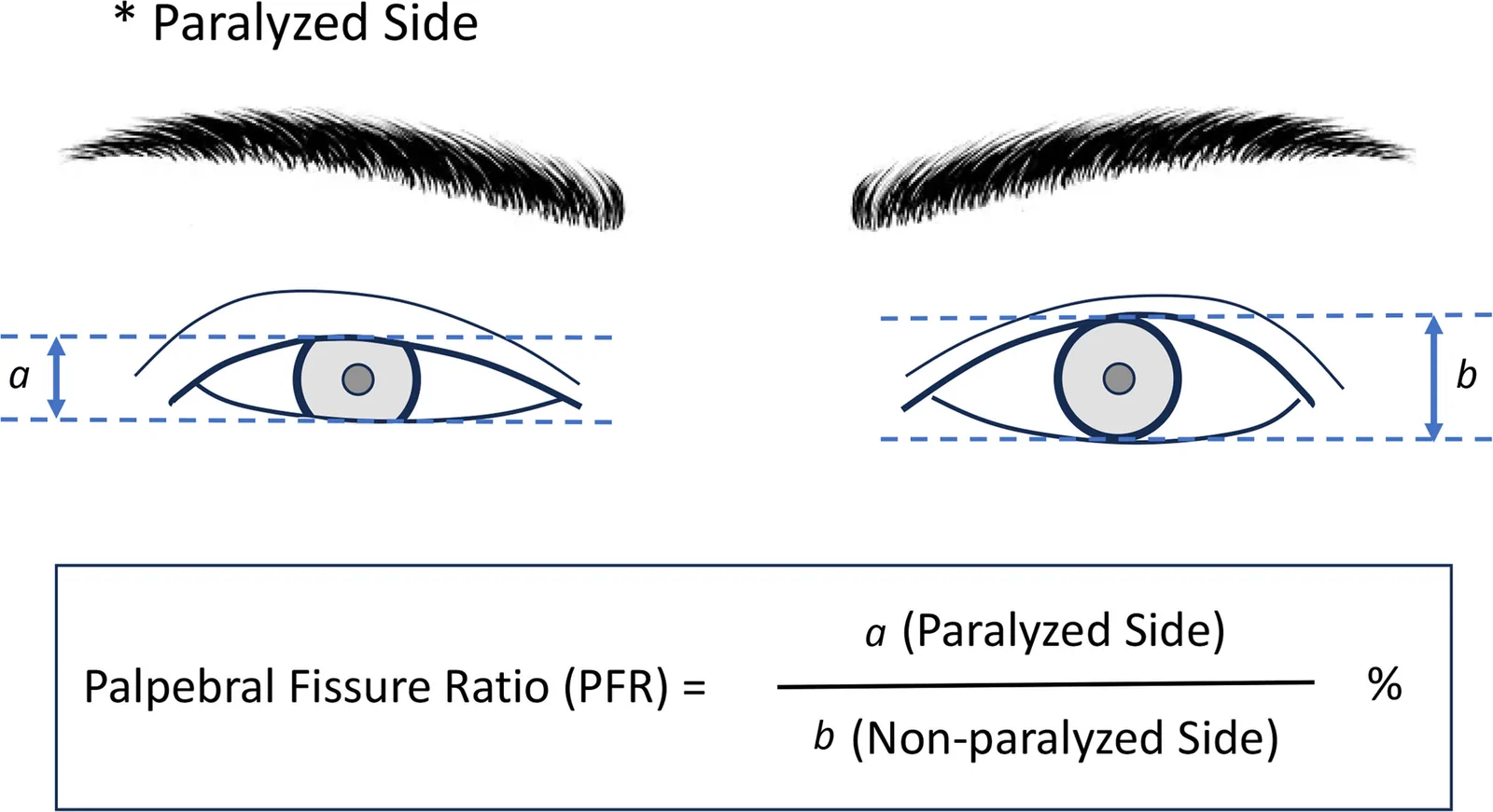
The severity of oral–ocular synkinesis was evaluated using the palpebral fissure ratio (PFR). PFR is defined as the ratio of the palpebral fissure width on the affected side to that on the unaffected side, expressed as a percentage (Fig. 1 ). To measure PFR, photographs of the patient were first taken at rest, without any oral movement, to calculate the PFR at resting position. Subsequently, the patient was instructed to perform specific oral movements by articulating [u:], [e:], and [pu:], and the corresponding PFR values were measured and recorded from the captured images. The average of these 3 values was calculated as the PFR with oral movement (mPFR). All photographs were taken by the first author, and PFR measurements were performed independently by multiple coauthors to minimize bias.
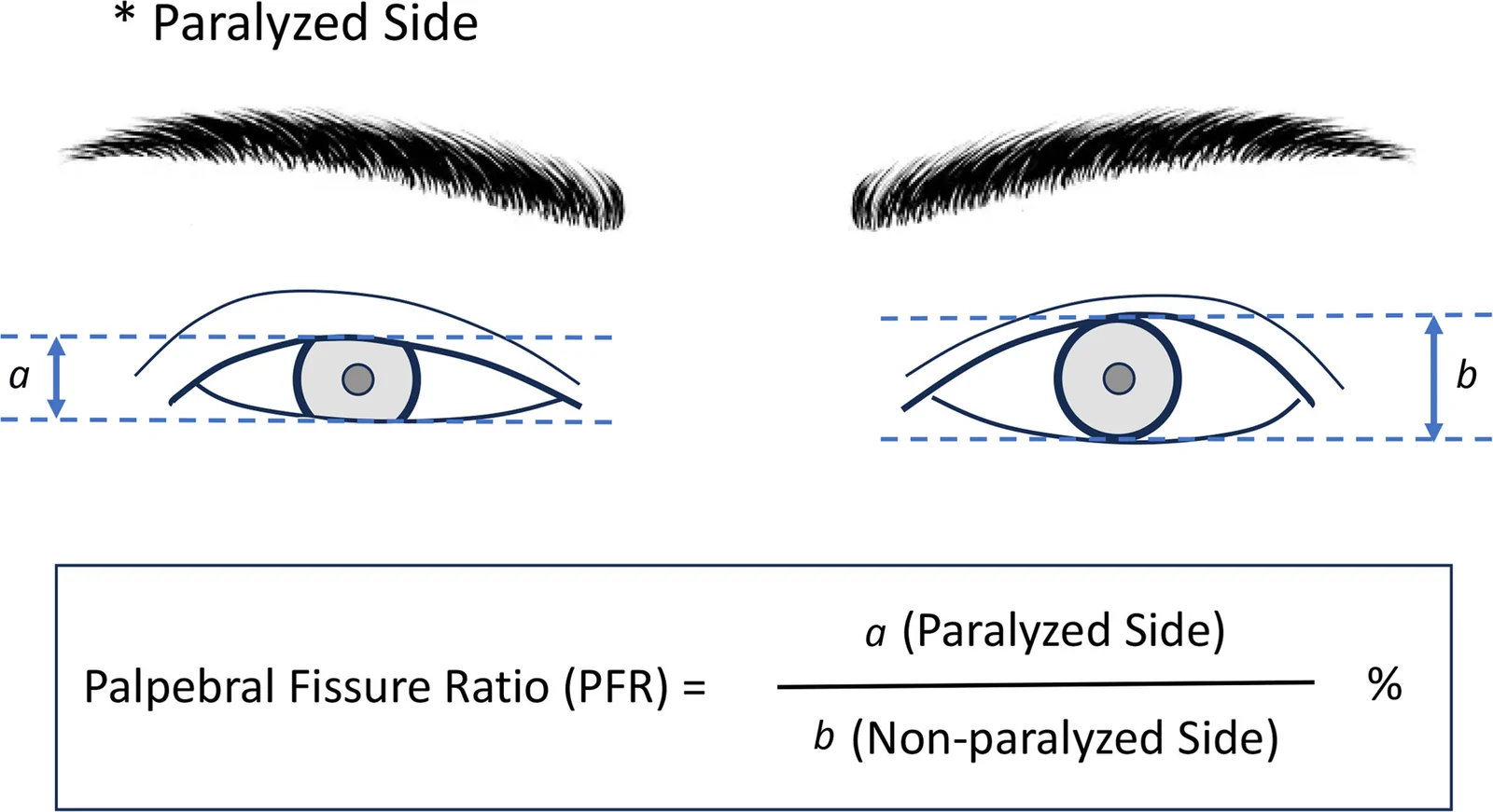
Fig. 1. PFR was calculated by measuring the palpebral fissure width at the level of the pupil center from photographs taken in the frontal view. The width of the palpebral fissure on the paralyzed side, denoted as “a,” was divided by the width of the palpebral fissure on the unaffected side, denoted as “b,” and the result was expressed as a percentage. *Indicates paralyzed side.睑裂比(PFR)测量示意:患侧睑裂宽度a除以健侧b,以百分比表示,用于量化口动时眼睑闭合程度。
Based on the mPFR values obtained, the severity of synkinesis was categorized into 4 distinct levels as given further. ( See Video [online] , which displays the clinical classification based on mPFR.)
Type I is characterized by orbicularis oculi muscle spasm during oral movement without a decrease in the PFR. Although patients may experience discomfort due to the unintended muscle spasm, the mPFR remains at 90%–100%, making it challenging to objectively assess the severity of the symptoms. BTX administration often provides effective symptom relief, and consequently, surgical intervention is generally not necessary.
The moderate type includes cases where the mPFR ranges from 30% to 89%, does not result in complete eyelid closure and does not cause visual field impairment (Fig. 2 ). What is unique to this type is that the mPFR typically returns to an almost normal range (typically 80%–100%) with BTX administration. However, since the effects of BTX are temporary, synkinesis typically recurs in about 3 months after BTX administration. We encourage patients to continue mirror biofeedback (MBF) therapy to prolong the effects of BTX for as long as possible.
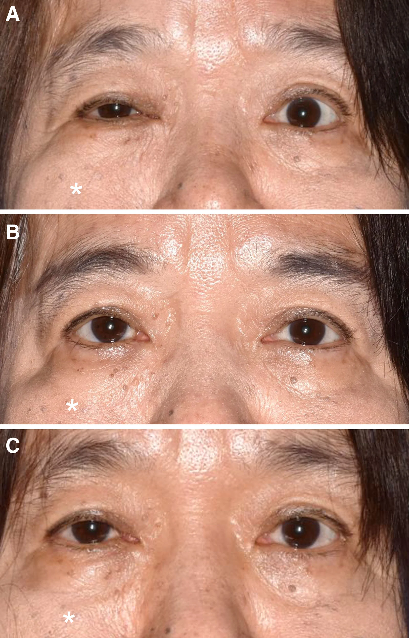
Fig. 2. Typical clinical course in a type II (moderate) patient. A, Before treatment, the mPFR was 54.6%, and moderate involuntary eyelid closure was observed during oral movement [pu:]. Notably, the upper eyelid did not fully cover the pupil, and no visual field impairment was present. B, One month after BTX administration, synkinesis had resolved, and the mPFR improved to 100%. C, Three months after BTX administration, as the effect of the toxin diminished, synkinesis recurred and the mPFR decreased to 71.3%. *The paralyzed side.II型(中度)典型病例:肉毒毒素注射1个月后联带运动缓解、mPFR接近正常,3个月后药效消退复现。
The most severe type is characterized by complete eyelid closure during mouth movement, corresponding to an mPFR of 0%–29% (Fig. 3 ). Many patients report unilateral visual field impairment caused by involuntary eyelid closure. Although these patients generally respond to BTX treatment, the intensity of muscle contraction often makes it difficult to achieve adequate therapeutic effects. Surgical denervation procedures, such as selective neurectomy or myectomy, are frequently required to obtain satisfactory results. Moreover, because this type is often accompanied by incomplete recovery of facial paralysis, adjunctive procedures such as blepharoplasty or brow lift may be necessary to correct brow ptosis and improve both functional and aesthetic outcomes.
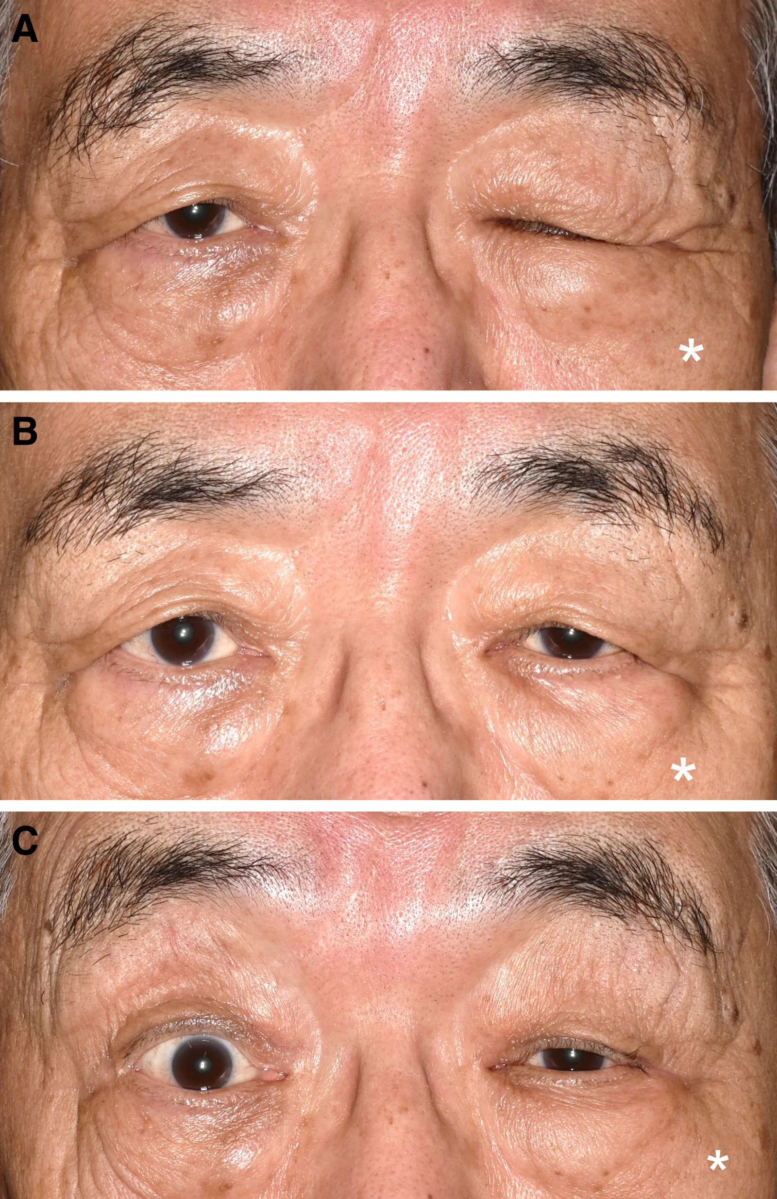
Fig. 3. Typical clinical course in a type III (severe) patient. A, Before treatment, the mPFR was 0%, with visual field impairment due to involuntary eyelid closure. B, One month after BTX administration, the mPFR improved to 77.5%, and the visual field impairment resolved. C, Three months after BTX administration, synkinesis recurred, and the mPFR decreased to 42.9%. Compensatory frontalis muscle activation (brow elevation) was observed in an attempt to maintain the visual field. *The paralyzed side.III型(重度)典型病例:治疗前口动时完全闭眼伴视野受损,注射后改善,3个月复现并见额肌代偿抬眉。
This type is characterized by significant fluctuations in symptoms attributed to synkinesis, varying considerably depending on the situation or timing. For objective classification, patients were defined as type IV, if (1) a discrepancy of minimum 20% in mPFR was observed between measurements obtained on different examination days, and (2) a paradoxical decrease in mPFR was observed following BTX administration, after excluding confounding factors such as ptosis or other BTX-related adverse effects (Fig. 4 ). Although the complaints may at times resemble nonspecific symptoms, careful listening and a supportive, patient-centered approach may, in some cases, lead to symptom relief without the need for invasive treatment.
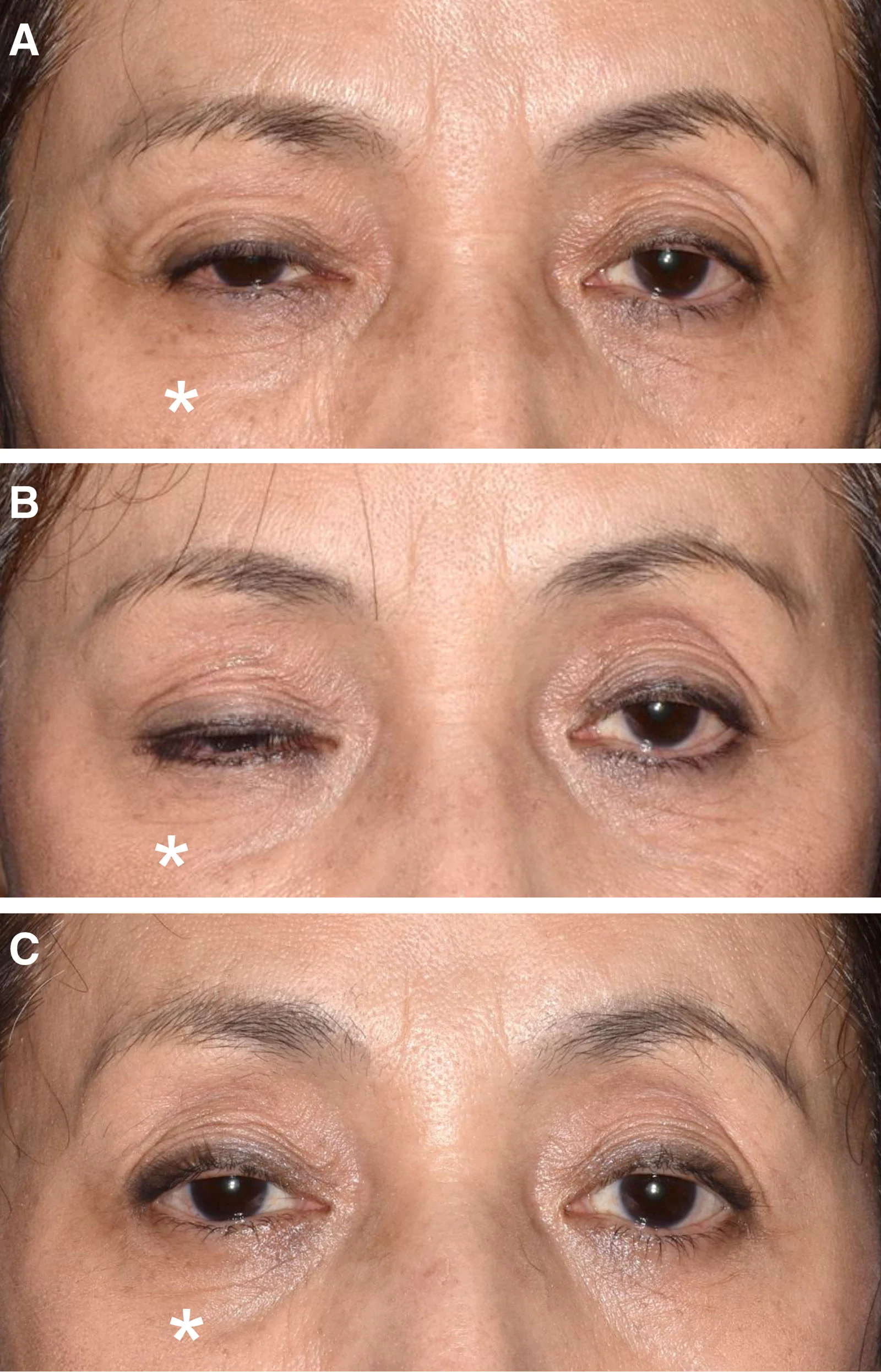
Fig. 4. Typical clinical course in a type IV (fluctuating) patient. A, Before treatment, the mPFR was 52.7%. B, One month after BTX administration, the mPFR decreased to 44.6%, accompanied by visual field impairment due to involuntary eyelid closure. C, Three months after BTX administration, the mPFR increased to 68.5%, with restoration of the visual field. This paradoxical course, showing no consistent physiological response to BTX, is characteristic of type IV. *The paralyzed side.IV型(波动型)典型病例:对肉毒毒素呈反常、不一致反应,mPFR随观测时点明显波动。补充视频 / 附件(原文未随文提供原图,可于出版方页面查看)gox-14-e8134-s001.mp4Video 1. This video demonstrates representative cases of each clinical type classified according to the palpebral fissure ratio with oral movement (mPFR). Pre- and post-botulinum toxin (BTX) findings are shown to illustrate differences in severity and treatment response. The video aims to facilitate intuitive understanding of the classification system and its relationship to treatment strategy.
As a noninvasive treatment for oral–ocular synkinesis, BTX therapy was proposed for all cases, except for patients with contraindications. Botox (GlaxoSmithKline K.K., Tokyo, Japan) for injection was administered at 5–6 injection points per eyelid, with 1.25–2.5 units per site, for a total of approximately 10–15 units per eyelid. The dosage was adjusted as necessary based on the patient’s age, sex, and physique. Patients were reassessed 1 month after BTX administration to evaluate its peak effectiveness. A follow-up at 3 months postinjection confirmed the waning effects of BTX and the recurrence of synkinesis, at which point repeat injections were administered.
Alongside BTX administration, all patients were instructed in MBF. They performed oral movements by articulating [u:], [e:], and [pu:] sequentially while using a mirror to recognize their oral–ocular synkinesis. Patients were advised to ensure that eyelid closure did not accompany oral movements during the exercises. They were encouraged to practice MBF for 10 minutes daily and to continue even after synkinesis was alleviated by the effects of BTX. 8
Selective neurectomy or myomectomy was performed in types II and III patients with refractory oral–ocular synkinesis unresponsive to combined BTX and MBF therapy. A 1-cm wide segment of the orbital portion of the orbicularis oculi muscle in the upper or lower eyelid was excised, ensuring the removal of misdirected nerves along with the muscle tissue. 9 , 10 To prevent the development of lagophthalmos, the tarsal portion of the orbicularis oculi muscle was carefully preserved during the procedure.
For patients with incomplete recovery from facial paralysis, defined as a Yanagihara score 7 of less than 32 and referral to our clinic within 6 months to 1 year after onset, nerve reconstruction incorporating multiple motor sources 11 was proposed (Fig. 5 ). A cross-facial nerve graft to the zygomatic branch innervating the orbicularis oculi muscle on the affected side was performed using the contralateral zygomatic branch as the motor source. In addition, the masseteric nerve on the affected side was transferred to the distal end of the zygomatic branch. Furthermore, a nerve graft was interposed between the main trunk of the facial nerve on the affected side and the ipsilateral hypoglossal nerve (cross-link surgery). 12
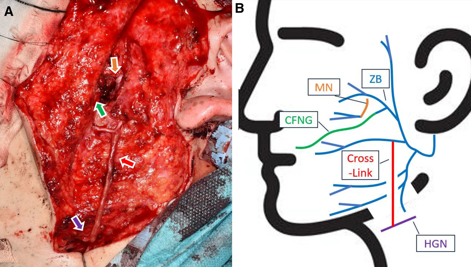
Fig. 5. Nerve reconstruction. A, Intraoperative photograph, and (B) schematic illustration. The zygomatic branch of the facial nerve, which innervates the orbicularis oculi muscle of the lower eyelid, is coaptated to the masseteric nerve (orange arrow), and its proximal stump is reinnervated using a cross-facial nerve graft (green arrow) from the contralateral zygomatic branch. For restoration of oral commissure movement, the main trunk of the buccal branch of the facial nerve (red arrow) is connected to the hypoglossal nerve (purple arrow) through an interposed nerve graft using end-to-end neurorrhaphy (cross-link surgery).神经重建示意:颧支与咬肌神经吻合并以对侧跨面神经移植再支配,口角颊支与舌下神经经移植桥接。
Treatment outcomes were evaluated by comparing pre- and posttreatment mPFR values in each case. Patient-reported numerical rating scale (NRS) scores (10 = intact state before synkinesis onset; 1 = first visit before treatment) were also recorded to assess subjective symptom improvement. Both mPFR and NRS were assessed at baseline (pre-BTX), 1 month post-BTX, and 3 months post-BTX, with final outcomes defined as those after completion of 3 BTX sessions or at 1 year postoperatively.
The study included 10 patients: 4 men and 6 women, with a mean age at onset of 69.6 years (Table 1 ). According to our classification system, the distribution of cases was as follows: type I, n = 1; type II, n = 5; type III, n = 2; and type IV, n = 2. The underlying causes of facial paralysis were Bell palsy in 8 cases, and Ramsay Hunt syndrome in 2 cases. Initial electroneurography values ranged from 4.0% to 100%, with a mean of 22.6%. The time from onset to the initial visit to our department ranged from 7 to 31 months, with a mean duration of 12.3 months. The Yanagihara scores ranged from 20 to 40, with an average score of 33.2.
Table 1. Patients Characteristics and Treatment Outcomes
| Patient No. | Age, y | Sex | Etiology of Paralysis | Electroneurography at Onset, % | Onset of Paralysis, mo | Yanagihara Score, 0–40 | Classification | Treatment Strategy | mPFR, % | BTX, Unit | NRS, 1–10 | Note |
|---|---|---|---|---|---|---|---|---|---|---|---|---|
| Pre-BTX | Post-BTX (1 mo) | Post-BTX (3 mo) | Final | Pre-BTX | Post-BTX (1 mo) | Post-BTX (3 mo) | Final | |||||
| 1 | 29 | F | Hunt | 11.0 | 7 | 32 | I | BTX | 100 | 100 | 100 | 100 |
| 2 | 66 | F | Bell | 5.8 | 10 | 36 | II | BTX+MBF | 54.6 | 98.2 | 71.3 | 75.3 |
| 3 | 70 | F | Bell | 39.0 | 8 | 34 | 58.3 | 104.8 | 85.6 | 74.0 | 10 | 1 |
| 4 | 29 | M | Bell | 4.0 | 11 | 34 | 66.7 | 94.4 | 73.1 | 79.6 | 12.5 | 1 |
| 5 | 61 | F | Bell | 7.0 | 31 | 34 | 80.5 | 107.5 | 93.6 | 87.9 | 10 | 1 |
| 6 | 31 | M | Bell | No record | 11 | 20 | 85.7 | N/A | N/A | 84.7 | N/A | 1 |
| 7 | 72 | F | Hunt | 17.0 | 8 | 26 | III | Surgical intervention | 28.5 | 50.5 | 48.3 | 64.4 |
| 8 | 75 | M | Bell | 10.0 | 12 | 36 | 28.6 | 77.5 | 42.9 | 70.4 | 12.5 | 1 |
| 9 | 67 | F | Bell | 100.0 | 7 | 40 | IV | Supportive therapy | 52.7 | 44.6 | 68.5 | 82.6 |
| 10 | 44 | M | Bell | 10.0 | 18 | 40 | 85.2 | 74.7 | 71.8 | 89.5 | 15 | 1 |
| Average | 54.4 | 22.6 | 12.3 | 33.2 | 64.1 | 83.6 | 72.8 | 80.8 |
BTX was administered to 9 patients, excluding 1 who had undergone prior nerve reconstruction. Improvement in mPFR and NRS was observed in 7 of these cases (Fig. 6 ). In the remaining 2 cases—both classified as type IV—mPFR worsened following BTX treatment. The dosage of BTX ranged from 10 to 15 units, with an average of 11.7 units per eyelid.
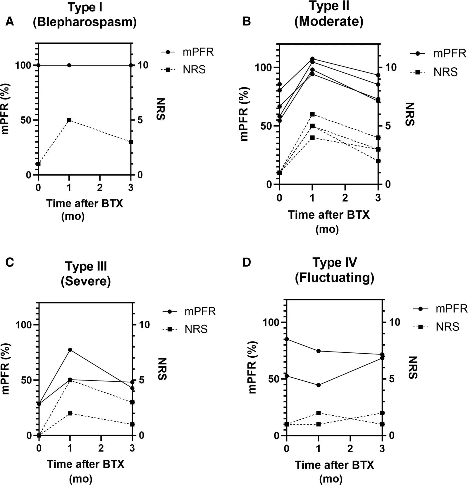
Fig. 6. Effect of BTX. Changes in the mPFR and the NRS following BTX administration are plotted for each patient and sorted by classification type. Note that in types I–III (A–C), both mPFR and NRS show temporary improvement after BTX injection but gradually decline by 3 months, indicating recurrence of oral–ocular synkinesis. In contrast, type IV (D) shows an inconsistent response to BTX.肉毒毒素疗效汇总:I–III型mPFR与NRS在注射后短暂改善、3个月渐降;IV型反应不一致。
Among the 5 patients classified as type II (moderate), combined treatment with BTX and MBF resulted in 2 patients (40%)—patients 2 and 3 in Table 1 —becoming BTX-free. Both cases initially presented with moderate oral–ocular synkinesis with pretreatment mPFR values in the 50% range. BTX was administered every 3 months, and patients were instructed to perform MBF exercises during the intervals. After the third session, mPFR was reassessed 6 months after the final BTX injection—when the effect of BTX had completely worn off—and remained in the 70% range for both patients (Fig. 7 ). These findings suggest a sustained improvement in synkinesis through the combined BTX and MBF therapy.
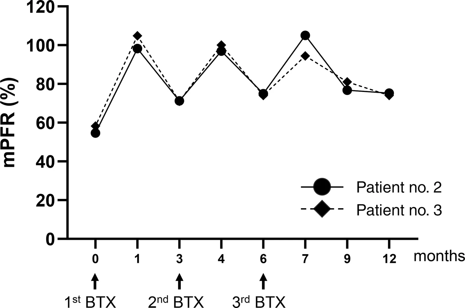
Fig. 7. Combination therapy with BTX and MBF. In 2 patients, who continued combination therapy with BTX and MBF for 12 months, sustained improvement in oral–ocular synkinesis was achieved. The mPFRs for the 2 patients at the start of treatment were 54.6% and 58.3%, respectively. Even 6 months after the third BTX injection—when the effect of BTX had worn off—mPFR remained improved at 75.3% and 74.0%, respectively.肉毒毒素联合镜像生物反馈(MBF)持续12个月者,即便停药后mPFR仍维持改善。
Selective neurectomy was performed in 1 case (patient 8 in Table 1 ). At the initial visit, the patient exhibited severe synkinesis with an mPFR of 28.6% and was classified as type III. Although BTX administration temporarily improved the mPFR to 77.5%, the effect was not sustained. Following surgery, the mPFR improved to 70.4%, and the NRS score also decreased to 4.
Two patients (patients 6 and 7 in Table 1 ) presented with incomplete paralysis, defined as a Yanagihara score of less than 32. Both were referred to our clinic within 6 months to 1 year after onset and exhibited moderate or severe synkinesis (types II or III). Based on these findings, nerve reconstruction incorporating multiple motor sources 11 was performed. In particular, patient 7 demonstrated a marked improvement in synkinesis following nerve reconstruction (Fig. 8 ).
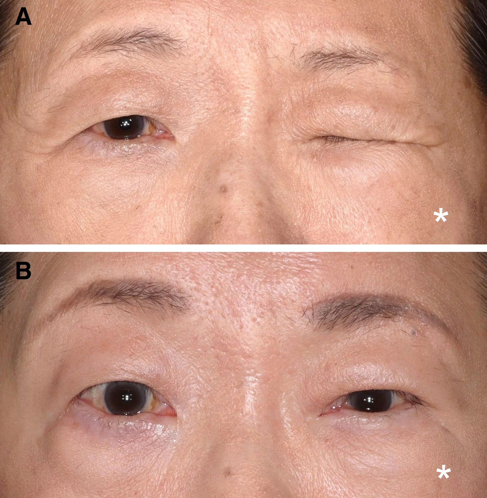
Fig. 8. Representative case of cross-link surgery. A 72-year-old woman developed severe oral–ocular synkinesis following Ramsay Hunt syndrome. Cross-link surgery was performed 8 months after the onset of facial paralysis. A, Preoperatively, pronounced involuntary eyelid closure was observed during oral movement [pu:]. B, At 11 months postoperatively, the mPFR had improved to 64.4%. *The paralyzed side.跨面神经吻合(cross-link)手术代表病例:Ramsay Hunt综合征后重度联带运动,术后11个月mPFR改善。
Although the House-Brackmann and Sunnybrook Facial Grading Scales are widely used to assess synkinesis after facial nerve paralysis, they lack clearly defined objective criteria. In addition, they evaluate overall facial muscle movement and are not well suited for detailed assessment of oral–ocular synkinesis. 13 In contrast, palpebral fissure width, a standard parameter in oculoplastic surgery, provides a more targeted measure. To objectively assess severity, we adopted the PFR, which quantifies percentage asymmetry between the affected and unaffected sides. 14
Our PFR-based classification is directly linked to treatment strategies, providing a practical and clinically relevant framework (Fig. 9 ). It comprises 3 categories: type I (mPFR, 90%–100%), type II (mPFR, 30%–89%), and type III (mPFR, 0%–29%). The cutoff values were preliminarily defined based on the present cohort, as no prior external evidence exists. The proposed classification is preliminary, and given the limited sample size, further validation in larger, independent cohorts is required to confirm its robustness and clinical applicability.
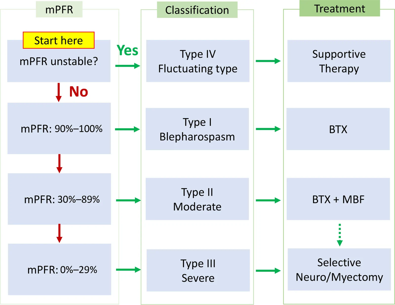
Fig. 9. Clinical classification based on mPFR and corresponding treatment strategy for oral–ocular synkinesis. Calculated the mPFR from standardized photographs and classified the severity of oral–ocular synkinesis to guide treatment selection. Mild-to-moderate cases are initially treated with BTX and/or MBF, whereas severe or refractory cases may require selective neuro-/myomectomy; patients with incomplete facial paralysis within 1 year of onset may additionally benefit from nerve reconstruction.基于mPFR的临床分型与对应治疗策略总览图。BTX is a widely accepted first-line treatment for synkinesis due to its efficacy, minimal invasiveness, and reversible effects, which typically diminish within 3 months. 15 We routinely administer BTX to all new patients and assess their response, as symptom improvement supports the diagnosis of synkinesis, whereas persistence or worsening suggests limited benefit from surgical intervention. In particular, patients with atypical presentations (eg, type IV) may not follow the typical clinical course, and surgical treatment may carry uncertain efficacy and risk of additional nonspecific complaints. Thus, BTX also serves as a useful diagnostic tool. However, its simplicity may lead to indiscriminate use by nonspecialists, potentially resulting in complications such as blepharoptosis, oral drooping, or resistance. 16 To address these issues, Miller and Hadlock 17 proposed the “beyond Botox” concept introducing a treatment ladder that includes selective neuro-myomectomy, nerve reconstruction, and muscle transfer. Nevertheless, the lack of clear criteria for treatment selection highlights the need for an objective classification system, which motivated us to develop a PFR-based severity classification.
A key feature of our proposed treatment strategy is the combination of BTX and MBF. Although selective neurectomy or myomectomy can provide substantial relief by removing the offending nerves or muscles, these procedures carry a risk of lagophthalmos. 9 , 18 Therefore, such procedures are considered a last-resort option for refractory synkinesis, and conservative approaches should be fully explored before surgical intervention is considered. Recently, several studies have highlighted the therapeutic potential of combining BTX with MBF in established synkinesis. 8 , 19 , 20 Although MBF was originally proposed to prevent misdirected axonal regeneration during recovery, modifying established aberrant neural circuits remains challenging. However, temporary chemodenervation with BTX may provide a window for central reprogramming, allowing symptom relief to persist even after its effects diminish. 8 In our series, 2 of 5 (40%) type II cases showed sustained improvement beyond the duration of BTX effect. Nevertheless, given the limited sample size, the efficacy of BTX combined with MBF requires further validation in larger studies.
When nonsurgical treatments fail to provide sufficient relief, selective neurectomy or myomectomy may be considered. In our experience, this applies to type II patients with persistent symptoms after 3 sessions of BTX with MBF therapy, as well as to type III patients, who often exhibit strong muscle contractions that make MBF difficult to perform; in such cases, these procedures may be considered as an early treatment option. Another operative option for synkinesis is nerve reconstruction. Given the strong association between incomplete paralysis and the development of synkinesis, 16 , 21 , 22 nerve reconstruction that simultaneously addresses the underlying paralysis and pathological synkinesis may represent a desirable treatment strategy in carefully selected patients. We consider the appropriate surgical indication and timing to the cases presenting with synkinesis and persistent incomplete paralysis within 1 year of onset. This 1-year threshold is based on the risk of irreversible degeneration in the facial mimetic muscles due to prolonged denervation. Even if successful nerve regeneration is achieved through reconstruction, the therapeutic benefit may be limited if the target muscles have already undergone irreversible degeneration.
To maximize the therapeutic efficacy of nerve reconstruction, incorporating multiple motor sources is crucial. 11 Among the available techniques, we favor hypoglossal–facial nerve interposition grafting, known as cross-link surgery. 12 In this procedure, a nerve graft is interposed between the facial and hypoglossal nerves, effectively creating a cross-connection. The primary mechanism of action involves the inflow of axons from the hypoglossal nerve into the facial nerve through the graft. 23 By incorporating the hypoglossal nerve as an additional motor source, the functional load on the native facial nerve is reduced. This reduction in neural demand also decreases the relative output to aberrant nerve fibers, thereby contributing to an improvement in synkinesis.
A unique advantage of cross-link surgery is its minimal invasiveness to the native facial nerve. Unlike procedures such as cross-facial nerve graft or masseter nerve transfer, which require transection of facial nerve branches on the paralyzed side—posing a risk of transient postoperative deficits such as lagophthalmos—cross-link surgery is less disruptive. It involves only the creation of a small epineurial window, preserving the underlying nerve fibers. This technique enables improvement of synkinesis while maintaining the existing function of the facial nerve. However, the present study includes only 2 cases treated with nerve reconstruction, and therefore it is not possible to determine which factors—such as timing of intervention, baseline severity, or technical aspects of the procedure—most strongly contributed to the observed improvements in synkinesis. Although cross-link surgery is supported by a strong theoretical rationale, the present findings should be considered hypothesis-generating, providing preliminary clinical observations that warrant validation in larger, prospective studies.
The clinical classification system for oral–ocular synkinesis, based on the mPFR, provides a simple and objective method for assessing severity and guiding optimal treatment strategies. BTX and MBF may be effective in improving symptoms in types I and II cases, whereas surgical interventions such as selective neuro-/myomectomy or nerve reconstruction may be beneficial for type III and selected type II cases. Type IV, characterized by fluctuating mPFR, appears to be resistant to both BTX and surgical interventions, and may require a supportive approach. Importantly, in patients with incomplete paralysis within 1 year of onset, nerve reconstruction may not only improve synkinesis but also contribute to facial reanimation. This classification may help personalize treatment strategies and improve long-term outcomes; however, further validation in large-scale studies is warranted.
The authors have no financial interest to declare in relation to the content of this article.
声明:中文精读 · 仅供学术参考。内容来自公开文献检索,不代表本人观点,不构成诊疗建议。 医疗美容需在正规医疗机构由执业医师实施。
基于相同主题推荐 · 继续深挖
由6名意大利多学科专家(皮肤科、整形外科、神经科)基于文献回顾(PubMed 1973–2025)与临床经验形成的专家共识,针对incobotulinumtoxin A(Bocou
一项前瞻性、开放标签探索性临床研究纳入13名颈部皮肤松弛女性(平均48.5岁),单次采用STIIM UP方案(CaHA+HA+BoNT-A经22G钝针扇形注射至颈前皮下)。
研究类型:单中心回顾性研究,先以40例病历建立耳部美学老化分类(AEAC,0–3级),再以50例50–65岁健康女性行A型肉毒毒素(Russo Ear Lift Technique