螺旋PDO线材(JAMBER)鼻部填充的疗效、持久性与安全性:23例12个月超声与三维扫描回顾性研究
2026年10月12日33 min read
Plastic and Reconstructive Surgery Global Open
Plastic and Reconstructive Surgery Global Open
结论与实践要点
本研究为单中心回顾性研究,纳入2023年1月至2024年8月接受JAMBER PDO线材鼻部填充的23例患者(21–72岁,平均35.3岁;男8女15)。线材为40 mm管状螺旋PDO(3-0 PDO展开全长414 mm),经21G套管递送,平均鼻背植入2.85根,鼻小柱追加1–2根35 mm弹性线材以改善鼻尖突度。随访1、2、3、6、9、12个月,以超声测量线材直径与软组织厚度,以LifeViz Mini三维扫描仪测量鼻体积与高度。
主要结局:线材直径由1个月时1.113 mm渐降至12个月时0.857 mm,6个月内维持>1 mm具统计学意义,12个月仍可检出线材;周围软组织厚度自1.213 mm增至1.403 mm(12个月增厚27.55%),提示胶原与肉芽组织形成。三维扫描示鼻体积1个月0.771 mL、12个月0.552 mL,鼻高度1.387 mm降至1.005 mm,仍高于基线。12个月内无感染、移位、外露或皮肤坏死,亦无线材取出。文中组织学为术后2周时点观察,线材占位中央区尚未完全长入胶原,仅反映早期反应,不能外推长期结局。
局限性与提示:样本小(n=23)、单中心回顾性、无对照,证据等级有限;该技术难以选择性填充鼻背不规则凹陷,全线材植入产生均匀对称增量;与填充剂不同,PDO线材缺乏透明质酸酶样逆转手段,对操作精度与患者筛选要求高,作者认为其螺旋结构不具备填充剂误入血管所致风险的解剖学基础。在规范操作与影像引导下,该线材或可成为鼻部微创填充的可选路径,结论尚待多中心、大样本随机研究验证。
Background:Polydioxanone threads, specifically 40-mm tubular JAMBER (JB), 414-mm coiled around a 23G cannula, have emerged as a safe alternative for facial volume augmentation. This study evaluated the efficacy, safety, and longevity of JB threads for nasal augmentation, tracking changes in nasal height and volume across 12 months.
Methods:A retrospective analysis included 23 patients (mean age 35.3 y; 8 men and 15 women) treated with JB threads from January 2023 to August 2024. Patients underwent ultrasound (Samsung RS85, Aplio i700, Konica Minolta HS2) and 3-dimensional scanner imaging (LifeViz Mini) at intervals of 1, 2, 3, 6, 9, and 12 months. An average of 2.85 JB threads were inserted in the nasal dorsum, complemented by 1.75 threads in the columella for tip projection.
Results:The JB thread diameter decreased from 1.113 mm at 1 month to 0.857 mm at 12 months, reflecting gradual absorption. Soft-tissue thickness increased from 1.213 to 1.403 mm, indicating collagen formation. Nasal volume initially rose to 0.771 mL at 1 month and stabilized at 0.552 mL at 12 months. Similarly, nasal height increased to 1.387 mm at 1 month and remained at 1.005 mm by 12 months. No complications, such as infection or extrusion, were observed.
Conclusions:JB threads provide effective and long-lasting nasal augmentation. Compared with fillers and implants, they offer a safer, minimally invasive alternative with no risk of vascular complications. Optimal outcomes require precise technique and patient selection. Further studies are warranted to validate these findings.
Question: Can coiled polydioxanone threads provide a minimally invasive approach for nasal augmentation with sustained structural support?
Findings: In this retrospective study of 23 patients over 12 months, coiled PDO threads were associated with increases in nasal height and volume, along with progressive soft-tissue thickening, whereas thread diameter gradually decreased without major complications.
Meaning: Coiled PDO threads may represent a minimally invasive option for nasal augmentation with sustained structural and tissue effects.
Polydioxanone (PDO) threads are increasingly being used as an alternative to implants for facial lifting and wrinkle reduction. The tubular shape of thicker PDO threads is particularly suited for applications such as smoothing nasolabial folds and enhancing nasal projection. Although fillers were previously popular for nasal augmentation, numerous reports—over hundreds of cases—have documented serious complications, including blindness and skin necrosis, when fillers inadvertently enter blood vessels. As a result, some patients now prefer PDO threads over fillers. The aim of this study was to evaluate the effects of PDO thread insertion for nasal augmentation, providing insights into its potential as a safe and effective option for patients seeking nonsurgical enhancement.
JAMBER-RIDGE (JB) 40 mm is a thread composed of PDO coiled around a 21G, 70-mm cannula, with a 414-mm 3-0 PDO thread length. 1 JB is used as a volume augmentation restorative agent to enhance volume in areas such as the nasolabial folds, marionette lines, forehead, chin, and nasal dorsum. 1 The author has used JB specifically for nasal augmentation. The study focused on volumetric and height changes of the nose observed via 3-dimensional (3D) scanning and ultrasound over 12 months. Nasal augmentation has traditionally involved silicone implants, but the use of hyaluronic acid fillers, introduced in the 2000s, became popular despite risks such as skin necrosis and blindness. Consequently, patients now seek safer alternatives. 2 – 7 JB threads, being thick and coiled, can achieve significant aesthetic effects with just 2–4 threads. The advantage of JB is that it poses no risk of blindness or skin necrosis, unlike fillers.
This retrospective study evaluated the efficacy and safety of JAMBER PDO threads for nasal augmentation. Twenty-three patients, aged 21–72 years (mean age: 35.3 y; 8 men and 15 women), were treated between January 2023 and August 2024. Follow-up assessments were conducted at 1, 2, 3, 6, 9, and 12 months using ultrasound (RS85; Samsung Medison, Seoul, Korea; Aplio i700; Cannon, Japan; HA2; Konica Minolta, Japan) and 3D scanner imaging (LifeViz Mini; QuantifiCare, France). Consent was obtained from all patients before the procedure.
Before the PDO procedure, the muscle layer was identified using ultrasound, and a tumescent solution, prepared by mixing 30 mL of lidocaine and 0.3 mL of epinephrine in 100 mL of normal saline, was injected below the muscle layer to facilitate hydrodissection. An initial 1 mL of the solution was injected into the nasal spine using an insulin syringe, followed by 1 mL into the nasal tip after a 3-minute interval. For full nasal anesthesia, an additional 10 mL syringe with a 25G cannula was used to distribute the solution below the muscle layer across the nasal area. This ensured precise placement and improved patient comfort.
PDO threads were strategically inserted along the nasal structure. Threads were extended from the nasion to the nasal tip for dorsum augmentation and from the nasal tip to the columella and nasal spine for tip projection and stability. On average, 2–4 JB 21G, 40-mm (NEO Dr. INC, Gangwon-do, Korea) threads were used for the nasal dorsum, and 1–2 35-mm JAMBER-Tanxing (JB-T23) threads were inserted into the columella. The JB-T23 threads were compressed to 20 mm for enhanced elasticity, enabling immediate nasal tip projection.
Postprocedure, the entry point was covered with a hydrodressing (Duoderm thin) for 2 days. Patients were monitored at all follow-up intervals for potential complications, such as thread migration or skin thinning. Ultrasound was used throughout the procedure and follow-up to verify correct thread placement and tissue response.
Imaging techniques were used to evaluate outcomes. Sonography was used to measure thread diameter over time and assess soft-tissue thickness, including collagen formation. A 3D scanner was used to measure changes in nasal volume and height at the 5 most elevated points. Statistical analysis assessed the maintenance of thread diameter using 95% confidence intervals with a threshold of 1 mm. Results indicated that the thread diameter was statistically significantly maintained greater than 1 mm for up to 6 months.
In this study, no cases among the 23 patients required removal of the JAMBER PDO threads throughout the 12-month follow-up period. Complications such as infection, migration, or extrusion were not observed, highlighting the threads’ safety profile and tolerance within the nasal tissue. On average, 2.85 PDO threads were used for the nasal dorsum, and an additional 1.75 threads were placed in the columella for elevation and enhanced tip projection. This approach allowed for precise and balanced augmentation across the nasal structure (Table 1 ).
Table 1. Areas of the Nose Enhanced With JAMBER PDO Threads
| Area of Enhancement | PDO Thread Insertion Technique | Purpose | Ease of Insertion |
|---|---|---|---|
| Nasion to nasal tip | Threads inserted from nasion to nasal tip along the dorsum | Augments nasal dorsum for height and definition | Moderate: requires precision to follow the natural nasal contour |
| Nasal tip to columella | Threads extended from the nasal tip to the columella | Defines and projects the nasal tip | Moderate: ensures stabilization without migration |
| Columella to nasal spine | Threads inserted down to the nasal spine through columella | Enhances nasal tip support and stability | Hard: allows precise anchoring in the nasal spine |
In sonography, JB 21G, 40-mm threads were observed to be coiled, creating a central space that allows for collagen ingrowth. New collagen tissues were created around the threads even after the threads were absorbed. The longevity of the PDO treads determined how long the augmented height was maintained. Additionally, ultrasound observations showed significant collagen or granulation tissue formation around the exterior surface and internal cavity of the JB 21G, 40-mm threads, contributing to nasal augmentation. JB is a volumizing thread composed of a 3-0 PDO filament measuring 414 mm in its fully extended length, which is coiled to form a 40-mm implantable structure. The thread is delivered using a 21G, 70-mm cannula system. 1
The diameter of JB 21G, 40-mm threads measured 1.113 mm at 1 month, 1.102 mm at 2 months, 1.09 mm at 3 months, 1.009 mm at 6 months, 0.953 mm at 9 months, and 0.857 mm at 12 months. Although the diameter decreased over time, the threads were still present at 12 months. The SDs were 0.027, 0.026, 0.043, 0.087, 0.091, and 0.105, respectively. This reduction in diameter indicates the gradual absorption of the PDO material. Despite this absorption, the structure remains sufficient to support tissue growth and maintain some degree of augmentation (Table 2 ) (Figs. 1 – 4 ). ( See Video 1 [online] , which displays 40-mm PDO thread insertion into the nasal dorsum for nasal augmentation, and 35-mm length spring PDO thread insertion into the columella for nasal tip projection for a 24-y-old woman.) ( See Video 2 [online] , which displays the inserted PDO threads at 6 d, 3 mo, 9 mo, and 1 y for a 24-y-old woman.) ( See Video 3 [online] , which displays 3 PDO threads inserted in a 72-y-old woman, with follow-up at 1 d and 12 mo [Fig. 2 ].) ( See Video 4 [online] , which displays preoperative and 1-y postoperative results in a 24-y-old woman [Fig. 1 ].)
Table 2. Statistical Significance and Duration of PDO Thread Diameter Maintenance (n = 13)
| Month | Mean Diameter, mm | 95% CI Lower | 95% CI Upper |
|---|---|---|---|
| 1 | 1.113 | 1.098323 | 1.127677 |
| 2 | 1.102 | 1.087866 | 1.116134 |
| 3 | 1.09 | 1.066625 | 1.113375 |
| 6 | 1.009 | 1.000096 | 1.056294 |
| 9 | 0.953 | 0.903532 | 1.002468 |
| 12 | 0.857 | 0.799921 | 0.914079 |
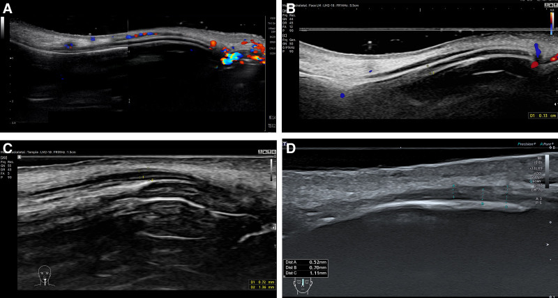
Fig. 1. Sonographic evaluation of PDO thread diameter and soft-tissue thickness on the nasal dorsum for a 24-year-old woman at 1 day and 3, 9, and 12 months. A, The diameter of the inserted PDO thread was 1.10 mm at 1 day. B, The diameter of the PDO was 1.30 mm, and new granulation tissue was created around the PDO at 3 months. C, The diameter was reduced to 0.89 mm at 9 months. D, The diameter was reduced to 0.70 mm at 12 months. Total thickness, including new granulation tissue, was thickened to 1.11 mm at 12 months.超声评估24岁女性鼻背PDO线材直径与软组织厚度:1天线材直径1.10 mm,3个月1.30 mm并伴新生肉芽组织,9个月降至0.89 mm,12个月0.70 mm;含新生组织的总厚度12个月达1.11 mm。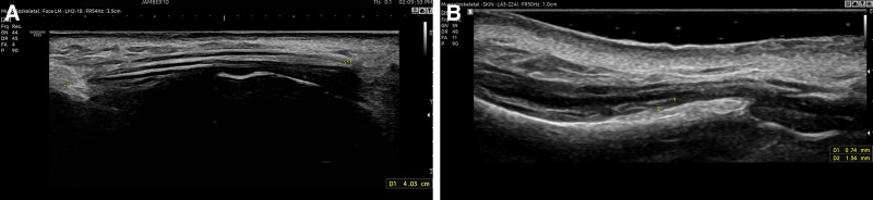
Fig. 2. Changes in PDO and soft tissue around the PDO in a 72-year-old woman at 1 day and 12 months. A, The total length of the inserted coiled PDO was 40.3 mm, and the diameter of the PDO thread was 1.10 mm at 1 day. The PDO threads were placed parallel under the muscle layer. B, At 1 year, the diameter decreased to 0.74 mm, but the total thickness, including PDO and soft tissue around the PDO, increased to 1.56 mm, suggesting that the degree of nasal elevation was well maintained despite the decrease in PDO diameter.72岁女性1天与12个月对比:植入线材总长40.3 mm,1年时直径降至0.74 mm,但含线材与周围软组织的总厚度增至1.56 mm,提示线材吸收后鼻部抬高仍获维持。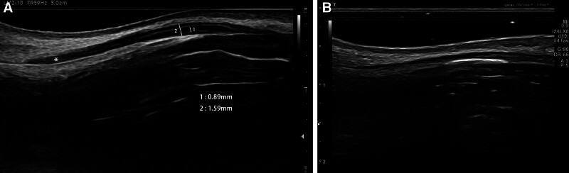
Fig. 3. New soft-tissue formation was found in sonographic evaluation for a 24-year-old woman at 9 and 12 months. A, After 9 months, new soft tissue (*) was created at the bar end of the thread. B, After 1 year, the diameter of the thread was reduced, but the new soft tissue was observed to have increased, showing newly formed soft tissue not only on the outside of the thread but also on the inside of the thread.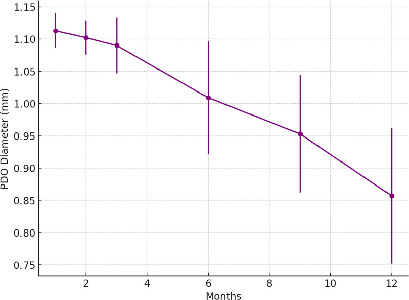
Fig. 4. Reduction in PDO thread diameter in sonography over time. The diameter of JB threads measured 1.113 mm at 1 month, 1.102 mm at 2 months, 1.09 mm at 3 months, 1.009 mm at 6 months, 0.953 mm at 9 months, and 0.857 mm at 12 months. Although the diameter decreased over time, the threads were still present at 12 months. The SDs were 0.027, 0.026, 0.043, 0.087, 0.091, and 0.105, respectively.超声下PDO线材直径随时间变化:1个月1.113 mm逐步降至12个月0.857 mm,各时点SD为0.027–0.105;统计分析(n=13)显示直径>1 mm维持至6个月。补充视频 / 附件(原文未随文提供原图,可于出版方页面查看)gox-14-e7500-s001.mp4Video 1. This video displays (front part) how an entry point was made by a 18G needle and two 40mm spring PDO threads were inserted into the nasal dorsum under the muscle layer. Since the tumescent solution was injected into the lower layer of the muscle in advance, PDO threads are easy to actually be placed under the muscle. (back part) It is a 35mm long spring PDO, and when compressed while inserting the thread, it is compressed to 20mm in length. After insertion into the columella, the tip of the nose protrudes due to the elasticity of the PDO.补充视频 / 附件(原文未随文提供原图,可于出版方页面查看)gox-14-e7500-s002.mp4Video 2. This video displays the ultrasound evaluation of coiled PDO threads used for nasal augmentation. At 6-days, the coiled PDO thread (414 mm, 3-0) was clearly visualized beneath the muscle layer, with an empty central lumen. Numerous surrounding blood vessels were identified, while the coil structure itself could not enter vessels, indicating a high safety profile. At 3-months, the PDO diameter remained stable (1.11 mm) with persistent central hypoechogenicity. At 9-months, partial PDO degradation was observed, with diameter reduction to 0.72 mm and marked perithread soft-tissue formation. At 1-year, mature hyperechoic soft tissue filled the PDO lumen, maintaining nasal height despite thread resorption.补充视频 / 附件(原文未随文提供原图,可于出版方页面查看)gox-14-e7500-s003.mp4Video 3. This video displays the ultrasound at 1-day demonstrated three inserted PDO threads with a total longitudinal length of 40 mm. No soft tissue ingrowth was observed, and the lumen appeared markedly hypoechoic. At 1-year, mature hyperechoic soft tissue was noted surrounding and lateral to threads, indicating ongoing intraluminal tissue formation. Nasal height was well maintained by newly formed soft tissue. Arterial mapping revealed numerous vessels coursing above the muscle layer, while PDO threads remained stably positioned beneath without migration. Unlike fillers, which may widen the nasal bridge due to absorption or migration, PDO thread augmentation preserved a sharp nasal contour with superior vascular safety补充视频 / 附件(原文未随文提供原图,可于出版方页面查看)gox-14-e7500-s004.mp4Video 4. This video displays (right lateral view) comparing the before and after views of the nose after one year, the height of the nose was still high. (warmth eye view) Comparing the nose before and after one year later, the height of the nose was still higher and there has been no migration, so the sharp shape of her nose was maintained well. In the case of thread nose augmentation, there was no migration, so it can prevent side effect, the formation of broad nasal dorsum. (left lateral view). The nasal height has increased and the improvement in the mock line has been maintained for more than one year.
The ultrasound observations indicated that the actual thickened soft tissue, due to collagen formation, was greater than the thickness of the JB 21G, 40-mm threads. The thickened soft tissue measured 1.213 mm at 1 month, 1.210 mm at 2 months, 1.267 mm at 3 months, 1.288 mm at 6 months, 1.341 mm at 9 months, and 1.403 mm at 12 months, showing an increase over time. The SDs were 0.240, 0.181, 0.215, 0.1349, 0.142, and 0.145, respectively. This increase suggests significant collagen formation and granulation tissue development around the threads. The consistent increase over time implies that the body responds positively to the presence of the threads by generating new tissue, which contributes to the sustained augmentation effect, even as the threads themselves diminish.
At 1 month, there was a 10.27% increase in soft-tissue thickness, indicating early collagen formation and tissue growth. The percentage increase continued to rise over the months, reaching a 27.55% increase at 12 months. This sustained growth suggests significant collagen formation and granulation tissue development around the PDO threads. The SDs reflect the variability in measurements, with the overall trend showing a consistent increase in tissue thickness. The ongoing increase in soft-tissue thickness from the baseline indicated that the body responds positively to the PDO threads, generating new tissue that contributes to the sustained augmentation effect. The data showed a statistically significant increase in soft-tissue thickness around the PDO threads over time, starting from the baseline of 0 months with an assumed PDO thickness of 1.1 mm. The most substantial increases occurred progressively over the months, highlighting the effectiveness of the PDO threads in stimulating collagen formation and tissue growth. This confirmed the threads’ ability to provide long-lasting support and augmentation in nasal procedures (Table 3 ) (Fig. 5 ) ( see Videos 3 and 4 [online] ).
Table 3. Increase in Soft-Tissue Thickness Around PDO Threads in Sonography Over Time
| Month | Mean Thickness, mm | SD | Percentage Increase, % |
|---|---|---|---|
| 1 | 1.213 | 0.24 | 0 |
| 2 | 1.21 | 0.181 | −0.24732 |
| 3 | 1.267 | 0.215 | 4.451772 |
| 6 | 1.288 | 0.1349 | 6.183017 |
| 9 | 1.341 | 0.142 | 10.55235 |
| 12 | 1.403 | 0.145 | 15.66364 |
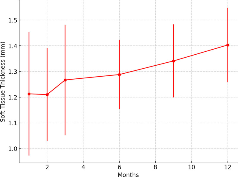
Fig. 5 Increase in soft-tissue thickness around PDO threads in sonography over time. The thickened soft tissue measured 1.213 mm at 1 month, 1.210 mm at 2 months, 1.267 mm at 3 months, 1.288 mm at 6 months, 1.341 mm at 9 months, and 1.403 mm at 12 months, showing an increase over time. The SDs were 0.240, 0.181, 0.215, 0.1349, 0.142, and 0.145, respectively.超声下线材周围软组织厚度随时间增加:1个月1.213 mm至12个月1.403 mm,提示持续胶原形成。
Using the QuantifiCare LifeViz Mini 3D scanner, changes in nasal volume and height were observed. Nasal volume increased to 0.771 mL at 1 month, 0.703 mL at 2 months, 0.682 mL at 3 months, 0.653 mL at 6 months, 0.619 mL at 9 months, and 0.552 mL at 12 months. The SDs were 0.220, 0.186, 0.179, 0.179, 0.178, and 0.141, respectively (Table 4 ) (Fig. 6 ) ( see Video 1 [online] ).
Table 4. Changes in Nasal Volume and Height in 3D Scanner Analysis Following PDO Thread Insertion
| Month | Mean Volume, mL | Volume SD | Mean Height, mm | Height SD |
|---|---|---|---|---|
| 1 | 0.771 | 0.22 | 1.387 | 0.28 |
| 2 | 0.703 | 0.186 | 1.19 | 0.186 |
| 3 | 0.682 | 0.179 | 1.122 | 0.179 |
| 6 | 0.653 | 0.179 | 1.062 | 0.179 |
| 9 | 0.619 | 0.178 | 1.057 | 0.178 |
| 12 | 0.552 | 0.141 | 1.005 | 0.141 |
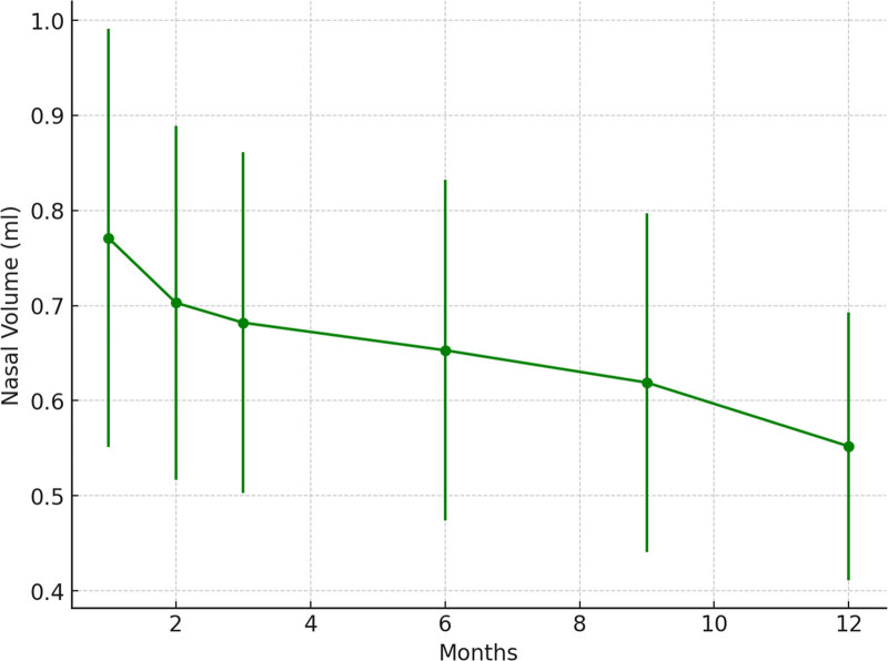
Fig. 6. Change in nasal volume in a 3D scanner following PDO thread insertion. Nasal volume increased to 0.771 mL at 1 month, 0.703 mL at 2 months, 0.682 mL at 3 months, 0.653 mL at 6 months, 0.619 mL at 9 months, and 0.552 mL at 12 months. The early rise in nasal volume reflects postprocedural swelling and the immediate presence of the threads, whereas the gradual decline corresponds to swelling resolution and PDO absorption, yet the 12‑month volume remaining above baseline indicates a sustained augmentation effect driven by new tissue growth.三维扫描鼻体积变化:1个月0.771 mL,12个月0.552 mL;早期升高与术后肿胀及线材占位有关,后期下降反映肿胀消退与线材吸收。
Nasal height changes observed with the 3D scanner were 1.387 mm at 1 month, 1.190 mm at 2 months, 1.122 mm at 3 months, 1.062 mm at 6 months, 1.057 mm at 9 months, and 1.005 mm at 12 months. The SDs were 0.280, 0.186, 0.179, 0.179, 0.178, and 0.141, respectively. The trend mirrors that of nasal volume, with early increases due to swelling and thread placement, followed by a stabilization phase. The maintained increase at 12 months demonstrates the efficacy of the threads in providing a sustained lift to the nasal structure.
The initial increase in volume was likely due to immediate postprocedural swelling and the initial effect of the threads. The subsequent decrease reflected the resolution of swelling and the natural absorption of the threads. However, the final volume at 12 months remained higher than baseline, indicating a lasting augmentation effect facilitated by tissue growth around the threads. The observed changes in nasal volume as measured by the 3D scanner showed a decrease over time, despite an initial increase. This trend can be explained by several factors: (1) Resolution of swelling: postprocedural swelling typically subsides over time, leading to a reduction in measured volume. (2) Absorption of PDO threads: as the PDO threads gradually absorb, the physical bulk they contribute diminishes, resulting in a lower volume measurement. (3) Redistribution of tissue: the initial tissue response, including collagen formation, might initially contribute to increased volume, but over time, the tissue may remodel and stabilize, leading to a more natural contour with less volume than immediately after the procedure.
Although volume decreased, the height increase observed in the ultrasound data suggests that the augmentation effect might be more about structural support and lifting rather than bulk volume addition. The PDO threads likely provided a scaffold for tissue support, enhancing nasal height and shape without significantly increasing overall nasal bulk. The increased soft-tissue thickness due to collagen formation around the PDO threads, as observed in ultrasound measurements, supported the sustained elevation effect. The structural changes from collagen may contribute more to the shape and contour rather than to overall volume, explaining why volume decreases but height and structural integrity remain enhanced. The decrease in nasal volume as measured by the 3D scanner, despite initial increases, can be explained by the resolution of postprocedural swelling, gradual absorption of PDO threads, and tissue redistribution. The increased height and tissue thickness observed in ultrasound measurements highlight the effectiveness of the PDO threads in providing structural support and lift, resulting in an aesthetically pleasing nasal contour without a significant increase in bulk volume (Table 4 ) (Fig. 6 ) ( see Videos 1–4 [online] ). Despite the reduction in nasal height as observed by the 3D scanner, the sustained increase in soft-tissue thickness around the PDO threads suggests that the augmentation effect is still present, just less pronounced over time. The balance between the gradual absorption of the PDO threads and ongoing collagen formation results in a stabilized, but slightly reduced, nasal height over the long term. The reduction in nasal height observed by the 3D scanner and reported by patients can be explained by the natural resolution of postprocedural swelling, gradual absorption of PDO threads, and the dynamic process of collagen remodeling and tissue settling. Although the immediate postprocedure height increase was more pronounced due to swelling and the physical presence of the threads, the long-term augmentation effect was maintained through collagen formation, resulting in a more natural and subtle enhancement. This underscores the importance of managing patient expectations and understanding the dynamic nature of tissue augmentation procedures (Table 4 ) (Figs. 7 – 12 ) ( see Videos 3 and 4 [online] ).
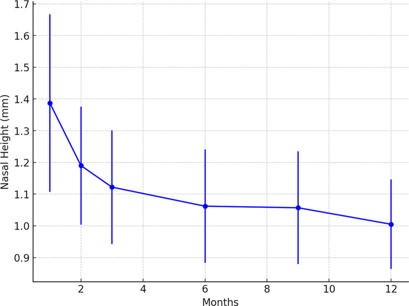
Fig. 7. Change in nasal height in a 3D scanner following PDO thread insertion. Nasal height changes observed with the 3D scanner were 1.387 mm at 1 month, 1.190 mm at 2 months, 1.122 mm at 3 months, 1.062 mm at 6 months, 1.057 mm at 9 months, and 1.005 mm at 12 months. The early postoperative increase in nasal height reflects swelling and thread placement, whereas the subsequent stabilization—with height still greater than baseline at 12 months—indicates a sustained lifting effect of the threads.三维扫描鼻高度变化:1个月1.387 mm,12个月1.005 mm;早期升高源于肿胀与线材放置,其后趋稳且仍高于基线。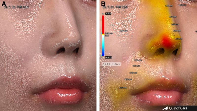
Fig. 8. Nasal height changes observed with the 3D scanner for a 24-year-old woman at 1 week. A, Before procedure. B, After 1 week, 2 JB-F PDO threads placed on the nasal dorsum and 1 JB-Tanxing thread on the columella produced a clear increase in nasal height and projection. The 3D scanner color map highlights these elevation changes, with red areas representing the greatest postoperative enhancement.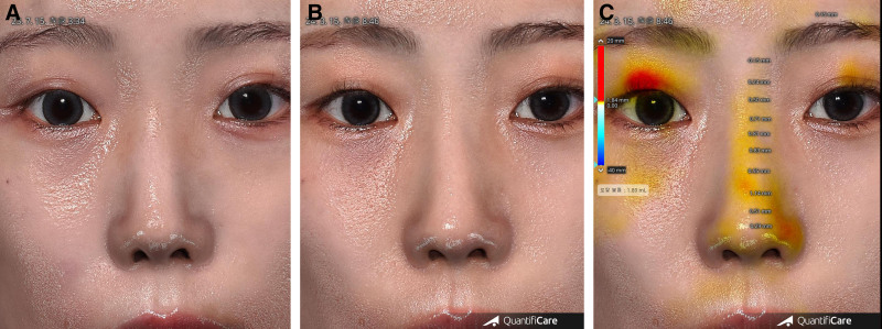
Fig. 9. Nasal height changes observed with the 3D scanner for a 24-year-old woman at 9 months. A, Preprocedure nasal appearance. B, Nasal appearance immediately after the JAMBER PDO thread procedure. C, At 9 months postprocedure, the 3D scanner shows sustained augmentation after placement of 2 JB-F threads along the dorsum and 1 JB-Tanxing thread in the columella.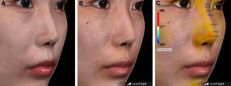
Fig. 10. Nasal height changes observed with the 3D scanner for a 24-year-old woman at 12 months (45-degree oblique view). A, Preprocedure nasal appearance. B, Nasal appearance at 12 months postprocedure. C, At the 12-month follow-up, 3D scanner measurements confirm persistent augmentation after placement of 2 JB-F threads in the dorsum and 1 JB-Tanxing thread in the columella.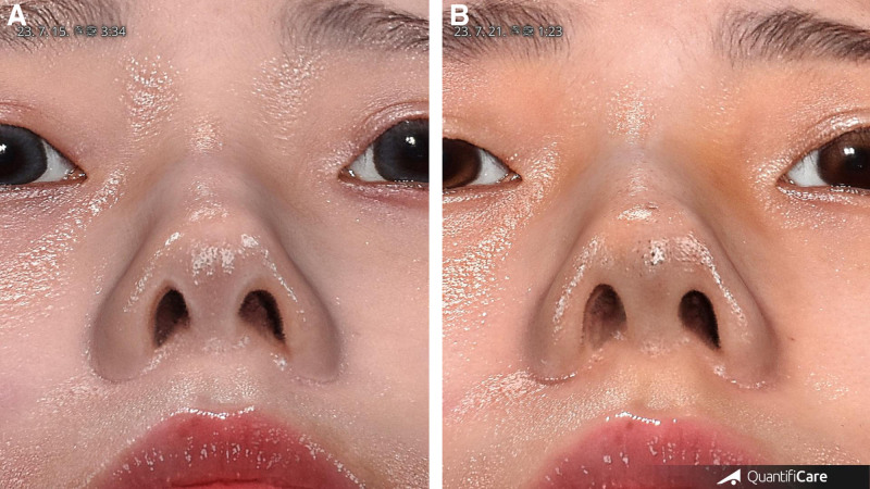
Fig. 11. Basal view of a 24-year-old woman at 1 week postprocedure. A, Preprocedure basal view. B, This early postoperative enhancement demonstrates the ability of PDO threads to provide structural support and refine nasal contours in a minimally invasive manner.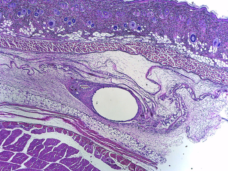
Fig. 12. Histological analysis of the 19G PDO thread placement site in nasal tissue at 2 weeks postinsertion (H&E Stain, 40x). The central white region represents the site of the PDO thread, which has yet to be fully infiltrated by collagen. Surrounding tissue displays an initial tissue response without significant collagen ingrowth in the core, although the diameter of the PDO remains the same for 6 months (P < 0.05). H&E, hematoxylin and eosin.术后2周鼻组织H&E染色(40×):线材占位中央区尚未被胶原完全浸润,核心区无明显胶原长入,属早期组织反应的组织学观察,不能直接推断长期临床结局。The advantages of JB, 21G, 40-mm threads include effective volume augmentation and long-lasting results. The threads are resilient to pressure and deformation due to their elasticity. The coiled structure of JB threads minimizes discomfort and the sensation of foreign material when facial expressions are made or the nose is moved. Compared with fillers or silicone implants, JB thread procedures are simpler and shorter in duration. Importantly, JB threads are much safer than fillers in terms of vascular complications, as there is no risk of skin necrosis or blindness. The results highlight the effectiveness of JB threads in nasal augmentation, providing an initial boost in volume and height due to their physical presence and the body’s response. Over time, collagen and granulation tissue maintain the augmented structure as the threads absorb. However, the study’s small sample size (23 patients) and its retrospective, single-clinic design may limit generalizability. Future multicenter studies with larger cohorts and randomized trials are needed to confirm the long-term efficacy and safety of JAMBER PDO threads. A key limitation of this technique is the inability to selectively augment areas of irregularity or depression along the nasal dorsum. The placement of threads along the entire length of the dorsum produces a uniform, symmetrical augmentation, which may not adequately address all contour concerns. This limitation should be considered when selecting patients and setting expectations for outcomes.
The absence of severe complications such as skin necrosis or blindness, which are common risks with fillers, highlights the safety of JB threads. Ultrasound-guided placement ensures correct insertion, reducing the risk of thread visibility or discomfort. In this study, no cases of thread removal, infection, migration, or extrusion were observed. If removal is necessary, a minor incision under local anesthesia, guided by ultrasound, can effectively extract the threads with minimal tissue trauma. Although JB threads reduce the risk of embolism compared with fillers, they lack an immediate reversal agent like hyaluronidase, requiring careful technique. The sustained increase in soft-tissue thickness and nasal height/volume over 12 months underscores their efficacy, supported by new tissue growth around the threads.
The JB thread procedure is simpler and quicker compared with traditional fillers and silicone implants. The coiled structure of the threads offers flexibility, comfort, and minimal invasiveness, making them an attractive option for nasal augmentation. JB threads provide a safe and effective alternative to traditional methods, with gradual thread absorption balanced by new tissue formation for sustained improvements in nasal volume and height. Advanced imaging techniques such as ultrasound and 3D scanning ensure precise placement and enhance patient satisfaction.
In this study, involving 23 patients who underwent nasal augmentation with JAMBER PDO threads, no actual complications were observed throughout the 12-month follow-up period. Specifically, there were no cases of infection, migration, extrusion, or skin necrosis. This finding underscores the safety profile of JAMBER PDO threads when applied with proper technique and monitoring. However, it is essential to remain vigilant for potential complications, such as infection, thread migration, or extrusion, as these risks, although theoretical in this study, are inherent to PDO thread procedures. Further studies with larger cohorts and extended follow-up are encouraged to confirm these safety findings.
JAMBER PDO threads demonstrate effective nasal augmentation, with increased nasal height and volume sustained during a 12-month period. The threads stimulate collagen formation, contributing to long-lasting aesthetic results.
Compared with traditional fillers, JAMBER PDO threads offer a safer alternative for nasal augmentation, particularly in reducing the risk of vascular complications such as embolism. However, their lack of easy reversibility requires careful planning and precision during placement.
This study suggests that JAMBER PDO threads are a viable and minimally invasive option for patients seeking nasal augmentation, providing durable results with minimal complications.
Further research with larger cohorts and extended follow-up is recommended to confirm long-term efficacy and to better understand the histological response over time.
The author has no financial interest to declare in relation to the content of this article. All funding for the study was provided by the author.
Patients provided written consent for the use of their images.
The author expresses thanks to Dr. Saran Wan of WanSiri Hospital for supporting this article.
声明:中文精读 · 仅供学术参考。内容来自公开文献检索,不代表本人观点,不构成诊疗建议。 医疗美容需在正规医疗机构由执业医师实施。
基于相同主题推荐 · 继续深挖
单中心回顾性研究纳入23例鼻部填充患者(年龄21–72岁,平均35.3岁,男8女15),随访12个月。
鼻部塑形疗效与安全性:12个月前瞻性研究/PMC13100342_fig1.jpg)
这项单中心前瞻性研究纳入24名中国女性受试者,接受CaHA凝胶鼻部注射(骨膜上/骨膜浅层,单侧单次总量≤2.0 mL,平均0.93±0.22 mL),全部完成12个月随访,无脱落。
前瞻性单臂研究(西班牙 Asturias 伦理委员会批准),39 例健康女性志愿者(23–68 岁,平均 47 岁,Fitzpatrick II–III),腹部/腰侧 25 例、大