形体雕塑手术的美学单元与黏附区:躯干及四肢切口规划的系统分类与六原则
2026年10月14日29 min read
Plastic and Reconstructive Surgery Global Open
Plastic and Reconstructive Surgery Global Open
结论与实践要点
本文是一篇将面部美容亚单位理论延伸至躯干与四肢的解剖学分类研究。作者基于 35 年实践经验与 3 例 MWL 患者资料,把躯干和四肢划分为若干美学单元(AU),并在 AU 边界识别出皮肤支持带的黏附区(ZA),提出“切口即瘢痕、瘢痕即疗效”的六原则规划框架:锚定 ZA、位于界面不跨越、隐匿于皮褶、肢体内侧、衣着下隐匿、规避关节屈伸线。
临床价值在于把以往依赖个人经验的切口设计转化为可教学、可复用的解剖坐标系,尤其对 MWL 后形体雕塑(腹壁成形、提升术、上臂成形等)的术前标记具有直接参考意义;男女前胸壁 ZA 走行差异的解释(乳房下皱襞 vs 胸肌下 ZA)对隆胸/男性乳腺手术切口选择同样有提示。需要说明的是,本文为描述性研究与专家意见,无客观瘢痕量表或对照比较,六原则的相对权重在不同术式中仍需术者权衡(作者亦承认“一个特征可能压倒另一个特征”)。将其作为术前标记的教学框架而非循证等级较高的疗效证据来使用,是恰当的定位。
Background:Aesthetic units (AUs) and zones of adherence (ZAs) have been previously noted in the face and torso. A systematic classification of common AUs and ZAs has not been previously described for the torso and extremities. Contour and scars are equally important in obtaining the best result. Therefore, the six principles to consider in body contouring surgery (BCS) are that the resulting scar be anchored at a zone of adhesion; at the interface of AUs; concealed within flexion or extension skin creases, or placed according to conventional usage and experience; positioned on the medial aspect of a limb; hidden when nude, topless, or in a variety of clothing such as one-piece and two-piece swimsuits, crop tops, short sleeves, etc; and positioned to avoid a contracture across a flexion joint crease or a dehiscence across the extension aspect of a joint.
Methods:Images of male and female massive weight loss (MWL) patients with a history of MWL were reviewed and analyzed.
Results:AUs and Zas are described and codified. An algorithm is provided to aid in planning incisions for BCS.
Conclusion:With the increase in BCS after MWL, a classification of AUs and ZAs is considered useful in planning surgery and optimizing the aesthetic result.
Question: The key problem is optimizing scar locations and preserving aesthetic units when performing body contouring surgery in patients after massive weight loss.
Findings: Aesthetic units, zones of adherence, and the medial aspect of the thigh or arm are used to guide incisions for body contouring surgery and provide patients with the optimal result.
Meaning: Planning body contouring surgery based on aesthetic units and zones of adhesion is a practical approach to optimize results.
In a letter to Robert Hooke in 1675, Isaac Newton made his most famous statement: “If I have seen further, it is by standing on the shoulders of Giants.” This statement is now often used to symbolize scientific progress and to give credit to those who have laid the foundations of our cognitive and practical skills. I would like to acknowledge the immeasurable knowledge and good sense that was handed down to me by Frederick McCoy, my program director and mentor at the University of Missouri Kansas City, without whom this article would probably not have seen the light of day.
Excluding fetal surgery, 1 a surgical incision results in a scar. The ideal scar is an invisible one. For a plastic surgeon, the choice of incision from the very first cut (direction and location) is not only the first step on a journey but akin to firing a cannonball. Once the ordinance leaves the barrel, trajectory and destination are, for all practical purposes, irrevocable and unchangeable. Similarly, a “good incision” often predicts a successful outcome, and a bad one, the opposite. Frederick McCoy would say, “if you aim for less than perfection you will hit it every time.” 2 This article is an attempt to provide a trajectory for that aim, in creating an atlas of aesthetic surgery for the human body.
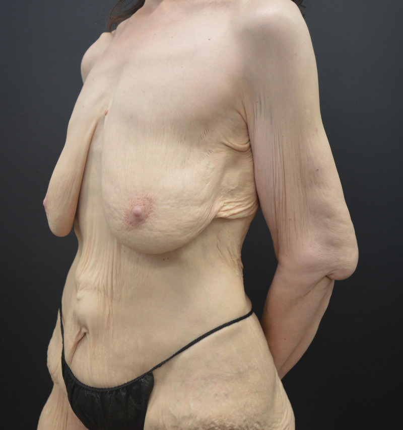
Burget and Menick 3 previously described aesthetic subunits of the nose. It is interesting that the alar sill, while not noted as such, is also a zone of adherence (ZA). Figure 4, Chapter 1 4 shows the consequences of “patching holes,” and the result of ignoring aesthetic units (AUs). Gonzalez-Ulloa et al 5 and Millard 6 have also discussed the head and neck in terms of AUs. The body may also be divided into AUs 7 – 14 and ZAs 15 (Tables 1 – 4 ). It is important to note that even in cases of massive obesity, the skin does not simply hang like a “onesie” garment (Fig. 1 ) but is tethered by these ZAs. 15
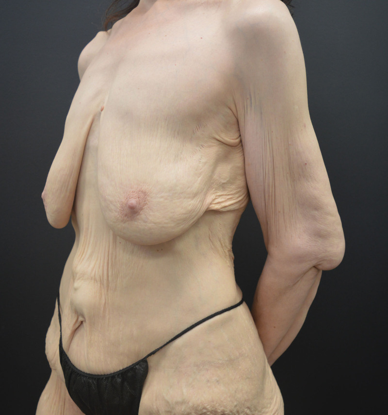
Fig. 1. A 56-year-old woman after 200-pound weight loss, oblique view.Choosing the ideal incision, particularly in body contouring surgery (BCS) and indeed, for all aesthetic and reconstructive procedures, is a choice between the good, the bad, and the ugly. This article is limited to below the clavicle and also excludes hands and feet. This does not imply that these anatomical regions are irrelevant, but for the purpose of this article that focuses on BCS, these anatomical parts are omitted.
“The superficial fascial system (SFS) produces many of the topographical landmarks of body-surface anatomy. The creases, plateaus, valleys, and bulges of our bodies are explained by the anatomy of the SFS and its relationships to skin, fat, and the musculoskeletal system. Understanding the anatomy and age-related pathologic changes of the SFS of the trunk and extremities may help explain body contour deformities and provide the [aesthetic] basis for surgical correction.”15
An AU may be determined in shape by an underlying muscle or group of muscles, such as the pectoralis major, rectus abdominis, or anterior leg compartment muscles, or may simply be the anterior or posterior anatomical aspect, such as the anterior and posterior thigh. AUs are bordered by other AUs. ZAs occur at the boundary of an AU but the converse does not apply. AUs are aesthetic building blocks and are applicable to all patients, nonobese, obese, and massive weight loss (MWL) patients alike (Tables 1 – 3 ). In this article, there is no attempt to subdivide AUs as proposed in liposculpture. 16 – 20 Breast subunits have also been previously described and are not further discussed. 7 – 12
Five terms discussed in this article are defined as follows:
Depending on the procedure performed, the principles for optimal planning of a scar for BCS are that the incision satisfies one or more of the following six criteria, which may apply with different emphases (Table 5 ); ( See Video [online] , which displays the body contouring AUs and zones.)
Ideal scars should, therefore, be:
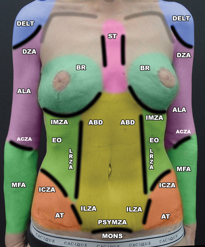
Fig. 2. AU distribution. Female, anterior torso and arm after LBL, brachioplasty, and mastopexy with curvilinear lateral chest extension. ACZA indicates antecubital FC ZA; BR, breast AU; DELT, deltoid muscle AU; DZA, deltoid insertion ZA; ICZA, iliac crest ZA; ILZA, iliac crest ZA; IMZA, inframammary fold ZA; LRZA, lateral rectus ZA; MFA, medial (ventral) forearm AU; MONS, mons pubis AU; PSYMZA, pubic symphysis ZA; ST, sternum AU.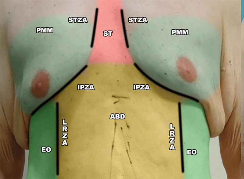
Fig. 3. AU distribution. Male, upper anterior torso after MWL. IPZA indicates caudal edge of PMM ZA; LRZA, lateral rectus ZA; STZA, sternum ZA.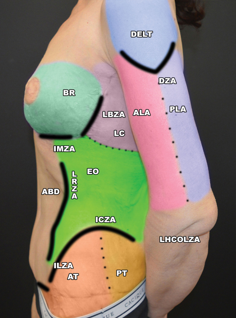
Fig. 4. AU distribution. Female, lateral torso and arm, after MWL, preoperative. ABD indicates rectus abdominus muscle AU; BR, breast AU; DELT , deltoid muscle AU; DZA, deltoid insertion/lateral arm ZA; ICZA, lateral chest AU; ILZA, inguinal ligament ZA; IMZA, inframammary fold ZA; LBZA, lateral breast ZA; LC, lateral chest AU; LHCOLZA, lateral humeral condyle to olecranon ZA; LRZA, lateral rectus ZA.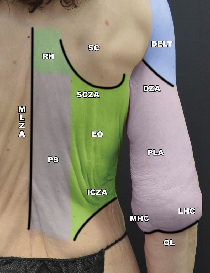
Fig. 5. AU distribution. Female, posterior torso, preoperative. DELT indicates deltoid muscle AU; DZA, deltoid insertion ZA; ICZA, iliac crest ZA; LHC, lateral humeral condyle; MHC, medial humeral condyle; MLZA, midline torso spinous processes ZA; OL, olecranon; PLA, posterior lateral arm; PS, paraspinal muscles AU; SC, scapula AU; SCZA, scapula ZA.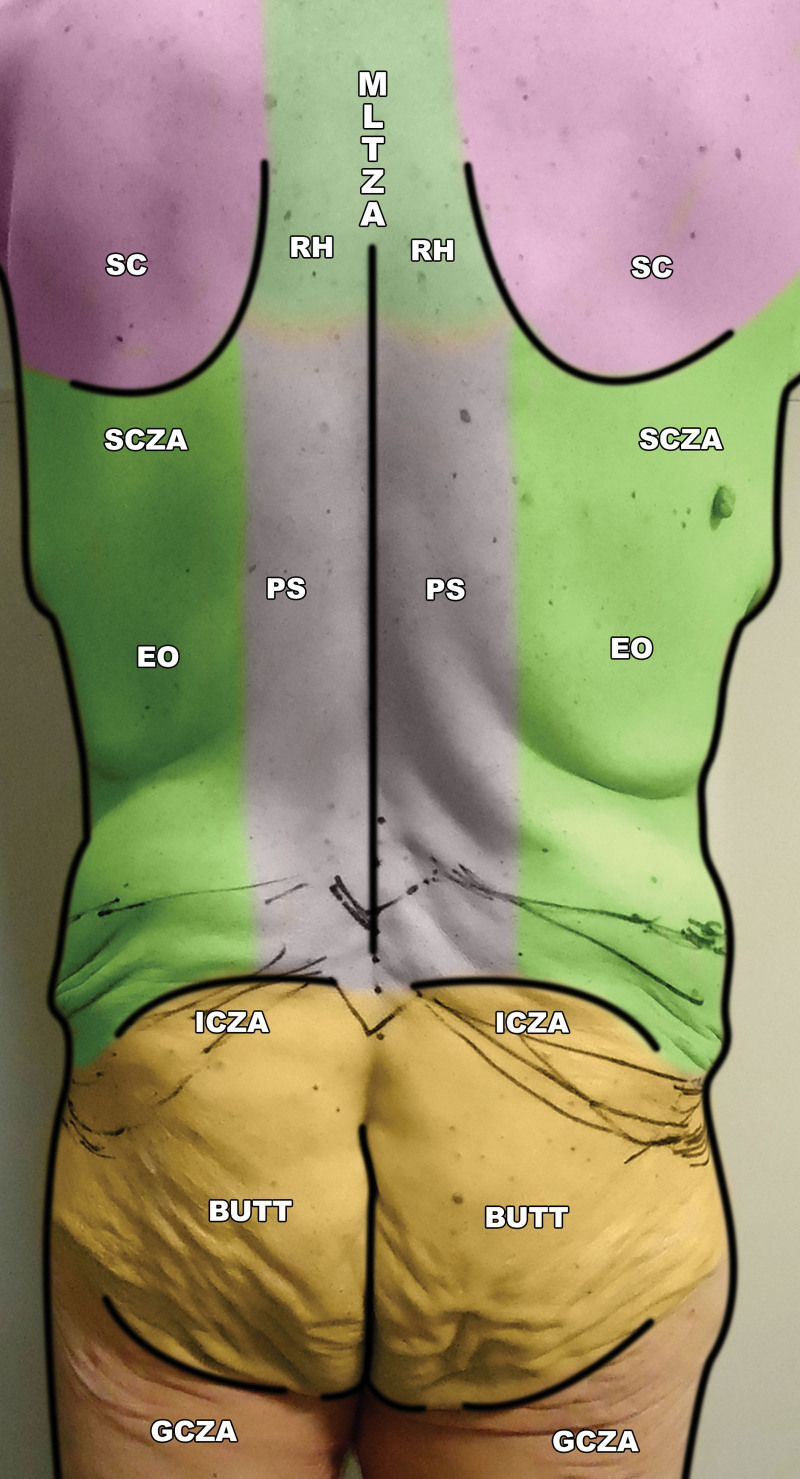
Fig. 6. AU distribution. Male, posterior torso, preoperative. BUTT indicates buttock AU; EO, external oblique muscle AU; GCZA, gluteal crease ZA; ICZA, iliac crest ZA; MLT ZA, midline torso ZA; PS, paraspinal muscles AU; RH, rhomboid muscles AU; SC, scapula AU; SCZA, scapula ZA.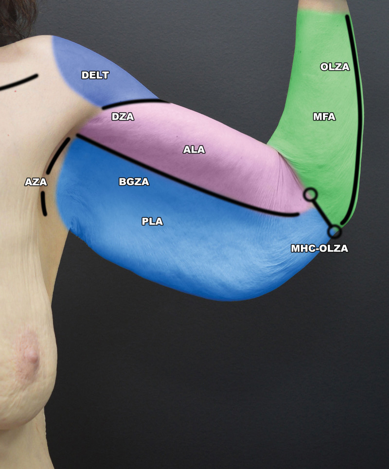
Fig. 7. AU distribution. Female left upper extremity anterior, preoperative. ALA indicates anterior lateral arm; AZA, axillary ZA; BGZA, bicipital groove ZA; DELT , deltoid muscle AU; DZA, deltoid insertion ZA; MFA, medial (ventral) forearm AU; MHC-OLZA, medial humeral condyle-olecranon ZA P;LA, posterior lateral arm; RH, rhomboid AU.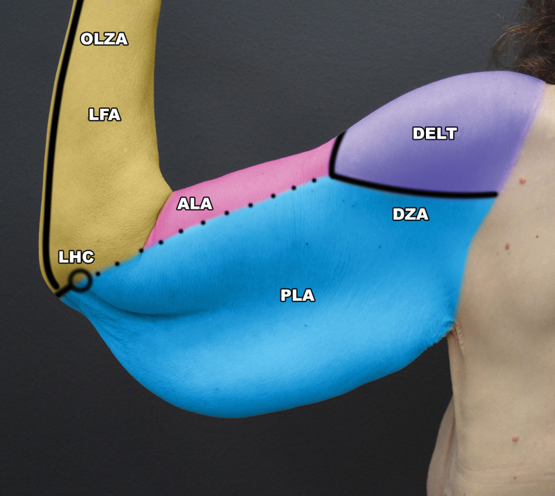
Fig. 8. AU distribution. Female left upper extremity posterior, preoperative. ALA indicates anterior lateral arm AU; DELT, deltoid muscle AU; DZA, deltoid insertion ZA; LFA, lateral forearm AU; LHC, lateral humeral condyle; OLZA, olecranon ZA; PLA, posterior lateral arm.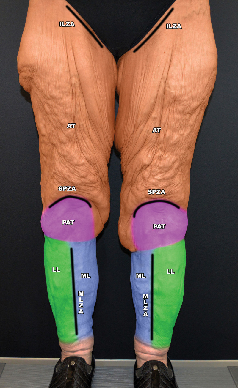
Fig. 9. AU distribution. Female lower extremity anterior, preoperative. AT indicates anterior thigh AU; ILZA, inguinal ligament ZA; LL, lateral leg AU;ML, midline anterior leg AU; MLZA, midline anterior leg ZA; PAT , patellar AU; SPZA, suprapatellar ZA.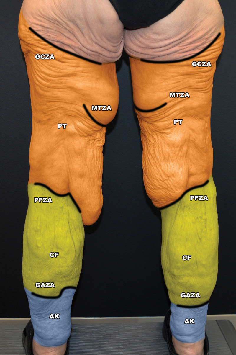
Fig. 10. AU distribution. Female, lower extremity posterior, preoperative. AK indicates ankle AU; CF, calf AU; GAZA, gastrocnemius ZA; GCZA, gluteal crease ZA; MTZA, medial thigh ZA; PFZA, popliteal fossa ZA; PT, posterior thigh AU.
This study is based on multiple patients in the United States and the United Kingdom who have presented for BCS during the author’s 35 years in practice. This article draws on the application of solid plastic surgery principles during this time. As Frederick McCoy would have said, many of the observations are “measured scientifically by eye.” Methods and results are limited by an analysis of three patients (two females and one male). There will obviously be variations between patients in the same way that there are anatomical variations of internal anatomy. Nonetheless, there is overall consistency, as evidenced by Lockwood’s study of 12 fresh cadaver segments and 20 body-contour patients. 15
Preoperative and postoperative male and female, anterior, posterior, and lateral views of the torso and upper and lower extremities were studied after MWL. AUs were identified that demarcated specific parts of the torso and extremities. AUs were defined as above. ZAs always demarcated boundaries of AUs but not vice versa (see above). No attempt was made to subdivide AUs further, as this was not felt to be relevant to planning BCS (Figs. 2 – 10 ). In the figures presented, AUs are filled with single blocks of color, and ZAs are identified as solid black lines. “Soft” AUIs without a ZA are identified as dotted black lines. AUs are labeled by their abbreviations, without any additional suffix, whereas ZAs are labeled with their abbreviations plus the additional ZA suffix, to avoid confusion with AUs. AUIs are not labeled. AUs (Tables 1 – 3 ) and ZAs (Table 4 ) are tabulated and illustrated (Figs. 2 – 10 ).
Our patient is a 56-year-old White woman with the height of 5’ 3” and maximum weight of 154 kg. Her current weight is 73 kg and body mass index is 28 kg/m 2 with history of self-induced weight loss of 81 kg over a period of 24 months and stable weight for 6 months. The patient lost a further 15 kg after consultation and before initial surgery. The patient was self-referred. She has no significant medical history. She is a nonsmoker and social drinker. In-patient surgery was performed at Greenwich Hospital, Greenwich, Connecticut. Preoperative view and postoperative views are shown 4.5 months after lower body lift (LBL) and 1 month after an extended brachioplasty contiguous with a mastopexy-breast autoaugmentation 27 – 29 (Figs. 11 , 12 ).
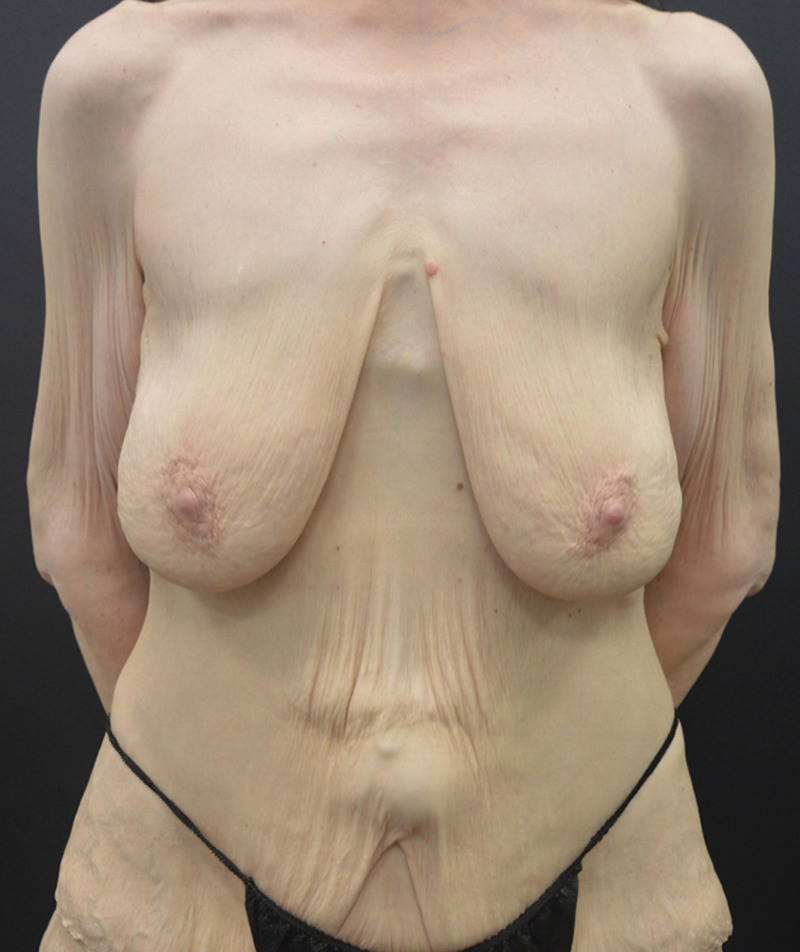
Fig. 11. A 56-year-old woman after self-induced MWL, preoperative front view.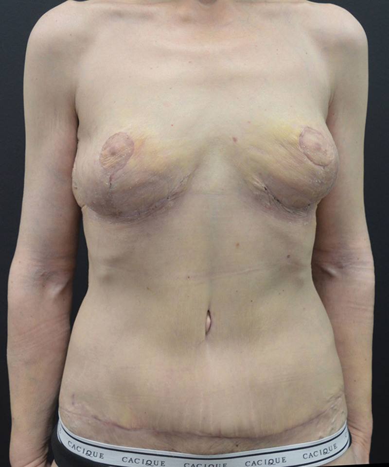
Fig. 12. A 56-year-old woman after self-induced MWL, 4.5 months after LBL, and 1 month after extended brachioplasty and mastopexy, postoperative front view.
Our patient is a 34-year-old White woman with the height of 5’ 6” and weight of 58 kg. Her body mass index is 20.7 kg/m 2 with a history of self-induced weight loss of 77 kg and stable weight for an undetermined period. Patient was self-referred. She has no significant medical or surgical history. She is a nonsmoker and nondrinker. In-patient surgery was performed at the Princess Grace Hospital in London, United Kingdom. Preoperative view and postoperative views are shown 3 months after extended abdominoplasty and 1 month after breast lift and breast autoaugmentation 27 – 29 with an upper body lift (UBL) (Figs. 13 , 14 ).
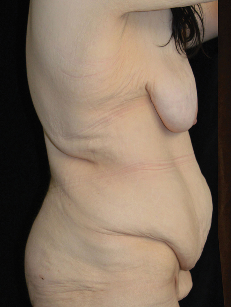
Fig. 13. A 34-year-old woman, after self-induced MWL, preoperative right-side view.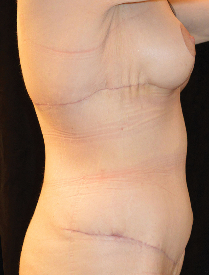
Fig. 14. A 34-year-old woman, 3 months after extended abdominoplasty and 1 month after breast lift and breast autoaugmentation, postoperative right-side view.Table 1. AUs of the Upper Extremity
| AU | Description |
|---|---|
| DELT | Deltoid muscle |
| ALA | Anterior lateral arm |
| PLA | Posterior lateral arm |
| MFA | Medial (ventral) forearm |
| LFA | Lateral forearm |
Table 2. AUs of the Anterior, Lateral, and Posterior Torso
| AU | Description |
|---|---|
| ST | Sternum |
| PMM | Pectoralis major muscle |
| BR | Breast |
| LC | Lateral chest |
| ABD | Rectus abdominis muscles |
| EO | External oblique muscle |
| MONS | Mons pubis |
| SC | Scapula |
| RH | Rhomboid muscles |
| PS | Paraspinal muscles |
| BUTT | Buttock |
| PER | Perineum |
Table 3. AUs of the Lower Extremity
| AU | Description |
|---|---|
| PT | Posterior thigh |
| AT | Anterior thigh |
| PAT | Patellar |
| ML | Midline anterior leg: (anterior crest of tibia and medial subcutaneous surface) |
| LL | Lateral leg (overlying anterior compartment) |
| CF | Calf: medial and lateral gastrocnemius muscles |
| AK | Ankle: inferior to bellies of gastrocnemius muscles/attachments to Achilles tendon |
Table 4. ZAs
| ZA | Anatomy |
|---|---|
| Upper extremity | |
| AX | Axilla |
| D | Deltoid insertion/lateral arm |
| BG | Bicipital groove |
| MHC-OL | Medial humeral condyle to olecranon |
| LHC-OL | Lateral humeral condyle to olecranon |
| AC | Antecubital FC |
| UL | Subcutaneous border of ulna |
| Anterior torso | |
| ST | Sternum |
| IM | Inframammary fold |
| IP | Caudal edge of PMM |
| LB | Lateral breast |
| LR | Lateral rectus |
| IC | Iliac crest |
| IL | Inguinal ligament |
| PSYM | Pubic symphysis |
| Posterior torso | |
| SC | Scapula |
| MLT | Midline torso spinous processes |
| GC | Gluteal crease |
| IPR | Ischio-pubic ramus |
| Lower extremity | |
| GT | Ilio-tibial tract/ greater trochanter |
| MT | Medial thigh (mid-medial thigh) |
| SP | Suprapatellar |
| PF | Popliteal fossa |
| IKN | Inferior knee (medial) |
| ML | Midline anterior leg (anterior crest of tibia and medial subcutaneous surface) |
| LL | Lateral leg: origin of peroneal muscles (lateral border of fibula). |
| GA | Gastrocnemius: caudal termination of medial and lateral bellies at Achilles tendon transition |
Table 5. Body Contouring Procedures: Choice of Scar Incisions, Based on Procedure and Clothing Considerations
| Surgical Site | Procedure | Scar Direction/Location | Visibility Considerations | Clothing Suggestions |
|---|---|---|---|---|
| Upper arm | Proximal brachioplasty | Transverse in axilla or with short “T” shaped extension along bicipital groove | Axilla and upper medial arm | Top with sleeves |
| Sleeveless outfit | ||||
| Bikini top | ||||
| Arm | Brachioplasty | Longitudinal with “V” break in axilla | Medial side of arm | Sleeveless or sleeved top |
| Arm and lateral chest | Extended brachioplasty | Vertical or curvilinear into IMF | Medial side of arm and lateral chest | Sleeveless or sleeved top |
| Elbow | Extended brachioplasty | Medial side of elbow | Medial side of elbow | Sleeveless or sleeved top |
| Lateral chest | Extended brachioplasty | Vertical | Beneath bra/crop top | Crop top |
| Curvilinear: concave superiorly | Backless dress or one-piece swimsuit | |||
| Flank lift | ||||
| Back | UBL | Horizontal | Beneath bra/crop top | Crop top |
| Beneath T-shirt | Avoid backless dress | |||
| Avoid extension to posterior midline | ||||
| Anterior chest (male) | Gynecomastia excision | Transverse at inferior border of PMM | Shirtless | Tight T-shirt |
| Avoid extension to anterior midline if possible | “Muscle” tank top | |||
| Breast (female) | Mastopexy | Wise pattern mastopexy | Avoid extension to midline if possible | Sports bra |
| Autoaugmentation | Lateral chest extension (horizontal) | Crop top | ||
| May extend horizontally: (1) with or without flank-lift; (2) in a curvilinear fashion into the apex of the axilla, with or without a brachioplasty; and (3) with or without UBL | ||||
| Lateral chest extension (curvilinear) | ||||
| Abdomen (female) | Abdominoplasty | Horizontal or high lateral | Beneath swimsuit | Thong |
| Extended abdominoplasty | Two-piece | |||
| One-piece | ||||
| LBL | ||||
| Abdomen (male) | Abdominoplasty | Horizontal | Beneath waistline of swim short | Swim shorts, that is, trainer type or full thigh |
| Extended abdominoplasty | ||||
| LBL | ||||
| Back (female) | LBL | Curvilinear-superior edge of buttock | Beneath swimsuit | Two-piece swimsuit |
| One-piece swimsuit | ||||
| Back (male) | LBL | Low posterior in men | Beneath swimsuit | Swim shorts |
| Gym shorts | ||||
| Thigh (proximal) | Upper MTL | Perineum/medial thigh junction | Perineal/thigh crease | Two-piece |
| One-piece | ||||
| Risk of caudal migration | Shorts | |||
| Anchor lift | ||||
| Thigh to knee | Vertical MTL | Vertical | Medial thigh | Swim shorts, that is, trainer type or full thigh (male) |
| Medial knee | ||||
| Bikini bottom (female) | ||||
| Yoga pants (female) | ||||
| Calf | Calf-lift | Vertical | Medial | Yoga pants |
AUs, ZAs, AUIs, ECs, and FCs are identical between men and women, except for the anterior chest wall, as reflected by the female curvilinear inframammary fold ZA, infra-pectoral ZA with the most caudal point at the breast meridian versus the male IPZA, which has an oblique medial-inferior to latero-superior direction.
With increasing demand for BCS after MWL, both contour and resulting scars must be considered in obtaining the optimum result. The six principles in optimal planning of an incision for BCS have been stated above. Each of these principles has varying degrees of importance, depending on the procedure (Table 5 ).
AUs have been described from the cartographic viewpoint. 13 Others have discussed AUs as they pertain to liposuction. 16 – 20 Gusenoff et al 14 stressed the importance of AUs in the treatment of pseudogynecomastia. With regard to incisions, Courtiss et al 21 commented that “certainly a prime method by which a patient judges the competence of his surgeon is by the appearance of the resultant scar.” Webster 22 suggested that wrinkle lines should be followed when making skin incisions. “The simplest rule for making incisions in the most favorable direction is to follow the natural wrinkle lines. These are usually recognizable on the face, the neck, the wrist, the axilla, the groin, or the back of the knee.” 22 Kraissl 23 further reviewed wrinkle lines with respect to the face and body, explaining differences in the scapular region between his lines and those of Langer. 24 – 26 Wilhelmi et al 30 stated that Kraissl lines rather than Langer lines (LLs) may be the best guides for elective incisions of the body. However, excluding joints, in the lower extremities, LLs reconcile with modern surgical approaches (Fig. 21 from Kraissl article). 23 Kraissl incisions for the abdomen and back reconcile with current body contouring incisions and are similar to LL except in the midline of the back where Kraissl lines are vertical as compared with the horizontal lines of Langer (Figs. 17, 18 from Kraissl article). 23 Relaxed skin tension lines of the face have been described by Borges and Alexander. 31 Incision lines have been further analyzed by Paul. 32
When planning incisions for BCS, it is important to appreciate the topography of AUs, ZAs, FCs ECs, joints, and skin tension lines. 21 – 33 One feature may override or mitigate against another. For example, the posterior LBL incision in the male is often lower than the iliac crest ZA, to ensure concealability beneath a swimsuit or a pants waistband, whereas in women, the incision should follow the iliac crest ZA along the superior curvilinear border of the buttock AU, which is coincidentally in a desirable position for concealability beneath a thong-style bikini bottom. See the LBL posterior scar in a woman (Fig. 2 by Rohrich et al 34 ), which is not curvilinear and lies above the AUI, and the LBL posterior scar in a man, (Fig. 13 by Lockwood 35 ), which demonstrates a low horizontal scar. Parenthetically, many articles of clothing highlight AUs and ZAs such as the “thong-style” bikinis and work-out “muscle” T shirts. ( See figure, Supplemental Digital Content 1 , which displays “muscle” T-shirt outlining scapula AUs, http://links.lww.com/PRSGO/C624.). The ideal incision for gynecomastia excision after MWL, also described as pseudo-gynecomastia, 14 is at the AUI between the PMM, abdomen (ABD), and EO AUs (Table 2 ). For women, inframammary fold (IMF) incisions permit the wearing of a crop top. In the female breast, it is important to avoid the upper medial quadrant in terms of scar visibility and to use the IMF as one border of an AU reconstruction. 7 A lower abdominoplasty scar permits wearing of high- or low-waisted pants. In contrast, a “fleur-de-lis” vertical scar precludes wearing an outfit such as a crop top, which exposes the midriff. Gynecomastia excision with a free nipple graft and a transverse scar through the center of the breast 36 and the “boomerang” scar 37 technique transgress the integrity of the PMM AU. High posterior curvilinear incisions 38 in an LBL prevent concealment of the scar below the waistband of pants, swim shorts, or a bikini bottom. UBL incisions, which cross the midline of the back will prevent wearing a low-backed dress or one-piece swimsuit. 39
Results of reconstructive procedures are published in the contemporary plastic surgery literature, which ignore AUs and create a patchwork quilt-like appearance. 40 The concept of AUs, as described by Burget, Spear, and others, 3 – 15 is further elaborated in this article, in the context of torso and limb surgery (Tables 1 – 3 ). ZAs, previously described, 15 are cataloged and classified (Table 4 ). The concept of the interface between an AU and ZA (AUI) is defined. Clothing preferences are reviewed with respect to scar placement and AUs. This classification is intended to act as a basis for planning BCS and for measuring outcomes based on scar position and visibility. Finally, an algorithm is presented, which outlines the six primary decision points in planning incisions for different BCS procedures. The six decision points are represented as a horizontal row with columns of procedures below. A single procedure may occur in multiple columns since more than one decision point may affect the choice of incision (Fig. 15 ). ( See Video [online] , which displays the body contouring AUs and ZAs together with the principles of planning BCS.)
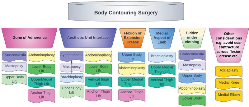
Fig. 15. Body contouring surgery. Decision points in planning incisions.补充视频 / 附件(原文未随文提供原图,可于出版方页面查看)gox-11-e5093-s001.mp4Video 1. This video shows the body contouring aesthetic units and zones.
This article describes a practical, comprehensive description of AUs and ZAs of the trunk and extremities. This is a metaphorical “quantum theory” for aesthetic and reconstructive surgery, combining multiple concepts and anatomical landmarks into one simple unifying theory that applies to all elements of the practice of plastic surgery of the torso and extremities. This “atlas of aesthetics” is for planning and execution of BCS and also for other aesthetic and reconstructive surgery of these body parts. AUs, ZAs, and AUIs are not limited to the torso and extremities, but also occur in the head, neck, hands, and feet, and a description of these may serve as subject matter for a publication by this or another author.
This article, with its presentation of AUs, ZAs, FCs, and ECs, and the accompanying figures may also serve as a useful addition to the curriculum for plastic surgeons in residency and fellowship training, as well as a guide for practicing plastic surgeons. At a minimum, it will stimulate a conversation and an exchange of ideas. Other authors may disagree or may choose to further improve on this analysis, as is the nature of scientific discourse. As plastic surgeons, we live and breathe the “aesthetic canons” of the human body, and it is inculcated into our training and practice that we be constantly mindful as to what will create a good, bad, or ugly result, with a view of avoiding, at all costs, the second and third options. Again, let us not forget the words of Dr. McCoy, past president of the ASPS (1976), “if you aim for less than perfection you will hit it every time.” Perfection must be our goal.
The author has no financial interest to declare in relation to the content of this article.
This paper would not be of its current high quality without the unpaid efforts and recommendations of the anonymous reviewers of PRS Global Open to whom I express my sincere gratitude. I am always wiser and better informed after reading their comments (positive and negative), and the final draft is literally more readable and substantial after taking their recommendations into consideration.
Ethical Compliance: All procedures performed in studies involving human participants were in accordance with the ethical standards of the institutional and/or national research committee and with the 1964 Helsinki Declaration and its later amendments or comparable ethical standards.
声明:中文精读 · 仅供学术参考。内容来自公开文献检索,不代表本人观点,不构成诊疗建议。 医疗美容需在正规医疗机构由执业医师实施。
基于相同主题推荐 · 继续深挖
上睑下垂手术的报道成功率很大程度上取决于“成功”如何定义,而非单纯反映手术本身的效果。
不同人群面部年轻化:专家共识与 AART 方法学临床应用经验/PMC13558784_fig1.webp)
这是一项基于改良 Delphi 法的专家共识(马来西亚6名具8–10年生物刺激剂经验的医师),并附4例 PLLA-SCA(Sculptra)临床应用个案,属专家意见与病例系列,并非
单中心、单臂前瞻性病例系列(证据等级 III),纳入 2024 年河北医科大学第一医院 786 例男性乳腺发育内镜腺体切除术患者(773 例双侧、13 例单侧),平均随访 96.5