下睑成形术后黑眼圈加重的机制与自体脂肪移植矫正:32 例回顾性研究
2026年10月15日26 min read
Plastic and Reconstructive Surgery Global Open
Plastic and Reconstructive Surgery Global Open
结论与实践要点
该文把「LLB 术后黑眼圈加重」这一常被归为患者预期问题的现象做了机制化解释:去脂后失去原有的光学补偿,泪沟凹陷阴影完全暴露。这提示 LLB 术前评估应纳入泪沟畸形与眶下空虚评估——只去脂、不处理韧带拴系与容量,可能让外观「越做越显老」。
矫正策略上有两点可复用:① 双平面(TTL 上皮下 + 下方前颌骨/前颧间隙)小量多次移植,单侧 1.3–1.6 mL、每点不超过 0.02 mL,坐位正面光源下以「睑–颧平滑过渡、无 demarcation 线」为终点;② 入路改良——鼻翼基底部外侧入路轨迹近乎垂直穿过 TTL,比传统平行于韧带的侧入路更易穿透致密韧带,同时避开睑缘薄皮肤区、降低轮廓不规则风险。病例 4 还揭示:残余外侧眶脂未处理者脂肪移植效果打折,需先去除残余脂肪。证据级别:单中心回顾性 + 病例展示,满意度为主观指标,长期体积保留率未量化。
Background:Lower eyelid blepharoplasty (LLB) is a widely performed cosmetic procedure. Many studies have investigated its postoperative complications; however, most have focused on lower eyelid retraction and malposition, with little attention given to the exacerbation of dark circles. This study aims to analyze the mechanisms underlying this complication and to assess the feasibility and effectiveness of autologous fat grafting as a corrective approach.
Methods:Thirty-two patients who developed dark circle exacerbation following LLB underwent autologous fat grafting between May 2023 and May 2025. The procedures included micro-fat harvesting, purification, and precise injection into the infraorbital region to restore volume and alleviate dark circle severity. Patient-reported improvement in dark circles was assessed at the final follow-up visit using a 5-point Likert scale.
Results:Postoperative questionnaires showed that the mean dark circle improvement score was 4.19 ± 0.69. In addition, 93.7% of patients perceived improvement in the eyelid–cheek junction, and 68.7% observed improvement in the appearance of the pretarsal roll (aegyo-sal).
Conclusions:Autologous fat grafting seems to be a safe and effective approach for managing dark circle exacerbation following LLB, demonstrating high rates of patient-reported improvement. This technique offers a valuable therapeutic option for this challenging postoperative complication.
Question: What causes dark circles to worsen following lower eyelid blepharoplasty, and can autologous fat grafting effectively correct this complication?
Findings: This study retrospectively analyzed 32 patients who developed dark circle exacerbation after lower eyelid blepharoplasty. All patients underwent micro-fat grafting targeting the infraorbital and premaxillary regions. At 10–12 months follow-up, more than 85% of patients reported significant improvement in pigmentation and contour with minimal complications.
Meaning: Autologous fat grafting effectively corrects postblepharoplasty dark circles by restoring volume, smoothing the eyelid–cheek junction, and improving skin pigmentation.
The development of infraorbital dark circles is influenced by multiple factors. Pigmented dark circles arise from excess melanin deposition in the lower eyelid skin, whereas vascular dark circles result from thin, translucent skin with limited subcutaneous fat, making the underlying muscle and vascular network more visible. 1 Another major subtype is primarily caused by anatomical irregularities at the eyelid–cheek junction, such as prominent orbital fat pads or depressions like the tear trough and infraorbital hollow. These anatomical variations disrupt the normal distribution of light, creating shadows along the tear trough and relative highlights on adjacent convexities. 2 Based on this description, we refer to this subtype as “structural dark circles,” though some authors call it “dark shadows.” 3 Clinically, most patients present with mixed-type dark circles, reflecting a combination of these underlying mechanisms rather than an isolated cause.
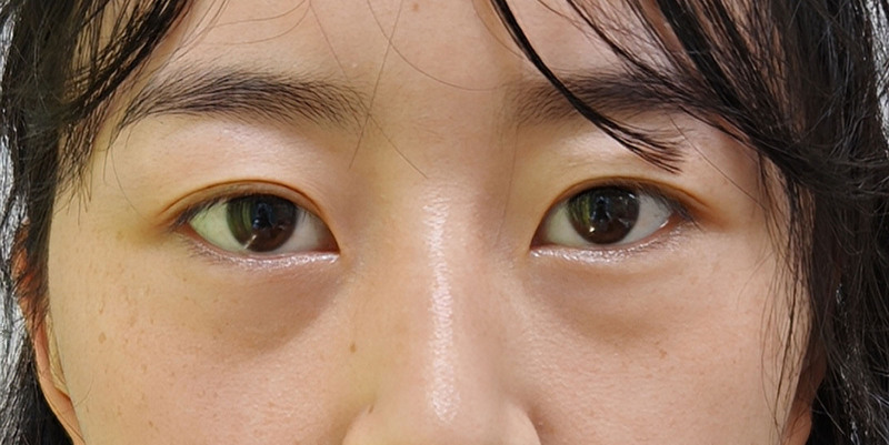
Lower eyelid blepharoplasty (LLB) is one of the most performed aesthetic procedures. Given its popularity, many studies have investigated its complications. However, most of this research has focused on lower eyelid retraction and malposition, with few addressing the exacerbation of dark circles. 4 This raises the question: how does LLB contribute to the worsening of dark circles in some cases? First, in patients with prominent eye bags, herniated orbital fat often creates a convex surface above the tear trough, generating a localized highlight that visually offsets the adjacent shadow. When this fat is reduced or no longer protruding—such as after surgical removal—the area above the tear trough may merge into a continuous shadow. Without augmentation of the tear trough and infraorbital hollow, this loss of structural convexity leads to an extended shadow along the eyelid–cheek junction, making the dark circles appear broader and more pronounced (Fig. 1 ). This observation is supported by previous anatomical analyses showing that facial fat descent and volume depletion, compounded by ligamentous tethering, contribute to orbital rim hollowing and deepening of the tear trough, thereby worsening the shadowing associated with periorbital dark circles. 5 Second, patients are often unable to clearly distinguish between eye bags and dark circles, yet they often desire treatment for both—particularly in cases with prominent pigmented type, which often require more superficial interventions targeting the cutaneous or subcutaneous layers to achieve optimal outcomes. 6 If this concern is overlooked and only a conventional LLB is performed, the patient’s expectations may remain unmet, potentially leading to dissatisfaction.
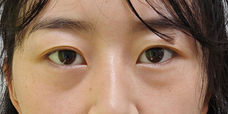
Fig. 1. Frontal view illustrating how dark circles may appear more pronounced after LLB. Reduced orbital fat prominence eliminates the compensatory highlight, resulting in a broader and more continuous infraorbital shadow.示意图:LLB 后眶脂高光消失、眶下阴影范围扩大导致黑眼圈显重的光学机制。As a filler with both volumizing and regenerative properties, autologous fat has been widely used in periorbital rejuvenation. 7 , 8 This study aims to systematically evaluate the effectiveness and safety of autologous fat grafting for treating dark circle exacerbation following LLB.
This study was designed as a retrospective study without a control group and was conducted at The People’s Hospital of Baoan Shenzhen between May 2023 and May 2025. Eligible participants had previously undergone LLB and subsequently developed worsening of infraorbital dark circles, with at least 10 months of postoperative follow-up after fat grafting. Dark circle exacerbation after LLB was defined as patient dissatisfaction, confirmed by surgeon evaluation of periocular appearance, including photographs. Patients with a history of periocular trauma, unrelated surgery, incomplete follow-up records, or prior injection of unknown substances after LLB were excluded. All patients agreed to undergo autologous fat grafting based on clinical recommendations.
Institutional approval was obtained from the Human Subjects Review Board before the initiation of the study. All patients signed written informed consent forms permitting the use of their clinical images, in accordance with the ethical principles of the Declaration of Helsinki (1975), as revised in 2013.
Adipose tissue was harvested from the lower abdominal region using a 3.0-mm blunt cannula with multiple side ports, following infiltration with tumescent solution and standard sterile technique. The aspirated fat was then processed following a modified Coleman protocol, which involved horizontal centrifugation at 1200 × g for 3 minutes. 9 After separation of excess fluid and oil, the refined micro-fat was transferred into 1-mL syringes for subsequent grafting.
Autologous fat grafting was performed based on 2 key principles: (1) injecting fat while advancing the cannula through ligaments such as the tear trough ligament (TTL) or the orbicularis retaining ligament (ORL); and (2) minimizing the injection volume per site. These strategies were intended to ensure even fat distribution and minimize the risk of nodularity.
The initial entry point was placed laterally at the infraorbital rim, slightly above the lowest point of the tear trough. After local infiltration with epinephrine-diluted lidocaine, a blunt cannula (approximately 1 mm in diameter) was introduced through the lateral attachment of the ORL. This provided direct access to the preseptal space, located between the orbital septum and orbicularis oculi muscle. The cannula was then advanced medially and inferiorly until resistance from the TTL was encountered. With controlled force, the cannula was passed through the ligament into the premaxillary space beneath it. Micro-fat was deposited in small aliquots per pass to achieve uniform volume restoration and contour correction (Fig. 2 ). If the concavity extended into the submalar region, micro-fat was also grafted into the adjacent prezygomatic space lateral to the premaxillary space. However, care was taken to avoid overfilling this area, as excessive fat grafting may impair periorbital lymphatic drainage by compressing the lymphatic pathways within the prezygomatic region. 10 Total injection volume for the deep plane typically ranged from 0.8 to 2 mL per side, with priority given to augmenting the premaxillary compartment. ( See Video 1 [online] , which displays an illustration of the deep-plane fat grafting approach for correction of post-LLB dark circles.)
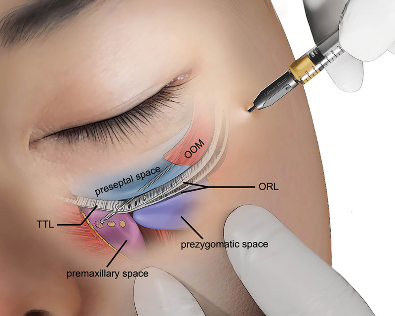
Fig. 2. Illustration of deep-plane fat grafting for correction of post-LLB dark circles. A blunt cannula passes through the lateral ORL into the preseptal space and is advanced medially and inferiorly across the TTL to reach the premaxillary space. OOM, orbicularis oculi muscle.深平面脂肪移植示意:钝针经外侧 ORL 进入隔前间隙,穿过 TTL 至前颌骨间隙。The secondary entry point was marked approximately 1.5–2 cm lateral to the alar base, vertically aligned with the lowest point of the tear trough depression. After the blunt cannula entered the superficial layer of subcutaneous fat, it was advanced toward the tear trough. Upon encountering the TTL or ORL, controlled force was used to traverse the ligament, and fat was injected in small aliquots into the subcutaneous plane (Fig. 3 ). To avoid postoperative bulging, injections were kept away from the lid margin, where the skin becomes thinner and the risk of contour irregularity increases. Approximately 0.5–1 mL of fat was placed into the subcutaneous space above the ligament. ( See Video 2 [online] , which displays the illustration of the superficial fat grafting technique used for correction of post-LLB dark circles.)
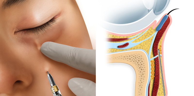
Fig. 3. Superficial fat grafting technique for correction of post-LLB dark circles. A cannula is introduced near the alar base and advanced within the superficial subcutaneous fat layer toward the tear trough region, with the sagittal schematic illustrating the superficial trajectory above the orbicularis oculi muscle.浅层脂肪移植示意:经鼻翼基底部外侧入路在皮下层向泪沟穿行。At the end of the procedure, patients were seated upright under frontal illumination to evaluate contour and symmetry; the final volume was individualized and guided by attainment of a smooth lid–cheek transition, with natural elevation toward the pretarsal fullness and no visible demarcation line (Fig. 4 ). ( See figure, Supplemental Digital Content 1 , which displays preoperative and 8-mo postoperative frontal views of the patient corresponding to Fig. 4 , showing improvement of the dark circles exacerbated after LLB, https://links.lww.com/PRSGO/E718.)
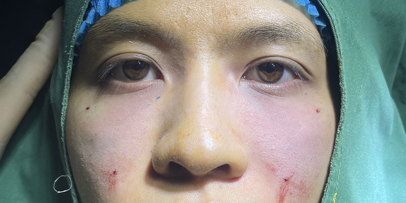
Fig. 4. Intraoperative frontal view demonstrating individualized end-point assessment under frontal illumination. Immediate postoperative appearance shows smooth lid–cheek transition without a visible demarcation line compared with the preoperative state.术中坐位正面光源下个体化终点评估:术前与即刻睑–颧过渡平滑对比。补充视频 / 附件(原文未随文提供原图,可于出版方页面查看)gox-14-e7561-s001.mp4Video 1. A blunt cannula was introduced through the lateral orbital retaining ligament to access the preseptal space. Gentle back-and-forth movements without resistance confirmed that the cannula was within the intended space. Once the tear trough ligament was identified, the cannula was carefully advanced through it into the premaxillary space. Micro-fat was injected in small aliquots per pass to ensure uniform volume restoration while avoiding overfilling.补充视频 / 附件(原文未随文提供原图,可于出版方页面查看)gox-14-e7561-s002.mp4Video 2. A secondary entry point was made beside the alar base, vertically aligned with the lowest point of the tear trough. After entering the subcutaneous fat layer, the cannula was advanced toward the tear trough. Upon encountering the tear trough ligament or orbital retaining ligament, gentle pressure was applied to traverse it. To avoid contour irregularities near the lid margin, where the skin is thinnest, the operator frequently released the guiding hand to check the subcutaneous position of the cannula tip. Each pass delivered no more than 0.02 mL for even distribution.
Standardized frontal and lateral photographs were taken before surgery and at the final follow-up visit (10–12 mo postoperatively) under consistent lighting and positioning. Patient-reported outcomes were collected using a customized questionnaire administered at the final visit. The primary assessment focused on subjective improvement of dark circles, rated on a 5-point Likert scale ranging from 1 (very unsatisfied) to 5 (very satisfied). The questionnaire also included subjective evaluations of improvements in the eyelid–cheek junction and pretarsal roll, although these were recorded descriptively without numerical scoring.
A total of 32 patients (15 men and 17 women) seeking correction for dark circle exacerbation following LLB were included in this study. The mean age was 28.9 years. The mean interval between LLB and fat grafting was 7.8 months, and the mean postoperative follow-up was 11.5 months (range, 10–12 mo). Among them, 22 had undergone transconjunctival LLB, and 10 had undergone transcutaneous LLB.
At final follow-up, 15.6% of patients were neutral (score 3); 50.0%, satisfied (score 4); and 34.3%, very satisfied (score 5) with the improvement of dark circles. No patients reported dissatisfaction (scores 1–2). The mean dark circle improvement score was 4.19 ± 0.69. Additionally, 93.7% reported improvement in the eyelid–cheek junction, and 68.7% noted improvement in the pretarsal roll. Three (9.4%) patients underwent a secondary fat grafting session to further refine dark circle correction. Detailed patient characteristics and outcomes are summarized in Table 1 .
Table 1. Patient Demographics and Subjective Outcomes at Final Follow-up
| Patients, n | 32 |
|---|---|
| Sex (M/F) | 15/17 |
| Mean age, y | 28.9 |
| Mean interval from lower blepharoplasty to fat grafting, mo | 7.8 |
| Type of prior lower blepharoplasty, n | |
| Transconjunctival | 22 |
| Transcutaneous | 10 |
| Patients requiring secondary fat grafting, n | 3 |
| Mean follow-up, mo | 11.5 |
| Dark circle improvement score (1–5), n (%) | |
| Score 1 | 0 |
| Score 2 | 0 |
| Score 3 | 5 (15.6) |
| Score 4 | 16 (50.00) |
| Score 5 | 11 (34.3) |
| Mean dark circle improvement score, mean ± SD | 4.19 ± 0.69 |
| Perceived improvement in eyelid–cheek junction, n (%) | 30 (93.7) |
| Perceived improvement in pretarsal roll, n (%) | 22 (68.7) |
No serious complications, such as arterial embolism, skin necrosis, or infection, were observed in this cohort. Minor hematoma occurred in 2 patients during advancement of the blunt cannula through the TTL. It resolved spontaneously within 2 weeks. Prolonged swelling in the prezygomatic region was observed in 1 patient. It was attributed to lymphatic vessel compression in this region. The patient was advised to perform daily periorbital massage, and the swelling disappeared spontaneously within 2 months. No delayed complications, such as chronic inflammatory changes, nodulation, or fibrosis, were recorded during the follow-up period.
A 28-year-old male patient sought treatment for dark circles. He had undergone LLB 1 year earlier. To address the tear trough deformity and exacerbated dark circles, he underwent autologous fat grafting. A total of 1.5 mL of micro-fat was grafted per side, including 0.5 mL in the subcutaneous plane above the tear trough and 1 mL in the premaxillary space below the tear trough. At 10-month follow-up, the patient demonstrated marked improvement in the appearance of dark circles (Fig. 5 ).
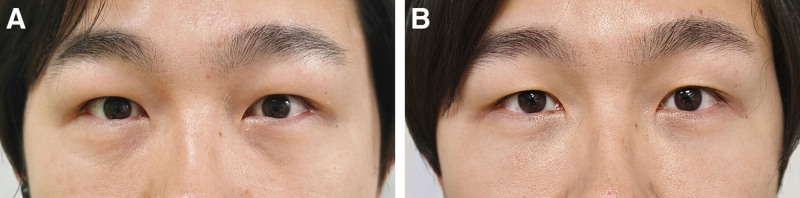
Fig. 5. Representative case of post–lower lid blepharoplasty dark circle correction treated with autologous fat grafting. A, Preoperative photograph showing dark circles exacerbated after LLB. B, Ten months postgrafting, improvement in the appearance of dark circles and the eyelid–cheek junction is observed.病例:术前黑眼圈加重 vs 脂肪移植术后 10 个月改善。
A 25-year-old female patient sought treatment for dark circles. She complained of having dark circles since childhood, which had worsened following LLB performed 6 months earlier. Clinical evaluation revealed tear trough deformity and prominent pigmented dark circles. To improve the appearance of dark circles, she underwent autologous fat grafting. A total of 1.4 mL of micro-fat was grafted per side, including 0.6 mL in the subcutaneous plane above the tear trough and 0.8 mL in the premaxillary space below the tear trough. At the 10-month follow-up visit, the patient demonstrated marked improvement in the appearance of dark circles (Fig. 6 ).
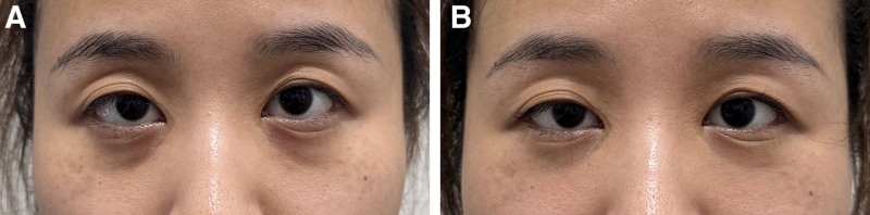
Fig. 6. Representative case of post–lower lid blepharoplasty dark circle correction treated with autologous fat grafting. A, Preoperative photograph showing dark circles worsened after LLB. B, Ten months after grafting, improvement in the appearance of dark circles is observed.病例:术前黑眼圈加重 vs 术后 10 个月改善。
A 22-year-old woman presented with worsening dark circles 6 months after LLB. She underwent autologous fat grafting, with approximately 1.6 mL per side: 0.8 mL was placed in the premaxillary and prezygomatic spaces below the tear trough, and 0.8 mL was injected subcutaneously above the tear trough. At the 12-month follow-up, marked improvement in the appearance of dark circles was observed, along with enhanced pretarsal fullness (Fig. 7 ).
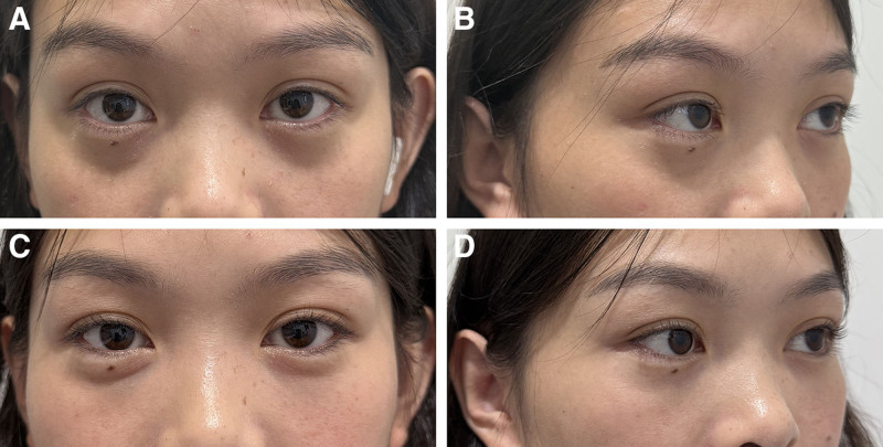
Fig. 7. Representative case of post–lower lid blepharoplasty dark circle correction treated with autologous fat grafting. A and B, Preoperative photographs showing dark circle exacerbation after LLB. C and D, Twelve months after grafting, both views demonstrate marked improvement in dark circles, pretarsal fullness, and the lid–cheek transition.病例:术前 vs 术后 12 个月正侧面改善。
A 28-year-old woman presented with worsening dark circles 8 months after LLB. She underwent autologous fat grafting with approximately 1.3 mL per side (0.5 mL above the tear trough and 0.8 mL in the deep spaces below the tear trough). At 3 months postoperatively, she reported marked improvement in the left side but remained dissatisfied with the right side, perceiving persistent “dark circles.” After a thorough explanation that the suboptimal outcome was due to residual right lateral orbital fat from the previous LLB, the patient underwent secondary transconjunctival removal of the residual right lateral fat and supplementary micro-fat grafting (0.3 mL) to the right prezygomatic space. At 11 months after the second intervention, substantial improvement in the appearance of dark circles and lower lid contour was achieved bilaterally, and the patient reported high satisfaction (Fig. 8 ).
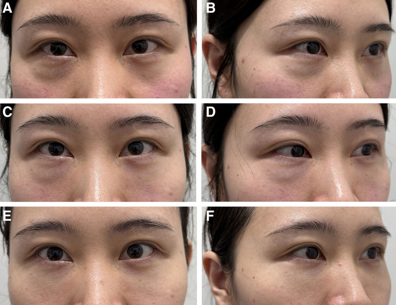
Fig. 8. Representative case of post–lower lid blepharoplasty dark circle correction treated with autologous fat grafting. A and B, Preoperative photographs showing dark circle exacerbation after LLB with residual right lateral fat herniation. At 3 months after initial grafting (C and D) and at 11 months after subsequent removal of residual fat with supplementary grafting, progressive improvement in dark circles and lower lid contour is demonstrated (E and F).病例:残余右侧外侧眶脂,初次移植 3 个月后补充手术。
This study explored the mechanism by which LLB may contribute to the exacerbation of dark circles and presented a corrective strategy with autologous fat grafting. Unlike conventional fat grafting confined to the tear trough groove, our approach used a crossing, region-based injection both above and below the trough, combined with simultaneous ligament release and micro-droplet placement.
If LLB focuses solely on orbital fat removal without addressing tear trough deformity and infraorbital hollowing, it may inadvertently exacerbate the appearance of dark circles. Tear trough deformity and infraorbital hollowing are thought to be significantly influenced by the tethering effects from the TTL and ORL. 11 In particular, the TTL binds the medial suborbital skin to the maxilla, and this tethering—when combined with thin, pigmented skin—has been recognized as a key contributor to the appearance of dark circles. 12 In our approach, we emphasized ligamentous release in conjunction with deep fat grafting into the premaxillary spaces. This combined strategy reduces tethering, facilitates upward repositioning of the tear trough base, and improves tear trough depression and infraorbital hollowing while attenuating shadow formation. Importantly, this technique allows for effective augmentation with less graft volume and reduces the risk of nodularity associated with overfilling.
To further improve the lid–cheek transition, we also addressed the superficial contour through subcutaneous augmentation above the TTL. Anatomical studies have shown that the lid–cheek junction is defined as a groove between the lower eyelid and the cheek, where the upper region is characterized by thin, fat-deficient preseptal skin, and the lower region by thicker skin with more abundant subcutaneous fat. 12 In youthful faces, this junction typically lies at a higher position but tends to descend with age. 13 In this study, we addressed this age-related descent by further releasing the tethering ligaments and augmenting the subcutaneous plane just above the tear trough. This approach helps elevate the lid–cheek junction toward a more youthful position and restore the natural convexity of the lower eyelid–cheek contour. Beyond contour restoration, fat grafting in this area may also improve skin quality, as the preseptal skin often lacks soft tissue and shows increased pigmentation. This effect may be partly explained by previous studies showing that adipose-derived stem cells within the grafted fat can suppress melanogenesis, 14 – 16 thereby contributing to the improvement of pigmentary changes.
Many authors have recognized the limitations of traditional LLB, particularly the persistence of tear trough deformity and infraorbital hollowing. To address these issues, adjunctive techniques such as TTL and ORL release, along with fat augmentation, have been widely adopted. Rohrich et al 17 introduced the “six-step lower blepharoplasty” technique, which involved stepwise ORL release followed by fractionated fat injection to enhance lid–cheek blending. Although their technique clearly separates the steps of ligament release and fat injection, our approach performs both maneuvers concurrently. Specifically, fat is injected immediately following focal ligament release within the same operative field, allowing real-time assessment of tear trough contour changes and minimizing the risk of overcorrection. Huang et al 18 reported a refined micro-fat grafting technique using an injection gun to deliver extremely small parcels (1/240 mL) across multiple planes, with repeated passes through the TTL to ensure even distribution. Notably, according to Khouri and Khouri, 19 fat parcels smaller than 17 µL (approximately 1/58 mL) are capable of complete revascularization and survival, thereby reducing the risk of nodular formation. Although the technique described by Huang 18 offers extreme precision, it requires specialized equipment that may not be available in routine practice. In our experience, approximately 0.02 mL per pass can be reliably achieved with manual pressure alone, eliminating the need for such devices and offering a practical alternative that is more accessible and adaptable in daily clinical settings.
Other authors have explored fat-derived products such as digested suspensions 20 or nanofat 21 for dark circles. Although these techniques benefit patients with superficial pigmentation and thin skin, they do not address deeper structural issues such as lid–cheek junction deformity, thereby limiting their applicability in post-LLB cases. Moreover, after emulsification, a substantial proportion of the obtained nanofat consists of free oil droplets. Previous studies have shown that such oil droplets may induce inflammation, fat necrosis, or even oil cyst formation, particularly in the periocular region. 22 To minimize this risk, we therefore selected micro-fat for the present study. Zhou et al 23 introduced a technique involving TTL release and reset, which showed promising improvement in tear trough deformity and dark circles. Nonetheless, their approach did not incorporate superficial fat supplementation, which may be more important for addressing pigmentary-type dark circles that require more superficial intervention.
Our previous study 24 described the treatment of structural dark circles using autologous fat grafting, in which fat was injected into the superficial layer via a lateral entry at the palpebromalar groove. This entry point has also been widely adopted by many authors due to its convenience. 18 , 25 , 26 However, this trajectory runs nearly parallel to the TTL, making it difficult to penetrate the dense and elastic structure. In the present study, the entry point was relocated to the lateral side of the alar base, creating a trajectory nearly perpendicular to the TTL, which facilitated easier penetration of the superficial portion of the ligament and allowed for more precise subcutaneous fat deposition (Fig. 9 ), particularly important when addressing pigmentary-type dark circles in addition to structural deficiencies.
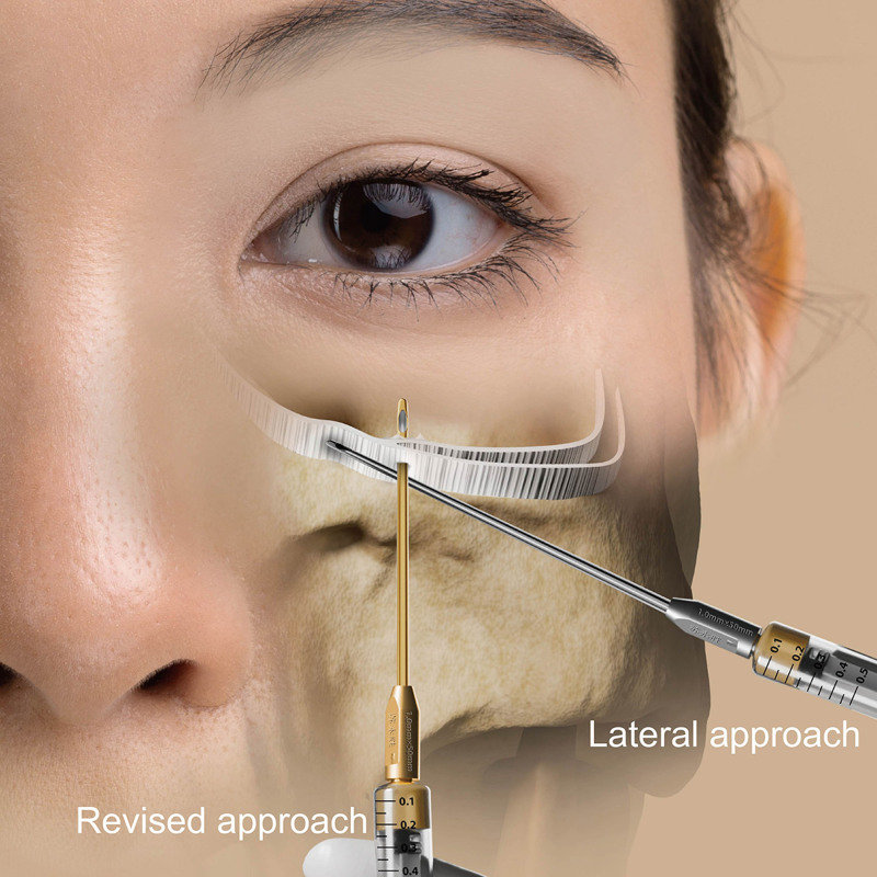
Fig. 9. Illustration comparing 2 injection trajectories relative to the TTL. The revised vertical approach from the alar base provides a more perpendicular path across the ligament, whereas the lateral approach advances more parallel to the ligament.两种入路与 TTL 关系示意:鼻翼基底部垂直入路更易穿透韧带。This study has several limitations. First, its retrospective design without a control group and the reliance on patient-reported satisfaction scores limit causal inference. Nevertheless, such patient-reported outcomes are widely used in aesthetic studies and still provide clinically valuable information. Second, standardized preoperative photographs from the initial LLB were often unavailable, as those procedures had been performed elsewhere; similarly, the exact surgical techniques previously performed (eg, fat removal versus repositioning) could not always be ascertained, limiting the ability to stratify outcomes by specific LLB approaches. Third, the cohort was predominantly young, reflecting the demographic characteristics of Shenzhen—a rapidly developing city with one of the youngest populations in China—which limits extrapolation to older patients with thinner skin and age-related changes. Fourth, differences in LLB approach—transconjunctival versus transcutaneous—may have influenced postoperative outcomes but were not specifically controlled in this study. More practical and validated measurement techniques may be incorporated in future investigations to enhance objectivity.
This study demonstrates that post-LLB dark circles can be effectively improved by our technique, which is both reliable and clinically applicable. In the future, combining our method with other adjunctive therapies—such as platelet-rich plasma or oil-free nanofat injection—may further enhance outcomes, particularly for pigmentary dark circles.
The author has no financial interest to declare in relation to the content of this article.
All patients provided consent for the academic use of their photographs.
声明:中文精读 · 仅供学术参考。内容来自公开文献检索,不代表本人观点,不构成诊疗建议。 医疗美容需在正规医疗机构由执业医师实施。
基于相同主题推荐 · 继续深挖
回顾性无对照研究(深圳市宝安区人民医院,2023-05 至 2025-05):32 例(17 女 15 男,平均 28.9 岁)下睑成形(LLB)后黑眼圈加重,接受自体微脂肪移植矫
回顾性大样本病例系列(2016年11月至2025年1月),共1999例连续手术,中位年龄36岁(IQR 31至42),中位BMI 29(IQR 26至32),最短随访6个月。
本研究为单中心前瞻性病例系列(200 例,2017—2024),平均随访 22 个月(范围 6—95 个月),评估经结膜延长下入睑成形术的长期美学与安全性结果。