浅层单极射频改善敏感皮肤屏障功能障碍的初步临床研究
2026年10月21日27 min read
Annals of Dermatology
结论与实践要点
敏感皮肤以屏障受损、神经高反应与炎症失调为特点(成人患病率约 40%–50%)。本研究采用浅层(shallow)模式可变脉冲单极射频(6.78 MHz,XERF),通过分段脉冲+受控间歇与冷却,选择性作用于乳头层/上网状真皮,以期调节表皮–真皮对话、改善屏障功能与血管反应。
主要人体临床数据为 10 例试点结果,统计效能与外推性有限;同步小鼠实验为临床前(动物)证据,不能直接等同于人体疗效,仅提供机制支持。第 8 周仍维持的改善提示或存在结构性重塑,但长期(>8 周)持久性、最佳维持方案与相对其他能量设备(如 IPL、低能量激光)的优劣仍需更大样本、设对照的研究确认。
BackgroundSensitive skin is a multifactorial condition characterized by impaired epidermal barrier function, neurosensory hyperreactivity, and inflammatory dysregulation, affecting approximately 40%–50% of adults worldwide. Despite its high prevalence, effective therapeutic options remain limited.
ObjectiveTo evaluate the efficacy and safety of a novel 6.78 MHz monopolar radiofrequency (RF) pulse pattern for improving both subjective symptoms and objective barrier function parameters in patients with sensitive skin.
MethodsIn this prospective pilot study, ten adults with sensitive skin underwent 2 sessions of shallow mode monopolar RF treatment at 4-week intervals. Assessments were performed at baseline, week 4, and week 8, including transepidermal water loss (TEWL), stratum corneum hydration (SCH), erythema index (EI), sebum production, melanin index (MI), and patient-reported symptom scores. Safety was assessed throughout the study. In parallel, a murine model was treated using identical shallow mode parameters to evaluate epidermal remodeling, collagen regeneration, and barrier-related molecular markers.
ResultsAll participants completed the study without serious adverse events. TEWL significantly decreased at weeks 4 and 8 compared with baseline ( p <0.001), while SCH, EI, and sebum levels showed significant improvement. Patient-reported symptom scores decreased from 65.57±5.49 at baseline to 41.91±4.66 at week 4, representing a 36.1% reduction ( p <0.001). Only mild, transient erythema was observed. Preclinical analysis demonstrated epidermal thickening, enhanced collagen remodeling, and upregulation of barrier-related markers.
ConclusionShallow-mode monopolar RF treatment improved subjective symptoms and objective barrier-related parameters in sensitive skin with excellent tolerability, supported by preclinical findings of upper-dermal remodeling.
Keywords: Monopolar radiofrequency、Sensitive skin、Skin barrier
Sensitive skin represents one of the most prevalent dermatological conditions, affecting approximately 40%–50% of the adult population worldwide, with higher prevalence in women (60%–70%) than in men (50%–60%) 1 . This condition is characterized by unpleasant sensory responses to stimuli that normally should not provoke such reactions, including environmental factors, topical products, and psychological stress 2 . The pathophysiology involves 3 interconnected mechanisms: impaired epidermal barrier function with elevated transepidermal water loss (TEWL) 3 , neurosensory hyperreactivity through upregulation of transient receptor potential vanilloid 1 channels 4 , and chronic subclinical inflammation mediated by increased expression of pro-inflammatory cytokines 5 . Recent studies on sensitive skin pathophysiology have revealed critical molecular alterations, including reduced expression of filaggrin and involucrin, essential proteins for maintaining epidermal barrier integrity 6 . Additionally, neurogenic inflammation plays a crucial role, with increased release of calcitonin gene-related peptide contributing to the characteristic burning and stinging sensations 7 . A recent study demonstrated that the intraepidermal nerve fiber density, especially that of peptidergic C-fibres, was significantly reduced in individuals with sensitive skin 5 . These findings suggest that effective therapeutic interventions must address multiple pathophysiological mechanisms simultaneously.
Monopolar radiofrequency (RF) devices have emerged as versatile tools in aesthetic dermatology, primarily recognized for their efficacy in skin tightening and rejuvenation 8 . These devices deliver controlled electromagnetic energy at specific frequencies, typically 6.78 MHz for medical applications, inducing selective dermal heating while preserving the epidermis through sophisticated cooling mechanisms 9 . The thermal effects trigger immediate collagen contraction followed by long-term neocollagenesis and elastin remodeling through activation of heat shock proteins and transforming growth factor-beta (TGF-β) signaling pathways 10 .
The introduction of dual-frequency RF platforms combining 6.78 MHz with 2.0 MHz represents a technological advancement in energy-based devices 11 12 . This dual-frequency approach enables more precise control of thermal energy distribution across different tissue depths. The 6.78 MHz frequency primarily targets superficial dermis (1–2 mm depth), while 2.0 MHz penetrates deeper (3–4 mm), creating a more uniform volumetric heating pattern 13 . Beyond combining these two frequencies, the system further offers three depth-selective treatment modes—generated through distinct waveform and pulse-pattern designs rather than frequency selection alone—allowing clinicians to tailor thermal engagement to specific tissue depths. Among these, the shallow mode delivers multiple pulses that focus on the superficial dermis, particularly the papillary and upper reticular layers. In this mode, 6.78 MHz monopolar RF is delivered through a finely segmented pulse structure that incorporates controlled off-time and cooling intervals within each treatment cycle. This pulse-delivery design is intended to regulate heat accumulation and limit excessive deep thermal spread, thereby favoring more selective thermal engagement of the papillary and upper reticular dermis 13 . Therapeutic efficacy is not determined solely by penetration depth; rather, it is determined by where thermal energy is deposited in relation to the intended clinical indication. Depth selectivity is therefore a critical determinant of treatment outcomes for specific indications. The papillary and reticular dermis host fibroblast populations with distinct programs that differentially influence epidermal support, adnexal structures, and matrix architecture 14 15 16 . Targeting the upper dermis, for example, is poised to modulate epidermal–dermal crosstalk and barrier-adjacent physiology 17 . In short, depth is not a goal in itself; the appropriate depth is the one that matches the intended biological target.
Despite the high prevalence and significant impact on quality of life, current therapeutic options for sensitive skin remain limited, consisting primarily of barrier repair moisturizers and anti-inflammatory topicals with variable efficacy 18 . The multifactorial pathophysiology suggests that monotherapy approaches may be insufficient, highlighting the need for treatments that simultaneously address barrier dysfunction, neurogenic inflammation, and vascular hyperreactivity. Based on the established biological effects of RF on skin physiology, we hypothesized that shallow-mode dual-frequency monopolar RF could provide therapeutic benefits for sensitive skin through multiple mechanisms. These include enhancement of epidermal barrier function through keratinocyte activation and increased synthesis of barrier-related proteins, modulation of neurogenic inflammation, and normalization of vascular reactivity through improved dermal microcirculation.
This pilot study aimed to evaluate the efficacy and safety of this novel therapeutic approach in patients with sensitive skin. Given the multifactorial pathophysiology of sensitive skin, this study primarily focused on barrier-related parameters and dermal remodeling associated with upper-dermal RF stimulation. In parallel, a preclinical murine study investigated the specific histological and molecular changes associated with epidermal barrier restoration.
This single-center, prospective, single-arm pilot study enrolled ten female adults classified as sensitive skin type by the Baumann Skin Type Questionnaire (BSTQ). Participants were classified as having sensitive skin if their sensitivity domain score was ≥30 on the BSTQ. Other BSTQ subtype classifications were not considered in the eligibility criteria. Ethical approval was obtained prior to study initiation, and all participants provided written informed consent before enrollment. Exclusion criteria were active inflammatory dermatoses; use of photosensitizing medications within 4 weeks; pregnancy or breastfeeding; history of keloid formation; previous facial RF treatment within 6 months; active infections in the treatment area; pacemaker or electronic implants.
Treatments were delivered using a novel pattern of noninvasive monopolar RF (NMRF) pulses (Shallow mode, XERF; Cynosure Lutronic, Goyang, Korea) equipped with temperature-controlled tip and integrated cooling. The shallow mode used in this study delivers 6.78 MHz monopolar RF only. Unlike conventional monopolar RF systems with fixed pulse patterns, this mode uses a more finely segmented pulse structure with controlled off-time and cooling intervals incorporated into each treatment cycle. This variable-pulse architecture is designed to modulate heat accumulation over time and to preferentially confine thermal effects to the papillary and upper reticular dermis, rather than promoting broader deep dermal heating. Shallow mode parameters were selected to preferentially target the superficial dermis and were delivered at intensity levels 5–6, producing an appropriate and controlled thermal effect at a depth of approximately 2–3 mm. Full-face treatment was conducted using ultrasound gel as a coupling medium, with continuous contact cooling to maintain epidermal temperatures below 40°C.
Participants received 2 treatment sessions at baseline (day 0) and week 4. All procedures were performed by a single experienced operator for consistency. Pre-treatment preparation included gentle cleansing with a non-ionic cleanser and photography under standardized conditions. No topical anesthetic cream was used prior to treatment. A 2–3 mm layer of ultrasound gel was used as the coupling medium. The RF handpiece was applied in a stamping pattern with continuous skin contact, while the device monitored the target temperature. Post-treatment care consisted of broad-spectrum sunscreen (SPF 50+) and a gentle moisturizer. Clinical assessments were performed at baseline, week 4, and week 8 (4 weeks after the second treatment). TEWL was measured using a Tewameter (Courage + Khazaka, Cologne, Germany), and stratum corneum hydration (SCH) was assessed using a Corneometer (Courage + Khazaka). Erythema index (EI) and melanin index (MI) were measured using a DSM II ColorMeter (Cortex Technology, Hadsund, Denmark), and sebum levels were evaluated using a Sebumeter (Courage + Khazaka). All measurements were performed under standardized environmental conditions (room temperature 20°C–24°C; relative humidity 40%–60%). Participants were acclimatized for at least 20 minutes prior to measurements. Measurements were obtained from standardized sites on both cheeks, and the mean values were used for analysis.
The study was conducted in accordance with the Declaration of Helsinki and the International Council for Harmonisation Good Clinical Practice guidelines. The study protocol was reviewed and approved by the Institutional Review Board (IRB) of Kangbuk Samsung Hospital (IRB No. KBSMC 2025-06-007-004).
Hairless mice (male, 5 weeks old; n=3 per group) were obtained from KOATECH (Pyeongtaek, Korea). After a 2-week acclimation period, mice were anesthetized with isoflurane prior to XERF treatment.
In the murine study, shallow-mode XERF treatment was delivered using the E10 tip at level 5 with 2 passes. The same shallow-mode waveform concept used in the clinical study was applied, although the protocol was adapted for the murine dorsal skin model rather than directly replicating clinical treatment conditions.
Animals were evaluated by comparing baseline untreated skin with post-treatment samples, and a separate sham-treated control group was not included in this study.
Skin tissues were harvested 1 month after treatment to assess remodeling-related histological and molecular changes rather than immediate post-treatment responses.
All procedures were approved by the Institutional Animal Care and Use Committee (IACUC) of Gyeongsang National University (IACUC No. GNU-250409-M0078) and were conducted in accordance with the National Research Council’s Guide for the Care and Use of Laboratory Animals. All experiments were reported in compliance with the Animal Research: Reporting of In Vivo Experiments (ARRIVE) guidelines.
Total RNA from treated cells was extracted with TRIzol Reagent (Thermo Fisher Scientific, Waltham, MA, USA) according to the manufacturer’s instructions. A total of 100 ng RNA was reverse-transcribed into cDNA, and one-twentieth of the reaction volume was used for quantitative mRNA analysis on a QuantStudio 3 system (Thermo Fisher Scientific). Probes for each gene were selected from Universal Probe Library (Roche, Basel, Switzerland) using ProbeFinder Assay Design Software. Primer sequences and probe numbers for each gene are provided below.
Primer sequences (5′→3′) were as follows: Filaggrin, forward GAA GAC TGG GAG GCA AGC TA and reverse GGT TTG GAG TGG GAT TGC TG; Involucrin, forward CAT CTG AGA CAG CAC CAG GA and reverse TGC TGC TTT TCT CCT GGA AT; Loricrin, forward GGA GGG GGC TAT TAC TCC TC and reverse CAC CTC CAC AGC TAC CAC CT; Tgfb1, forward TGA TAC GCC TGA GTG GCT GTC T and reverse CAC AAG AGC AGT GAG CGC TGA A; Has2, forward TGC AGT TTT GGT GAC GAC AG and reverse GCA TTG TAC AGC CAC TCT CG; and Has3, forward CAG TGG ACT ACA TCC AGA GGT G and reverse ACT CGA AGC ATC TCA ATG GT.
Ex vivo skin tissues were fixed in formalin, washed, dehydrated, and paraffin-embedded to generate donor blocks. Tissue microarrays (TMAs) were constructed by punching 3-mm cores from each block and arraying them into 6×6 recipient molds. TMA blocks were sectioned at 5 µm for histological and immunostaining analyses. Hematoxylin and eosin (H&E)-stained sections were analyzed using ImageJ (National Institutes of Health, Bethesda, MD, USA) to determine epidermal and dermal thickness. The former was measured from the basal layer to the stratum corneum surface; the latter from the dermo-epidermal junction (DEJ) to the subcutaneous fat boundary. We averaged measurements from 5 random sites across 3 sections per sample for statistical analysis.
For IF, sections were deparaffinized, rehydrated, and subjected to heat-induced antigen retrieval (97°C, 10 minutes; ab937, Abcam, Cambridge, UK), followed by blocking of nonspecific binding (X0909, Agilent, Santa Clara, CA, USA). Sections were incubated overnight at 4°C with primary antibodies against Aquaporin-3 (AQP3; ab125219), Collagen-17 (COL17A1; ab184996), Collagen-1 (COL1; ab270993), Collagen-3 (COL3; ab23445), Keratin-14 (KRT14; ab181595), and filaggrin (ab81468) (all from Abcam). Alexa Fluor ® 488- or 568-conjugated secondary antibodies were applied, and nuclei were counterstained with DAPI. Fluorescent signals were analyzed using a fluorescence microscope (Eclipse Ni-U, Nikon, Tokyo, Japan) at ×40–×400 magnification. Dermal elastic fibers were evaluated by Elastic Van Gieson staining (ab150667, Abcam). Histomorphometric quantification was performed in a blinded manner by an independent evaluator.
Data are presented as mean ± standard error of mean. Statistical significance was assessed using Student’s t-test in GraphPad Prism 9 (GraphPad Software Inc., San Diego, CA, USA), with p -value <0.05 considered statistically significant.
Ten female participants (aged 21–38 years) completed the study protocol. TEWL, SCH, EI, MI, and sebum production were evaluated at baseline, week 4, and week 8 (post-treatment). Friedman tests revealed significant overall differences across visits for all parameters, including TEWL (χ 2 (2)=18.20, p <0.001), SCH ( p =0.011), EI ( p =0.003), MI ( p <0.001), and sebum production ( p =0.002) ( Table 1 ). Pairwise Wilcoxon signed-rank tests with Holm correction showed that TEWL significantly decreased from baseline to week 4 (adjusted p =0.0059) and baseline to week 8 (adjusted p =0.0059), while no significant difference was observed between weeks 4 and 8 ( p =0.65). Similar post hoc analyses confirmed significant reductions in EI and significant increases in SCH, indicating improved skin barrier function and reduced erythema. MI values showed only minimal changes across visits. Sebum production also decreased significantly over time. Mean sensitive-skin symptom scores decreased from 65.57±5.49 at baseline to 41.91±4.66 at week 4, representing a 36.1% reduction ( p <0.001, Cohen’s d=4.85). Individual improvements ranged from 23.5% to 48.1%, with all participants demonstrating meaningful reductions (≥20% improvement). Treatment tolerability was excellent. Common immediate post-treatment effects included mild erythema in all participants (lasting 2–6 hours), warmth sensation in 8 participants (1–4 hours), and minimal edema in 3 participants (<12 hours). The transient erythema resolved spontaneously and was not reflected in EI measurements, which were obtained at scheduled follow-up visits. No serious adverse events, burns, pigmentary changes, or scarring were observed. The mean pain score during treatment was 3.2±1.1 on a 0–10 scale. Representative clinical photographs are shown in Fig. 1 . A representative participant exhibited visible improvement in facial erythema and pore appearance after 2 sessions of NMRF treatment. Baseline images revealed scattered erythema and enlarged facial pores, whereas post-treatment images at week 8 showed marked attenuation of erythema and noticeable reduction in pore prominence, consistent with the quantitative improvements observed in EI, MI, and barrier-related parameters.
Table 1 Changes in TEWL, SCH, EI, MI, and sebum production across 3 visits (baseline, 4 weeks, and 8 weeks after radiofrequency treatment)
| Metric | Visit 1 | Visit 2 | Visit 3 | p -Friedman |
|---|---|---|---|---|
| EI | 11.28±2.64 | 10.14±2.51 | 10.20±2.49 | 0.003 |
| MI | 32.70±2.47 | 32.71±2.46 | 32.62±2.46 | <0.001 |
| SCH | 23.75±8.78 | 27.78±7.62 | 27.28±7.49 | 0.011 |
| Sebumeter | 4.60±4.98 | 3.45±4.33 | 3.48±4.26 | 0.002 |
| TEWL | 19.60±5.49 | 16.05±4.48 | 16.88±4.40 | <0.001 |
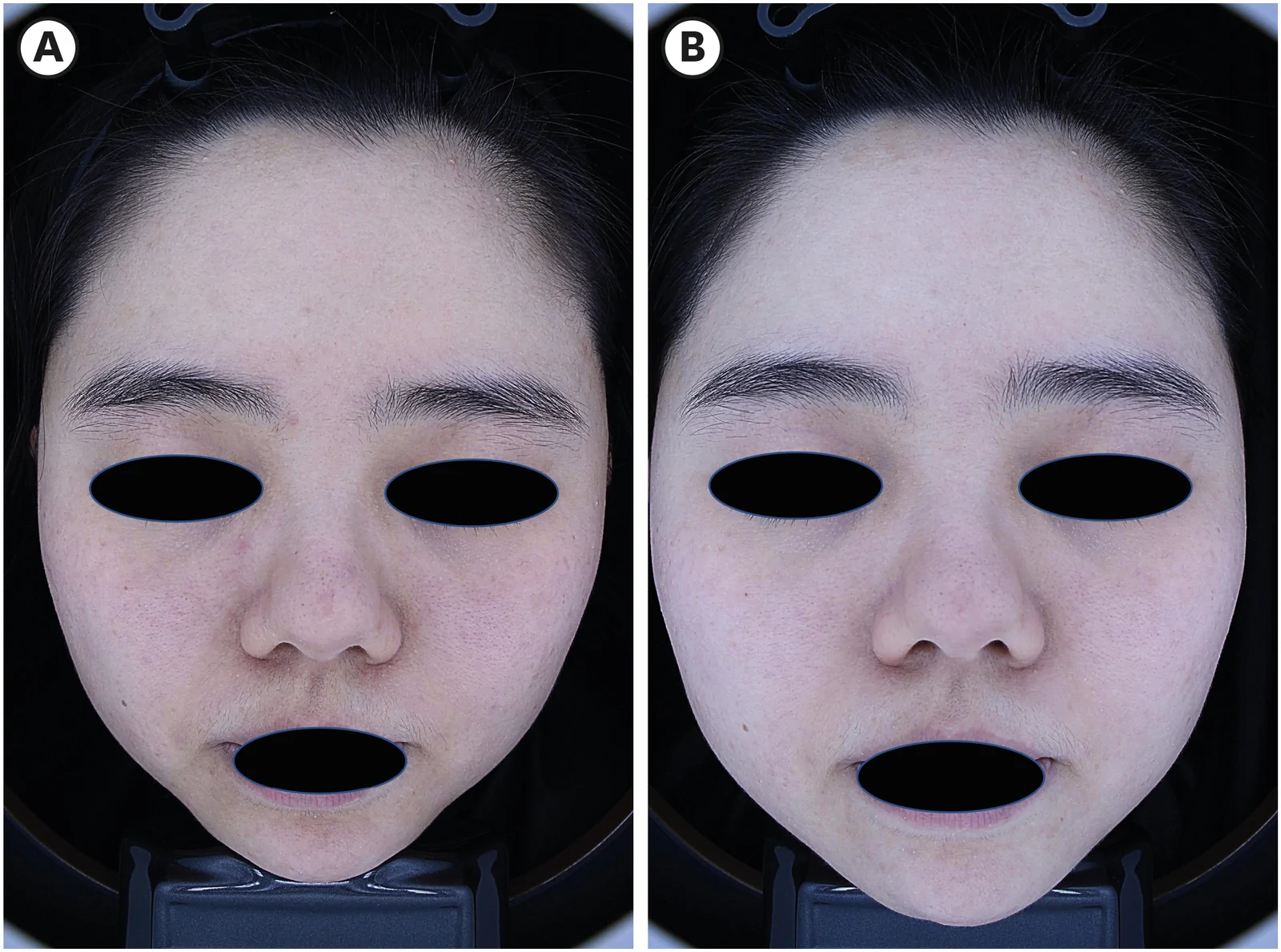
Fig. 1 Representative clinical case demonstrating the effects of NMRF in sensitive skin. Clinical photographs taken at baseline (A) and at week 8 following 2 NMRF treatment sessions (B) illustrate attenuation of facial erythema and improvement in pore appearance.NMRF: noninvasive monopolar radiofrequency.代表性临床案例:基线(A)与 2 次 NMRF 治疗后第 8 周(B)照片,示面部红斑减轻、毛孔外观改善。
To verify the histologic effects of NMRF, epidermal and dermal thickness were quantified on H&E-stained sections ( Fig. 2A ). Epidermal thickness was significantly greater in the NMRF group (12.677±0.949 µm) than at baseline (6.199±0.660 µm; p <0.05). Dermal thickness likewise increased significantly in the NMRF group versus baseline ( p <0.0001) ( Fig. 2B and C ).
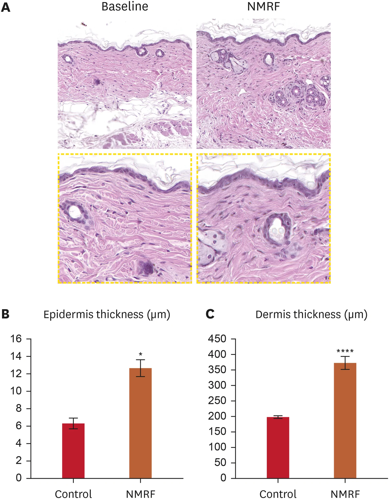
Fig. 2 Effect of NMRF on epidermal and dermal thickness. (A) Histological features of dorsal skin examined by hematoxylin and eosin staining (upper panel, 20× and lower panel, 49× magnification). (B) Epidermal and (C) dermal thickness was quantified. Data are presented as mean ± standard error of mean (n=10).NMRF: noninvasive monopolar radiofrequency.*p<0.05 vs. control group; ****p<0.0001 vs. control group.NMRF 对小鼠表皮与真皮厚度的影响:H&E 示治疗组表皮与真皮均较对照增厚(表皮 p<0.05,真皮 p<0.0001)。
Masson’s trichrome staining was used to assess collagen deposition. Controls showed a thin, loosely organized dermal collagen layer, whereas the NMRF group exhibited dermal thickening with intense dark-blue collagen staining ( Fig. 3A ). Quantification demonstrated increased collagen-stain density and collagen bundle thickening versus controls ( p <0.05), with a more regular fiber architecture at 1 month ( Fig. 3B and C ). To further evaluate collagen, COL1 and COL3 were examined by IF ( Fig. 3D ): COL1 signal was significantly increased in the NMRF group, consistent with histology ( p <0.05), while COL3 showed no significant change. In addition, TGF-β1—a key mediator of NMRF-induced remodeling—was elevated in the NMRF group ( Fig. 3E ).
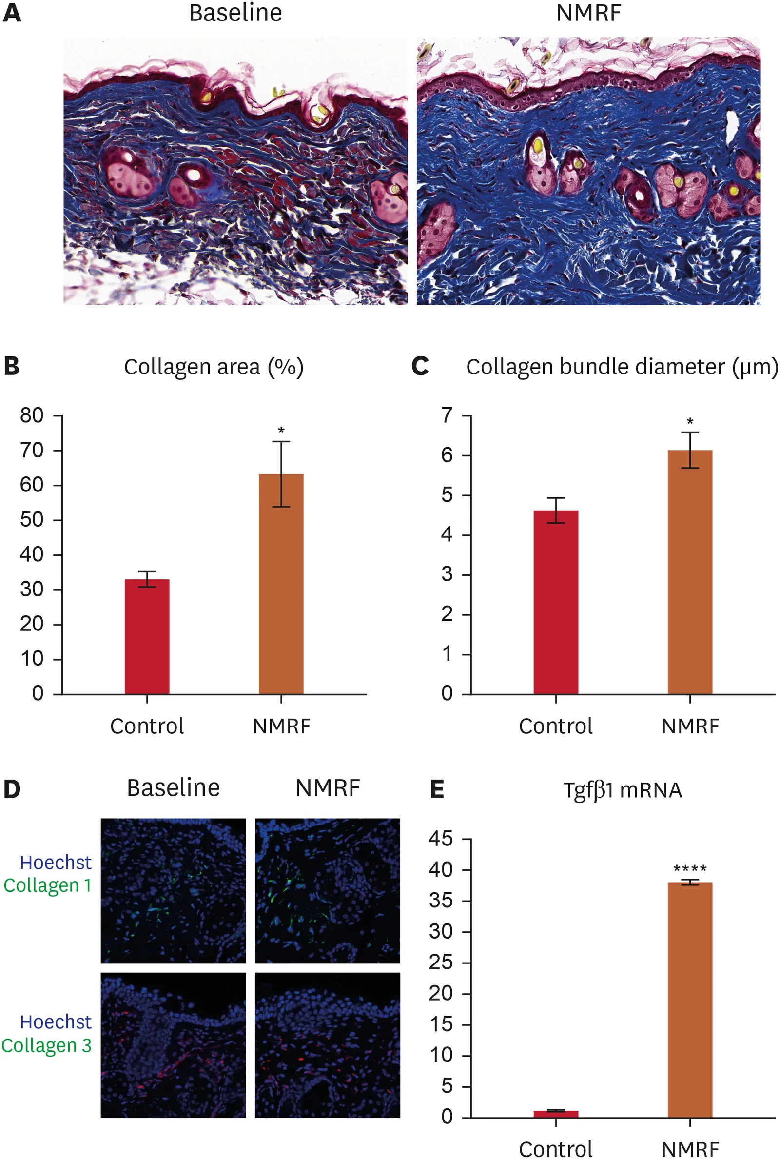
Fig. 3 Effect of NMRF on collagen deposition. (A) MT staining of dorsal skin at 1 month post-treatment (30× magnification). (B) Collagen-positive area and (C) collagen bundle diameter were quantified from MT-stained sections. (D) Immunofluorescence was performed to detect the expression and location of Col1 (upper) and Col3 (lower) in mouse dorsal skin. (E) Relative Tgfb1 mRNA levels measured by reverse transcription quantitative polymerase chain reaction. Data are presented as mean ± standard error of mean (n=4).NMRF: noninvasive monopolar radiofrequency, MT: Masson’s trichrome.*p<0.05 vs. control; ****p<0.0001 vs. control.NMRF 对胶原沉积的影响:Masson 三色示真皮胶原增加,COL1 显著上调(p<0.05),COL3 无显著变化,Tgfb1 升高。
This NMRF employs 3 depth modes achieved by distinct waveforms and pulse structures. The shallow mode preferentially targets the upper dermis/papillary dermis. In the mouse study, we applied the shallow mode and evaluated outcomes at 1 month.
Reverse transcription-qPCR showed significant upregulation of key barrier genes—Filaggrin, Involucrin, and Loricrin—after NMRF versus controls ( p <0.05; Fig. 4A-C ), supporting enhanced epidermal differentiation and barrier function. IF ( Fig. 4D and E ) further demonstrated increased cytokeratin 14 and higher AQP3 expression, consistent with improved epidermal maturation. In line with these findings, HAS2 and HAS3 mRNA levels were also elevated after treatment ( Fig. 5A and B ). Collectively, these data indicate that shallow-mode NMRF augments epidermal barrier integrity and hydration at the molecular level.
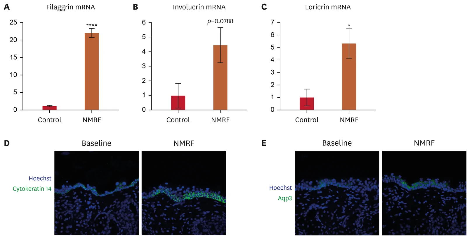
Fig. 4 Effect of NMRF on expression of skin barrier related gene. Total RNA was prepared from skin tissue, and the mRNA expression levels of (A) Filaggrin, (B) Involucrin, (C) Loricrin were determined by real-time reverse transcription polymerase chain reaction. Data are presented as mean ± standard error of mean (n=3–4). (D, E) Skin sections were prepared from dorsal skin harvested on Day 30 and were analyzed by immunofluorescence with an anti-cytokeratin 14 or Aqp3 antibody.NMRF: noninvasive monopolar radiofrequency.*p<0.05 vs. control; ****p<0.0001 vs. control.NMRF 对屏障相关基因的影响:qPCR 示 Filaggrin、Involucrin、Loricrin 显著上调(p<0.05),免疫荧光示 KRT14、AQP3 增加。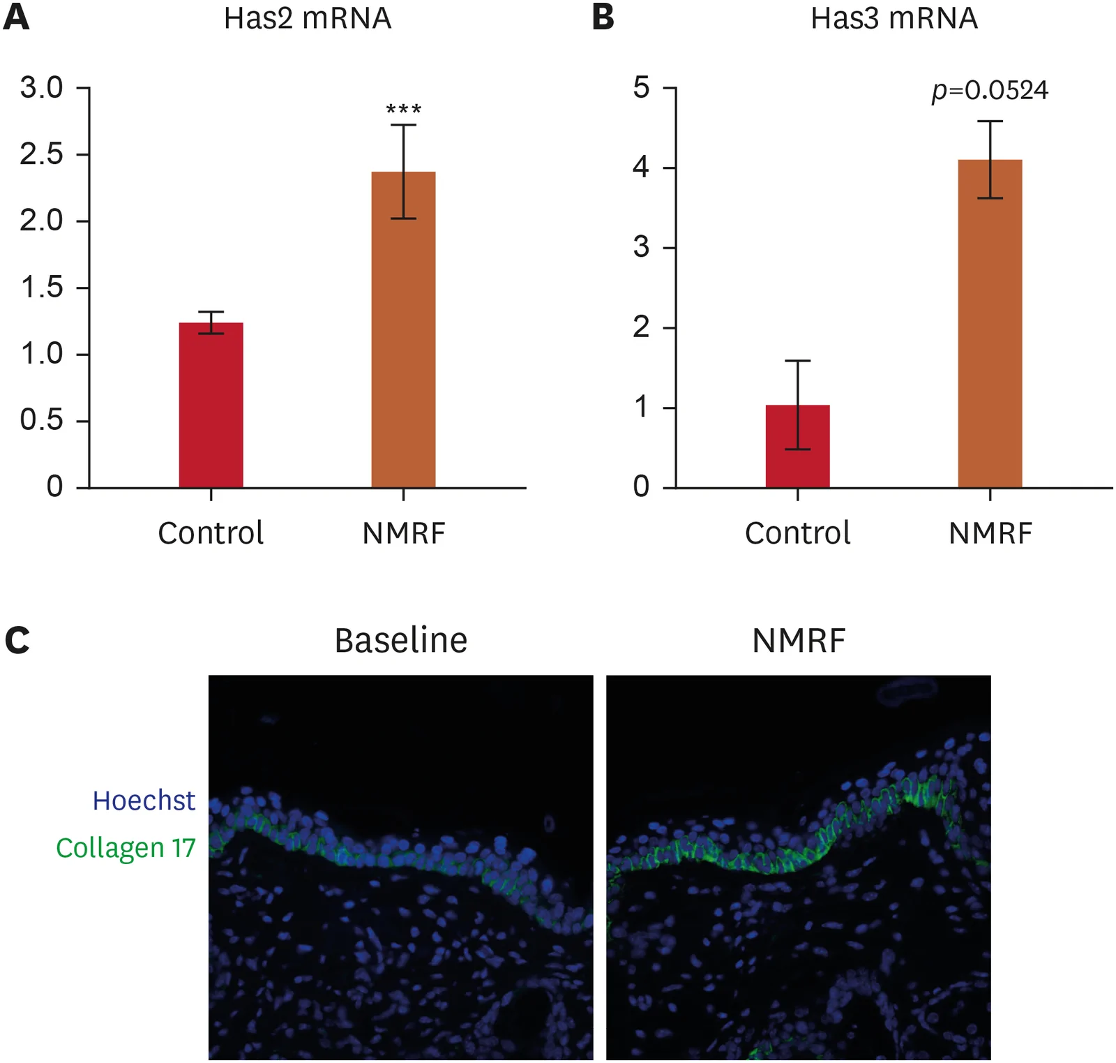
Fig. 5 Effect of NMRF on expression of hydration- and DEJ-associated markers. Total RNA was prepared from skin tissue, and the mRNA expression levels of (A) Has2, (B) Has3 were determined by real-time reverse transcription polymerase chain reaction. Data are presented as mean ± standard error of mean (n=2–4). (C) Representative immunofluorescence images showing COL17A1 (green) localization at the DEJ following shallow-mode NMRF treatment. Nuclei were counterstained with 4′,6-diamidino-2-phenylindole (blue). Increased DEJ-localized COL17A1 expression indicates enhanced hemidesmosomal integrity and epidermal–dermal adhesion.NMRF: noninvasive monopolar radiofrequency, DEJ: dermo-epidermal junction.***p<0.001 vs. control.NMRF 对水合与 DEJ 相关标志的影响:Has2/Has3 mRNA 升高,COL17A1 在 DEJ 表达增加,提示真皮–表皮黏附增强。
COL17A1 (BP180) is a transmembrane collagen component of keratinocyte hemidesmosomes that maintains DEJ stability by mediating epidermal–basement membrane adhesion 19 20 . IF showed a significant increase in DEJ-localized COL17A1 signal in the NMRF group versus controls ( Fig. 5C ). These findings suggest strengthened hemidesmosome-mediated adhesion and improved DEJ integrity, consistent with the observed increases in barrier- and hydration-related markers. Accordingly, shallow-mode NMRF appears to stabilize DEJ microarchitecture and enhance barrier function.
This prospective pilot study provides preliminary evidence that a novel 6.78 MHz RF pulse pattern, referred to as the shallow mode, can safely and effectively improve subjective symptoms and objective indicators of barrier dysfunction in patients with sensitive skin. The improvements were multidimensional, including a 36.1% reduction in symptoms, enhanced barrier function, and decreased vascular reactivity, with an excellent safety and tolerability. Changes in TEWL and SCH suggest that RF effectively enhances epidermal barrier integrity. The persistence of benefit at week 8, 4 weeks after the last treatment, suggests induction of durable structural remodeling rather than transient effects. The reduction in the EI indicates decreased vascular reactivity and inflammation, likely mediated through multiple RF-induced pathways. By preferentially engaging the papillary dermis, the shallow mode may promote fibroblast activation and microvascular circulation, promoting extracellular-matrix remodeling, barrier restoration, and skin homeostasis without provoking inflammatory flares. The substantial reduction in subjective symptoms, particularly stinging (42% reduction) and burning (40.4% reduction), suggests significant neuromodulatory effects. This is in line with previous studies showing that RF-induced thermal stimulation desensitizes C-fiber nociceptors through sustained depolarization and a subsequent refractory period, similar to the mechanism of capsaicin-induced desensitization 21 . Notably, the MI showed only minimal changes over time, suggesting that shallow-mode RF treatment does not induce clinically relevant pigmentary alterations. This finding supports the favorable safety profile of the procedure with respect to post-inflammatory hyperpigmentation.
Current therapeutic options for sensitive skin primarily consist of topical barrier-repair formulations and anti-inflammatory agents, with variable efficacy and frequent treatment failures. Compared to other energy-based devices for sensitive skin, RF may offer several practical advantages. Intense pulsed light has shown efficacy in erythematous sensitive skin but is associated with a higher risk of post-inflammatory hyperpigmentation, particularly in darker skin types 22 . Low-level laser therapy demonstrates anti-inflammatory effects but typically requires multiple weekly sessions over 8–12 weeks 23 . In this study, shallow-mode NMRF showed favorable clinical outcomes with good tolerability after only 2 treatment sessions.
These clinical observations were further supported by a depth-targeted preclinical investigation. Although traditionally associated with deep reticular tightening, shallow parameterization enables selective engagement of the papillary and upper reticular dermis, layers with distinct biological roles. Papillary fibroblasts promote epidermal morphogenesis and DEJ homeostasis, distinguishing them from deeper fibroblast lineages primarily responsible for tensile matrix synthesis. Guided by this rationale, we tested a shallow-mode RF protocol in a murine model optimized for upper dermal engagement. At one month post-treatment, histological analysis revealed dermal remodeling with concurrent epidermal thickening and enhanced DEJ integrity. These dermal remodeling and DEJ structural changes may influence epidermal barrier function through dermal–epidermal crosstalk, providing a mechanistic link between upper-dermal RF stimulation and the observed improvements in barrier-related clinical parameters.
Notably, COL17A1 expression—a marker for DEJ cohesion and epidermal-dermal communication—was elevated. These results suggest that targeted stimulation of papillary fibroblasts may contribute to structural and barrier-related changes in the epidermis, aligning with our clinical findings of improved TEWL and SCH. Interestingly, while COL1 expression was significantly increased following NMRF treatment, no significant change in COL3 was observed. Additionally, the absence of COL3 upregulation may be related to the timing of tissue analysis. COL3 expression is typically transient and peaks during early remodeling phases, whereas our analysis at 1 month post-treatment likely reflects a later remodeling stage in which COL1 predominates. Collectively, these preclinical findings suggest that shallow-mode NMRF may influence epidermal and DEJ-related remodeling associated with upper-dermal thermal engagement beyond conventional lifting or tightening effects. Importantly, the distinguishing feature of the shallow mode is not the use of 6.78 MHz monopolar RF alone, but the variable-pulse delivery architecture that incorporates segmented pulses with controlled off-time and cooling intervals. This configuration enables more refined control of thermal diffusion and superficial dermal targeting, which could be particularly relevant for indications such as sensitive skin, where modulation of epidermal–dermal crosstalk and barrier-adjacent physiology may be more beneficial than deeper volumetric heating. This depth-aware strategy may help standardize protocols to optimize both structural tightening and surface physiology restoration in sensitive skin.
The findings of this study have several important clinical implications for managing sensitive skin: 1) Novel therapeutic paradigm: RF treatment addresses multiple pathophysiological mechanisms simultaneously, offering a more comprehensive approach beyond symptomatic relief alone. 2) Rapid onset of action: Significant improvements were observed within 4 weeks, faster than most topical treatments which typically require 4–8 weeks for noticeable effects. 3) Sustained benefits: The maintained improvement at week 8 suggests potential for longer intervals between treatments, improving convenience and cost-effectiveness. 4) Broad applicability: The excellent safety profile suggests wide clinical applicability. 5) Combination potential: RF treatment could potentially be combined with topical therapies for synergistic effects, though this requires further investigation.
Several limitations must be acknowledged. First, the single-arm, open-label design without a placebo control limits interpretation due to potential placebo effects, which can be substantial in subjective conditions. Second, the small sample size (n=10) limits statistical power and generalizability. Third, the 8-week follow-up period is insufficient to evaluate long-term durability and to define optimal maintenance protocols. In addition, this study did not include direct clinical and preclinical comparisons with conventional RF devices. Therefore, the relative efficacy of the proposed NMRF approach compared to existing technologies could not be determined. Future head-to-head studies will be necessary to further define the advantages of depth-selective RF treatment. Finally, mechanistic assessments such as human skin biopsies for molecular markers or confocal microscopy for structural changes were not performed in the clinical setting, although the preclinical findings provide a mechanistic basis supporting the observed epidermal and dermal benefits.
声明:中文精读 · 仅供学术参考。内容来自公开文献检索,不代表本人观点,不构成诊疗建议。 医疗美容需在正规医疗机构由执业医师实施。
基于相同主题推荐 · 继续深挖
前瞻性、单臂、单中心试点研究纳入 10 例敏感皮肤女性(21–38 岁,BSTQ 敏感度评分≥30),接受浅层模式单极射频(NMRF,6.78 MHz)全脸治疗 2 次(第 0、4
回顾性队列研究,纳入93例哥伦比亚患者(2023.6–2025.7,随访≥1年),平均52.9岁(SD 8.13),女85例(90%)、男8例(10%),平均BMI 26.8 kg
前瞻性单臂研究(西班牙 Asturias 伦理委员会批准),39 例健康女性志愿者(23–68 岁,平均 47 岁,Fitzpatrick II–III),腹部/腰侧 25 例、大