上颞皮肤转位(STS)辅助睑外翻修复的技术要点与初步临床系列
2026年10月25日24 min read
Journal of Clinical Medicine
结论与实践要点
术式定位与适应证:本文在综述睑外翻各类修复技术(外侧睑板条 LTS、Bick 缩短、睑缘缝合、皮片移植、SOOF 提升、垂直-水平旋转肌皮瓣等)基础上,提出上颞皮肤转位(STS)作为重度、复发或睑板型(tarsal)睑外翻的辅助技术,常联合 LTS 或 Bick 及选择性内翻缝合。STS 的核心是保留传统术式中常被丢弃的外侧睑前层皮肤,并将其向上颞方向转位,以提供外侧上方的矢量支撑。
关键技术点:于外侧眦上方眶缘切除三角形皮肤床,将下睑保留的前层皮瓣转位悬置于该床并以多针间断缝合;后层缝合固定至外侧眶结节(Whitnall 韧带/眶结节)。相较单纯 LTS/Bick,STS 通过降低后层张力获得更稳固的对合,但操作时间约增加 5 分钟,出血与瘀斑风险略增。其「向外侧上方矢量促进愈合稳定」属力学/解剖学推断,并非在体临床终点,价值在于解释支撑机制。
初步临床结局与并发症防范:资深术者以 STS 联合改良 Bick(可选内翻缝合)用于 23 例患者(4 例双侧),随访 1–6 个月,按 Berry-Brincat 分级均达「满意或更好」,睑位良好、症状改善,无再手术;部分患者有轻度残留外翻。STS 较单纯 Bick 外侧皮肤瘢痕稍多,但患者多不介意;相比睑缘缝合它保留周边视野,相比 SOOF 提升更简便且适用于抗凝患者。需注意 FTSG 存活率报道可达近 100%、继发挛缩更低,瘢痕性病例应联合皮片。
证据等级与局限:本系列为单中心、术者经验的初步病例系列,随访短(1–6 个月)、且患者因满意而较少返院长期随访,长期有效性尚未确立;文中亦提示部分病例回顾认为应补加内翻缝合/皮片。临床落地建议:STS 宜作为复发性或睑板型睑外翻的备选辅助,术式选择仍应依据病因(退行性/麻痹性/瘢痕性)与严重度个体化,并待更长随访验证。
Background: Ectropion is a common eyelid problem and is defined as eversion of the eyelid margin and typically involves the lower eyelid. The main acquired causes of ectropion include involutional, cicatricial, paralytic, and mechanical. A severe manifestation of ectropion is tarsal ectropion, where much of the tarsal conjunctiva is visible, often with keratinization of the conjunctiva. causes. Common techniques for ectropion repair include horizontal tightening of the lid with lateral tarsal strip or Bick procedure, lateral tarsorraphy, inverting sutures and the sub-orbicularis oculi fat lift. However, all surgical techniques are prone to ectropion recurrence. We review the techniques for ectropion repair and describe a novel adjunctive technique called the superotemporal skin transposition (STS), which is well suited for patients with recurrent or tarsal ectropion. Methods: The STS is combined with a lateral tarsal strip or Bick procedure. For the STS, all of the anterior lamellae of the lateral lower lid is retained. The posterior lamellae is sutured to the lateral orbital tubercle. A triangular bed of skin is excised superotemporally, and the lower lid anterior lamellae is transposed and secured with multiple sutures. The STS can be combined with inverting sutures, or skin graft for cicatricial cases. Results: We used the STS with Bick procedure and optional inverting sutures on 23 patients, 4 of whom required bilateral ectropion repair. At 1–6 month followup all patients achieved satisfactory outcomes with a well-positioned eyelid and improved symptoms. The STS had more lateral cutaneous scarring than with a Bick procedure alone, but patients did not find this objectionable. No reoperations were required. Conclusion: The STS is a straightforward and useful adjunct for patients with severe, recurrent or tarsal ectropion. Further studies are needed to determine the long-term efficacy of this technique.
Keywords: oculoplastic surgery、superotemporal skin transposition、tarsal ectropion、recurrent ectropion、eyelid
Ectropion is eversion of the eyelid margin and predominantly occurs within the lower lid. Ectropion can result in corneal exposure, tearing conjunctival erythema, and eye irritation. The most commonly acquired causes of ectropion are involutional, cicatricial, paralytic and mechanical. Tarsal ectropion refers to marked eversion of the lower lid, which may be associated with keratinization of the palpebral conjunctiva. We review the indications, benefits and limitations of the established surgical technique for ectropion repair. These procedures include the lateral tarsal strip (LTS) procedure and Bick procedure to correct horizontal lid laxity, tarsorrhaphy or inverting sutures for disinsertion of the lower lid retractors, and skin grafts for cicatricial ectropion. The indications for and steps of superotemporal skin transposition (STS) as an adjunct for the treatment of severe or tarsal ectropion are described. Advanced techniques such as the sub-orbicularis oculi Fat (SOOF) lift for cicatricial ectropion or midface descent, and the vertical-to-horizontal rotational myocutaneous flap for cicatricial ectropion are briefly reviewed.
Table 1 presents a series of tarsal ectropion repair procedures, including their indications, specific techniques, and rates of success in conjunction with their follow-up duration, which vary across studies [ 1 , 2 , 3 , 4 , 5 , 6 ]. In this section, we discuss common surgical techniques for ectropion.
Table 1 Summary of commonly used techniques for managing tarsal ectropion by indication and success rates with corresponding follow-up durations.
| Series | Technique | Indication | Success Rate—Proportion (%) | Follow-Up Duration in Months—Mean (Range) |
|---|---|---|---|---|
| Kwon et al., 2015 [ 4 ] | LTS + 5 mm Lat. Tarsorraphy | Paralytic | 22/22 (100%; 3 patients did not report satisfactory cosmesis) | 16.5 (12–73) |
| Wang et al., 2022 [ 6 ] | Inverting sutures + mod Bick | Involutional | 31/34 (91%) | 4.5 (3–11) |
| Berry-Brincat et al., 2013 [ 1 ] | Inverting sutures ± LTS | Involutional | 18/20 (90%) | 3.6 (2–15) |
| Neavyn et al., 2012 [ 5 ] | Inverting sutures ± LTS | Involutional, Recurrent | 6/7 (86%) | 2 (1–4) |
| Chung et al., 2007 [ 3 ] | Midface lift + LTS | Cicatricial, Involutional, Paralytic | 17/22 (78%) | 12 (2–38) |
| Chang et al., 2006 [ 2 ] | Augmented Tarsal Strip | Paralytic | 13/14 (93%) | 21 (6–30) |
Horizontal lid-tightening techniques for ectropion include the LTS, Bick procedure, Kuhnt–Szymanowski and occasionally medial canthoplasty [ 7 ]. A video illustration of LTS is documented elsewhere [ 8 ]. The procedure begins with a lateral canthal incision to provide access, followed by an inferior cantholysis to release the lower canthal tendon. If medial or punctal ectropion is present, a medial spindle procedure can be performed as an adjunct [ 9 ]. In this step, a diamond-shaped section of the eyelid retractors and conjunctival tissue is excised just below the punctum, and then closed with an inverting suture that is externalized through the skin inferiorly. The tarsal strip is measured to ensure the appropriate length, after which the epithelium is debrided to prepare the tarsal strip. Next, the anterior and posterior lamellae are divided, followed by separating the tarsal strip from the lower eyelid retractors and conjunctiva. The anterior lamella is excised so that lash follicles are removed from the tarsal strip. The tarsal strip is then reattached to the periosteum at the lateral orbital tubercle with subsequent reshaping of the lateral canthus such that its proper angle is recovered. The use of a specialized needle, such as a m4-0 Vicryl S-2 semicircular needle, is advantageous for securing a precise bite of the inner rim of the lateral orbital wall, which aids in achieving optimal lower lid positioning. This technique helps achieve the lid globe apposition for optimal procedural outcomes. A 5-0 non-absorbable prolene suture may be advisable for recurrent ectropion, to decrease recurrence. In patients with tarsal ectropion, reverse Quickert sutures can be used as an adjunct [ 10 ]. This approach uses double-ended 5-0 chromic sutures, passed from below the tarsus and exiting near the lid–cheek junction, to plicate the retractors and enhance lower eyelid positioning. These sutures are typically placed in pairs, with the tension adjusted based on the degree of lid margin ectropion, often with a preference for slight overcorrection to ensure stability in both entropion and ectropion cases. Finally, the skin is sutured closed [ 7 ].
Medial canthoplasty is another horizontal tightening procedure that may be used in cases of ectropion with medial canthal tendon laxity. This surgical technique begins with an incision made inferior to the lacrimal canaliculus, which continues medially to the medial canthus and runs superior to the tendon. Next, the surgeon passes a mattress suture with 4-0 Prolene into the tarsus at the nasal aspect and subsequently passes it through the periosteum inferior to the insertion of the superficial arm of the medial canthal tendon [ 11 , 12 , 13 ].
Bick’s shortening is a horizontal tightening technique similar to the lateral tarsal strip. This procedure involves the full-thickness excision of a triangular segment of eyelid tissue near the lateral canthus of the affected eyelid. The extent of tissue resection is carefully tailored to the severity of the eyelid laxity. Following the excision, the edges of the resected eyelid tissue are precisely realigned to generate the necessary horizontal tension. Sutures are then placed through the tarsus to secure the newly adjusted eyelid position, with meticulous closure of the skin layers and optional closure of the conjunctiva and muscular layers as needed to optimize healing [ 14 ]. Compared to traditional surgical techniques such as LTS, Bick’s shortening method can be simpler to perform and has demonstrated superior anatomical and functional outcomes in ectropion management [ 14 ]. However, the technique has been criticized for potentially causing rounding and medial displacement of the canthal angle [ 14 ].
The Kuhnt–Szymanowski procedure involves shortening the horizontal lid with a full-thickness pentagonal wedge resection combined with a lateral lid incision approximately 3 mm from the lid margin. The authors perform the Bick and LTS procedures in preference to the Kuhnt–Szymanowski procedure, as the former are quicker and do not cause a lid margin notch.
Canthal suspension techniques include lateral canthal sling and tarsorrhaphy [ 12 ]. The lateral canthal sling procedure is employed to address cases of paralytic ectropion [ 15 ]. This condition can be further worsened by periocular aging or increased eyelid laxity, in which the lateral canthal tendon loosens, resulting in eyelid sagging and ectropion. The lateral canthal sling aims to provide support and restore tension in the lateral canthal tendon. In this technique, a small horizontal incision is made near the lateral canthus along the natural eyelid crease, exposing the lateral canthal tendon. The surrounding soft tissue is then dissected away to mobilize adjacent structures, including the tarsal plate and orbicularis muscle. An autologous or synthetic sling material is then threaded or looped around the canthal tendon and anchored to a stable structure around the orbital rim, often the periosteum of the lateral orbital rim for increased support. Finally, the sling is adjusted to achieve appropriate tension, realigning the lower eyelid in its proper anatomical position [ 15 , 16 ].
A small lateral tarsorrhaphy can help suspend the lid in patients with severe cases of paralytic ectropion and increased eyelid laxity, particularly when prior surgical management has failed or when eyelid exposure poses a risk to globe integrity [ 2 ]. Tarsorrhaphy involves partially suturing the upper and lower eyelid margins near the central or lateral canthus, with sutures that may be permanent or temporary depending on case severity. By decreasing the inferior scleral show, tarsorrhaphy reduces corneal desiccation and irritation. Tarsorraphy may serve as either temporary protection until more definitive reconstructive procedures are feasible or as a permanent solution when other management options are contraindicated or inadequate [ 2 ].
In some cases, ectropion may require skin grafts or Mustardé flaps. Particularly, in cases of cicatricial ectropion, which is often due to anterior lamellar tissue scarring, it may be necessary to remove the scarred area and support the anterior lamella deficiency with a skin graft. The upper eyelid, supraclavicular region, preauricular or postauricular areas and volar forearm are common hairless donor sites that may be harvested for the skin graft. Full-thickness skin grafts (FTSGs) include epidermis and dermis layers, whereas split-thickness skin grafts (STSGs) involve the epidermis and only a portion of the dermis. FTSGs are most commonly used to surgically manage cicatricial ectropion and provide a greater match to eyelid skin in terms of color and texture compared to STSGs. FTSGs demonstrate up to a 100% graft survival rate across several studies and are associated with lower rates of secondary contraction compared to STSGs; however, there is a theoretical risk of failure which may be influenced by individual patient characteristics [ 17 , 18 , 19 ]. STSGs are used less frequently for the repair of ectropion and are more likely to contract compared to FTSGs. Given the enhanced cosmetic results and reduced risk of secondary contraction, FTSGs are typically preferred over STSGs in cases of cicatricial ectropion repair [ 17 , 18 , 20 ]. Skin grafting begins with releasing the scarred tissue and subsequent harvesting of the graft from a donor site. The graft is shaped appropriately to the dimensions of the defect, secured into position with sutures and traction sutures. A pressure dressing is applied for 5–7 days postoperatively.
Marked ectropion can also be managed through a sub-orbicularis oculi fat (SOOF) lift, particularly in cases of paralytic ectropion [ 21 ]. It is indicated in cases with age-related descent of the midface structures and severe eyelid laxity [ 21 ]. This procedure provides structural support to the lower eyelid by lifting the orbicularis muscle pad. It begins with an incision on the interior aspect of the lower eyelid, about 1 mm below the caudal margin of the tarsal and through the orbicularis oculi muscle to expose the SOOF. The SOOF is released from the surrounding structures and lifted into a position higher along the orbital rim, where it is secured with sutures to the periosteum [ 21 ]. This procedure can be augmented with a lateral canthal tightening procedure to further reinforce the eyelid position.
Finally, a technique for treating cicatricial or severe ectropion, described by Chang et al., is the vertical-to-horizontal rotational myocutaneous flap (VHRMF) based on the Tsai procedure. “A vertical myocutaneous flap is created from the anterior lamella of the vertical pedicle in the lateral third of the lower eyelid. Following a horizontal relaxing incision from the base of the flap, a vertical myocutaneous flap is created and rotated to horizontal” [ 22 ]. The advantages of this technique include reduced donor site morbidity and correction of horizontal lid laxity [ 22 ]. For patients with cicatricial ectropion, the cicatrix still has to be released, whereby midface lifts can be performed and anterior lamella graft may still be required. The VHRMF technique may surpass the comprehensive ophthalmologist’s technical repertoire and, in some cases, may be accompanied by a visible vertical scar [ 22 ].
Superotemporal skin transposition is a versatile surgical technique within the purview of the comprehensive ophthalmologist and general plastic surgeon that may be used as an adjunct for the repair of tarsal and recurrent ectropion from involutional or paralytic causes. When performed with skin grafts, it can also be applied to cicatricial ectropion. This technique conserves the anterior lamella skin that is normally discarded during traditional LTS or Bick ectropion repairs and provides a superotemporal vector to the eyelid repair. A triangular bed of skin over the lateral orbital rim superior to the lateral canthus is excised. A skin flap from the inferolateral lid is transferred superotemporally. The skin flap allows for better lid globe apposition and structural support. Although the STS may cause a vertical scar in the lateral orbit area, it is not seen frontally, as may occur with the VHRMF.
We illustrate the application of the superotemporal skin-transposition technique for managing paralytic ectropion, underscoring its advantages over conventional methods in providing enhanced support and esthetic results.
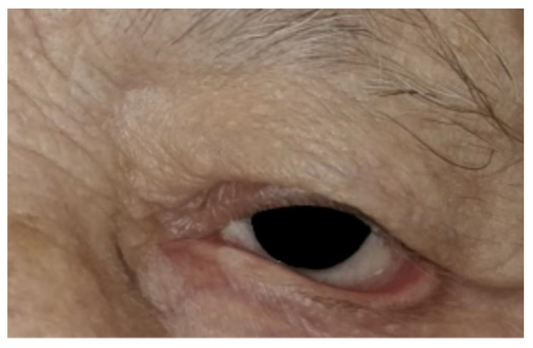
An elderly male patient presented with symptomatic left paralytic ectropion ( Figure 1 ). The patient reported issues with exposure-related symptoms, including irritation, tearing, and visual disturbances.
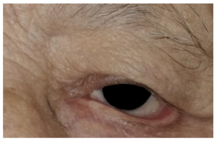
Figure 1 Preoperative appearance of the patient with ectropion in the left eye.左侧麻痹性睑外翻患者术前外观,示睑缘外翻与暴露相关症状。Following lateral canthotomy and cantholyis, the lateral lid was split into an anterior and posterior lamella. Unlike the LTS/Bick procedures, all the lateral lid skin was retained except at the lateral lid margin. The posterior lamella was shortened to create optimal tension and sutured to Whitnall’s ligament ( Figure 2 ).
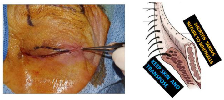
Figure 2 Initial incision and eyelid split—the eyelid was split with preservation of the skin, except at the lash margin, to expose the posterior lamella for later stabilization.初始切口与眼睑分层——保留皮肤(除睑缘睫毛区外)以显露后层,备后续固定于外侧眶结节。Next, the skin over the lateral orbital rim superior to the lateral canthus was marked using the preserved eyelid skin flap as a template. ( Figure 3 ) The superolateral skin was excised, and the preserved anterior lamella skin was transposed into the superotemporal bed. The transposed skin flap was positioned to fall into the natural contour, aiming for functional and esthetic restoration. Multiple interrupted sutures were placed to secure the transposed flap into the superolateral bed ( Figure 4 ). In this case, which involved paralytic ectropion, a brow lift and upper-lid gold weight were also inserted.
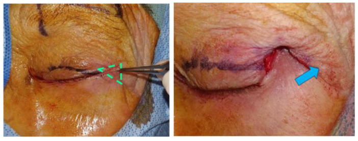
Figure 3 Preparation of superotemporal skin bed—an excision bed underneath the skin flap (green triangle) was created lateral and superior to the canthus to accommodate the transposed skin flap. The blue arrow illustrates the lateral and superior vector of STS that augments wound healing and stability.上颞皮肤床准备:于眦上外侧切除三角形皮肤床(绿三角)以容纳转位皮瓣,蓝箭头示 STS 向外侧上方的矢量方向。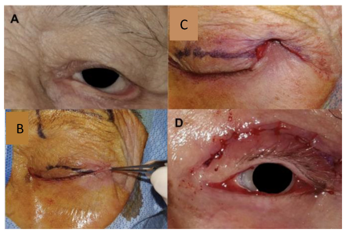
Figure 4 Summary of the steps in the superotemporal transposition procedure and the final appearance—(A) Pre-operative appearance in a patient with involutional and paralytic tarsal ectropion (B) The retained skin was used as a template for the amount of superotemporal skin to excise. Not visualized underneath, the posterior lamella is sutured to the lateral orbital tubercle. (C) The anterior lamella was transposed and suspended in the prepared superotemporal bed and secured with interrupted sutures. (D) The lower lid margin is well repaired. A gold weight and direct brow lift were also performed.STS 步骤与终态汇总:术前(A)、以保留皮肤为模板切除上颞皮肤(B)、前层转位悬置于上颞床并间断缝合(C)、下睑缘修复良好(D,同期行金片与上睑提肌悬吊)。The patient’s postoperative care included standard guidelines for ectropion repair, and monitoring for infection and healing.
The superotemporal skin transposition augmentation has benefits and drawbacks compared to the traditional surgical management options for ectropion ( Table 2 ) [ 1 , 2 , 4 , 5 ]. Compared to conventional techniques such as LTS or Bick’s shortening, adjunctive superotemporal skin transposition provides a stronger, more stable repair by reducing tension on the posterior lamella, allowing stronger adhesion. However, it requires a slightly longer procedure time with an increased risk of bleeding and ecchymoses, and the potential for mild lateral scarring or rhytid formation. When compared to the augmented LTS technique alone, the superotemporal transposition maintains better lid globe apposition. Compared to tarsorrhaphy, the superotemporal transposition offers improved cosmesis of the lid fissure and preserves peripheral vision, though it is more challenging to perform. Finally, when compared to the SOOF lift, the superotemporal technique is easier to perform and suitable for patients on anticoagulants. However, the SOOF lift does not have temporal scarring and serves as a valuable management option for cases involving significant midface descent or age-related laxity.
Table 2 Technique comparison: superotemporal skin transposition vs. other techniques for ectropion correction.
| Technique | Indications | Advantages | Disadvantages | Success Rate |
|---|---|---|---|---|
| Superotemporal Skin Transposition | Cicatricial, recurrent ectropion, and cases requiring robust lid stabilization | Provides a strong and stable repair by reducing tension on the posterior lamellaPreserves peripheral vision with cosmetically favorable resultsSafer for patients on anticoagulants | Longer operative timeRisk of lateral scarring and ecchymosisTechnically challenging | Initial follow-up (1–6 months): 100% satisfactory outcomes |
| Conventional LTS/Bick | Primarily involutional ectropion, moderate horizontal laxity cases | Effective for moderate horizontal laxity correctionHigh success rate in involutional ectropionSimplifies procedure with significant anatomical outcomes | Limited correction for severe cicatricial casesCan affect peripheral vision | 90–100% in several studies; reference [ 4 ] |
| Augmented LTS | Paralytic and severe involutional ectropion | Provides enhanced stability and support for the eyelid, especially in cases with severe laxityCan better preserve anatomical alignment and minimize postoperative complications like lid retraction | More technically complex than conventional LTSLonger operative and recovery time | Reported success rate: 93%; reference [ 2 ] |
| Tarsorrhaphy | Severe paralytic ectropion with risk of corneal exposure | Provides strong eyelid support in high-risk cases, reducing the risk of corneal exposureLimits eyelid opening, helping to protect the eye surface from desiccation | Significantly reduces peripheral visionMay negatively impact cosmetic appearance, often noticeable due to eyelid fusion | 85% in high-risk cases; reference [ 5 ] |
| SOOF lift | Cases with significant midface descent or age-related laxity | Enhances midface elevation and provides additional support to lower eyelidImproves esthetic appearance in patients with midface laxity | Not suitable for patients on anticoagulants due to bleeding riskTemporal scarring can occur, potentially affecting esthetics | Success rate varies; suitable as adjunct; reference [ 1 ] |
These points demonstrate the efficacy of the superotemporal skin transposition technique in managing tarsal ectropion. By providing stable lid positioning, improved functional outcomes, and a favorable aesthetic result, this approach is a viable adjunct to conventional surgical options. The senior author (EI) has used the STS with a modified Bick’s procedure and optional inverting sutures on 23 patients (see Figure 4 , Figure 5 and Figure 6 ). Four patients required bilateral ectropion repair. In the short-term follow-up period (1–6 months), all patients achieved outcomes that were rated as satisfactory or better on the grading scale used by Berry-Brincat, with a well-positioned eyelid with improved symptoms [ 1 ]. No reoperations were required, though some patients experienced mild residual eversion. Mild lower eyelid ectropion, unlike entropion, does not always necessitate surgical repair in patients with normal upper lid blink (i.e., non-paralytic cause), intact corneal sensation and a functional Bell’s phenomenon. Given these criteria, unstable or otherwise unsuitable patients may safely defer or avoid ectropion repair, provided that adequate eye lubrication and eye monitoring.
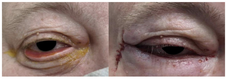
Figure 5 Pre- and immediate postoperative photos of a patient with involutional tarsal ectropion. The surgical technique was a Bick procedure and skin transposition without inverting sutures.退行性睑板外翻患者术前与术后即刻照片,术式为 Bick 联合皮肤转位(未行内翻缝合)。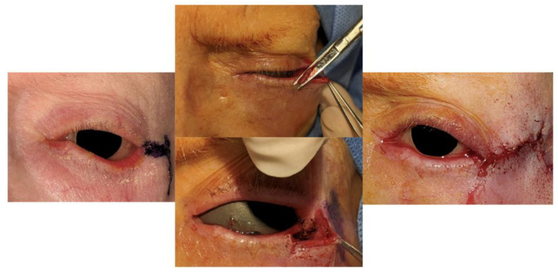
Figure 6 This patient with tarsal ectropion and skin cicatrix had residual punctal ectropion. In retrospect, inverting sutures and a skin graft could have been added.合并皮肤瘢痕的睑板外翻患者,残留泪点外翻;回顾认为应加用内翻缝合与皮片移植。
Tarsal ectropion is not uncommon, and all oculofacial surgeons should have a repertoire of surgical repair techniques based on the underlying etiology and severity of the case. This review highlights the effectiveness of these techniques and introduces an adjunctive superotemporal skin-transposition technique. This procedure is performed with horizontal lid tightening with the option of inverting sutures and offers enhanced lid stability compared to traditional procedures. The skin transposition requires about five additional minutes of operating time, and presents minor risks of scarring and ecchymoses. In our pilot case series, this method has demonstrated patient satisfaction and favorable outcomes in early follow-ups. However, as this series was conducted within the past 3.5 years, longer-term outcomes over several more years await investigation. In our experience, although patients were instructed to follow up if postoperative issues arose, all patients to date declined further follow-up given their satisfaction with the surgical outcome. Further studies are needed to establish the long-term efficacy of this procedure.
声明:中文精读 · 仅供学术参考。内容来自公开文献检索,不代表本人观点,不构成诊疗建议。 医疗美容需在正规医疗机构由执业医师实施。
基于相同主题推荐 · 继续深挖
本文综述睑外翻修复技术并提出上颞皮肤转位(STS)辅助术式,STS 常联合外侧睑板条或 Bick 缩短,保留外侧睑前层皮肤并向上颞方向转位。
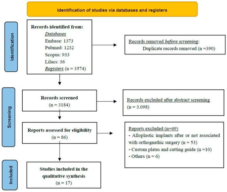
本文为遵循 PRISMA-ScR 的范围综述,系统检索 PubMed、Scopus、Embase、LILACS,共纳入 1998–2025 年发表的 17 项研究(6 项回顾性、2
本研究为韩国单中心回顾性系列,纳入 2017 年 1 月至 2024 年 5 月接受扩大深平面除皱、术后满 12 个月的 76 例患者,旨在预防并矫正 pixie ear 畸形。