Combined compound betamethasone and 595 nm pulsed dye laser improves hypertrophic scar and is associated with enhanced autophagy in myofibroblasts
2026年7月23日31 min read
Lasers in Medical Science 2026;41(1):152
Lasers in Medical Science 2026;41(1):152
The online version contains supplementary material available at 10.1007/s10103-026-04935-z.
关键词:Compound betamethasone / 595 nm pulsed dye laser / Hypertrophic scar / Autophagy
Hypertrophic scar (HS) is a cutaneous lesion resulting from excessive proliferation of newly formed connective tissue during repair after damage to the dermis and deeper tissues caused by trauma, surgery, burns, or other injuries [1]. Because scar formation can lead to pain, pruritus, and restriction of joint mobility, thereby impacting not only patients’ appearance but also their daily activities and psychological well-being. Consequently, treatment of HS has received widespread attention [2, 3]. The extracellular matrix (ECM) is crucial in scar formation due to its pathophysiological role [4]. Collagen, the primary ECM component, is found in HS at levels 2–3 times higher than in normal scars. Fibroblasts and their activated phenotype—myofibroblasts—are the principal cells responsible for ECM synthesis and secretion [4]. During normal wound healing, myofibroblasts promote wound closure through ECM secretion and are subsequently eliminated by apoptosis in the late stages of repair. In HS, however, myofibroblasts remain persistently activated and secrete excessive amounts of collagen, disrupting the dynamic balance between ECM synthesis and degradation [5, 6]. Therefore, inhibiting fibroblast and myofibroblast proliferation, reducing their collagen synthesis, or enhancing collagen degradation is essential for controlling scar formation and improving scar outcomes. Multiple studies have identified transforming growth factor-β1 (TGF-β1) as a central regulator of collagen and other ECM components produced by fibroblasts [7, 8]. TGF-β1 activates downstream Smad2/3 proteins to initiate transcription of fibrosis-related genes, including type I and III collagens and α-smooth muscle actin (α-SMA) [8]. Besides the TGF-β pathway, the integrin–FAK axis functions as a core medium of mechanical conduction, sensing mechanical cues and activating downstream signaling cascades such as PI3K/Akt, ERK, and mTOR to promote fibroblast proliferation, myofibroblast differentiation, and collagen deposition [9]. These signaling pathways are the subject of extensive investigation.
Autophagy involves the formation of autophagosomes, the fusion of autophagosomes with lysosomes to form autolysosomes, and the degradation of autophagosome contents [10, 11]. In normal circumstances, moderate autophagy enables cells to cope with adverse environments and maintain homeostasis. However, under pathological conditions, excessive autophagy can cause over-degradation of cellular components, leading to a form of type II programmed cell death termed “autophagic cell death [12–15]. Recent studies have linked autophagy to fibrosis, suggesting that autophagy could be a promising target for intervention in HS.
Topical pharmacotherapy, intralesional injections, laser therapy, pressure therapy, and surgery are all utilized in treating HS [16]. Intralesional glucocorticoids serve as the primary treatment for abnormal scars [17, 18]. Previous studies have indicated that glucocorticoids exert therapeutic effects on scars primarily via anti-inflammatory actions, inhibition of cell proliferation, promotion of extracellular matrix remodeling, suppression of angiogenesis, and immunomodulation of T cells [19, 20]. In laser therapy, pulsed dye laser (PDL) principally targets hemoglobin within blood vessels, resulting in occlusion of neovessels [21–24]. Its thermal effects can also inhibit fibroblast proliferation within HS [25]. However, these modalities have limited efficacy, and some scars recur after multiple treatments. Consequently, combined use of glucocorticoids injections and PDL has been increasingly adopted to improve outcomes. However, the underlying mechanisms of this combination therapy have not yet been reported.
In this study, we investigated the therapeutic effects of compound betamethasone injection combined with 595 nm PDL in a rabbit ear hypertrophic scar model and in hypertrophic scar–derived myofibroblasts. We specifically examined whether the efficacy of this combined therapy in improving hypertrophic scars is associated with enhanced autophagic activity in myofibroblasts. Our findings suggest that autophagy may contribute to this combined therapeutic strategy, offering a potential mechanistic insight that could help optimize clinical treatment approaches for hypertrophic scarring.
Five Japanese big-eared white rabbits (weight 2.5–3.0 kg, sex ratio 3 females to 2 males) were selected. After one week of acclimatization under standard housing conditions, the rabbits were anesthetized by intramuscular injection of Shutai 50. On each ear, four circular wounds, each 1 cm in diameter, were created with a trephine to the depth of the cartilage, spaced 1 cm apart, and the perichondrium was removed. 40 surgical wounds were generated in total. Four weeks after surgery, when stable hypertrophic scars had formed, the four scars on each ear were randomly assigned to control group, compound betamethasone injection (BI) group, pulsed dye laser (PDL) group, and combined treatment group (BI + P). Compound betamethasone was injected into scars until they became elevated and pale. The PDL (Vbeam2, Syneron & Candela, Boston, USA) parameters were: fluence 7.5 J/cm2null; spot size 7 mm; pulse duration 1.5 ms. For the combination therapy group, PDL was performed first, immediately followed by injection of compound betamethasone. Treatments were administered once every two weeks for a total of two sessions.
All rabbits were euthanized post-experiment using excessive anesthesia. The rabbit ear scars were excised and fixed in 10% neutral-buffered formalin overnight, followed by paraffin embedding. Three sections were prepared from each sample for staining and analysis. Tissues were sectioned at 5 μm and subjected to hematoxylin–eosin (HE) staining, Masson’s trichrome, immunohistochemical (IHC), and TUNEL assay. Due to the death of one rabbit and the loss of samples from another during the experiment, three rabbits were ultimately included in the analysis. The hypertrophic index (HI) was calculated. HI was calculated by determining the ratio between two perpendicular measurements: the distance from the cartilage to the highest point of the HS and the distance from the cartilage to the neighboring uninjured skin. Masson staining and the immunohistochemical markers p62 and LC3B were quantified using ImageJ 1.53e (NIH, USA). The Colour Deconvolution function was used to isolate the blue regions corresponding to collagen, and the IHC Toolbox plugin was used to extract the total areas of positive staining. Thresholds were then adjusted to fully encompass these regions, and measurements were recorded. For CD31, areas of microvessel aggregation were identified under low magnification and microvessel density was quantified at 200× magnification as microvessel count/0.739 to represent changes in microvessel number [26]. The primary antibodies used in IHC include p62/SQSTM1 (18420-1-AP, Proteintech), LC3B (18725-1-AP, Proteintech), and CD31 (ab182981, Abcam). The apoptosis rate for each field of view in the TUNEL assay was calculated as the proportion of positive cells (green fluorescence) to the total number of cells (DAPI). All sections were examined at 200× magnification. The analyzed field size was 0.4 mm2. For each section, five non-overlapping fields were randomly selected for image acquisition and analysis. Field selection adhered to strict randomization principles. All quantitative analyses were independently performed by two researchers who were completely blinded to the experimental groups, and image files were assigned randomized codes that contained no group-identifying information. The measurements obtained by the two operators were evaluated using the intraclass correlation coefficient and demonstrated a high degree of concordance. Our subsequent statistical analyzes will use each animal as an independent statistical unit for comparison between groups. All sections were examined under a microscope (Nikon, E100).
Tissues were obtained from five untreated patients with HS. Excised specimens were immersed in 50 mL centrifuge tubes containing a mixed antibiotic solution of 1% penicillin–streptomycin and transported to the laboratory. In a biosafety cabinet, the tissues were briefly immersed in 75% ethanol, washed twice with PBS containing 10% penicillin–streptomycin, and minced into 1 mm3 pieces with a scalpel. The tissue fragments were then placed in a digestion solution composed of 0.25% trypsin and DPBS (1:1) and incubated at 4 °C overnight. Subsequently, the tissue was further minced into a pulp-like consistency and transferred to centrifuge tubes containing collagenase I (C8140, Solarbio) for digestion at 37 °C. Following this digestion, the resulting tissue-containing suspension was filtered using a 70 μm nylon mesh. After centrifugation, the pellet was resuspended in complete DMEM/F12 medium supplemented with 15% FBS and 1% penicillin–streptomycin, and then transferred to culture flasks for incubation.
Hypertrophic scar-derived myofibroblasts were seeded in 96-well plates. Prior to irradiation, culture medium was removed and the wells were washed once with PBS, then add 100µL of PBS to each well to just cover the cell surface. A black sheet was placed beneath the plate to ensure uniform laser exposure of the cells. The vertical distance from the laser aperture to the cell monolayer was 1 cm. Remove the plate cover during irradiation. After irradiation, discard the PBS and replace it with complete growth medium for continued culture. Cells were returned to the incubator and cultured for an additional 24 h before further analyses. Non-irradiated cells served as the control group. PDL parameters were identical to those used in the animal experiments: spot energy density 7.5 J/cm2; spot diameter 7 mm; pulse duration 1.5 ms; single pulse.
Myofibroblasts isolated from HS tissue, specifically from 3 to 6 generations and during their exponential growth phase, were seeded into 96-well plates. Cells were exposed to varying doses of betamethasone dipropionate (HY-13571, MCE). Limit the final concentration of DMSO in the culture medium to 0.4% v/v. After 24 h, 10 µL of CCK-8 solution (C228166, YangGuangBio, China) was introduced to each well. The plates were subsequently returned to the incubator for an additional 4 h. The microplate reader measured absorbance at 450 nm. Cultured cells were divided into control, betamethasone dipropionate (BD), PDL, and combination (BD + P) groups, and cell viability was assessed as described above.
Myofibroblasts were seeded into 24-well plates and treated with betamethasone dipropionate for 24 h, with Rosup (S0033S, Beyotime, China) applied for 1 h as a positive control. After washing, cells were incubated for 30 min in serum-free medium containing 10 µM 2,7-dichlorofluorescein diacetate (DCFH-DA) (S0033S, Beyotime, China), and fluorescent images were subsequently captured using an inverted fluorescence microscope. Fluorescence intensities of each group were measured using a fluorescence microplate reader with excitation at 488 nm and emission at 525 nm.
Myofibroblasts cultured in 24-well plates were subjected to the experimental treatments. Subsequently, the cells were preserved using a 4% paraformaldehyde for half an hour. A proprietary blocking solution (C220702, YangGuangBio, China) was then applied for a 30 min incubation at ambient temperature. Following this, the cells were exposed to primary antibodies and left at 4 °C overnight. After thorough rinsing, corresponding secondary antibodies were added shielded from light, for 1 h. Nuclei were counterstained using an antifade mounting medium containing DAPI (C190401, YangGuangBio, China). Primary antibodies included anti-α-smooth muscle actin (α-SMA, ET160753, HUABIO), anti-vimentin (AF7013, Affinity), anti-COL1A1 (HA722517, HUABIO), and anti-collagen III (HA720050, HUABIO). Fluorescence images were captured using an inverted fluorescence microscope.
MDC can specifically label autophagic vacuoles. After grouped treatment of myofibroblasts seeded in 96-well plates, the cells were washed once with PBS. Then, MDC staining solution (C3018S, Beyotime, China) was added and the cells were incubated at cell culture incubator 15 min. Subsequently, the cells were washed three times with detection buffer and promptly examined using an inverted fluorescence microscope. Blue fluorescence was detected in the DAPI channel.
The cultured myofibroblasts were subjected to respective treatments, digested with 0.25% trypsin, centrifuged, and the supernatant discarded. Cells were then fixed in 2.5% glutaraldehyde at room temperature for 30 min and transferred to 4 °C for storage. After washing, dehydration and embedding, samples were cut into ultrathin sections of 60–80 nm. Autophagosomes within the cells were examined by TEM (JEM-1400, JEOL, Japan). Similarly, rabbit ear scar tissue was cut into 1 mm3 blocks, fixed in 2.5% glutaraldehyde at 4 °C in the dark, and subsequently processed and examined by TEM using the same protocol.
After treating myofibroblasts in grouped conditions for 24 h, followed by protein isolation using RIPA buffer supplemented with PMSF and phosphatase inhibitors. Protein quantification was done using a BCA assay kit. Subsequently, proteins (30 µg per lane) were separated by SDS-PAGE, electroblotted onto 0.45 μm PVDF membranes. Membranes were then blocked with 5% BSA and probed with primary antibodies overnight at 4 °C, followed by a 1-hour incubation with HRP-conjugated secondary antibodies at room temperature. Primary antibodies used included LC3 (14600-1-AP, Proteintech, 1:5000), p62/SQSTM1 (18420-1-AP, Proteintech, 1:25000), COL1A1 (HA722517, HUABIO, 1:2000), Collagen III (HA720050, HUABIO, 1:2000), GAPDH (10494-1-AP, Proteintech, 1:5000), and β-actin (81115-1-RR, Proteintech, 1:5000).
Data were presented as mean ± standard \documentclass[12pt]{minimal} \usepackage{amsmath} \usepackage{wasysym} \usepackage{amsfonts} \usepackage{amssymb} \usepackage{amsbsy} \usepackage{mathrsfs} \usepackage{upgreek} \setlength{\oddsidemargin}{-69pt} \begin{document}$$:(\stackrel{-}{x}\pm:s)$$\end{document}deviation (SD). All experimental data were analyzed using SPSS 25.0. Comparisons among multiple groups were performed by one-way analysis of variance (one-way ANOVA), and pairwise comparisons were further conducted using the Least Significant Difference (LSD) test. P<0.05 was considered statistically significant (*P < 0.05, **P < 0.01, ***P < 0.001).
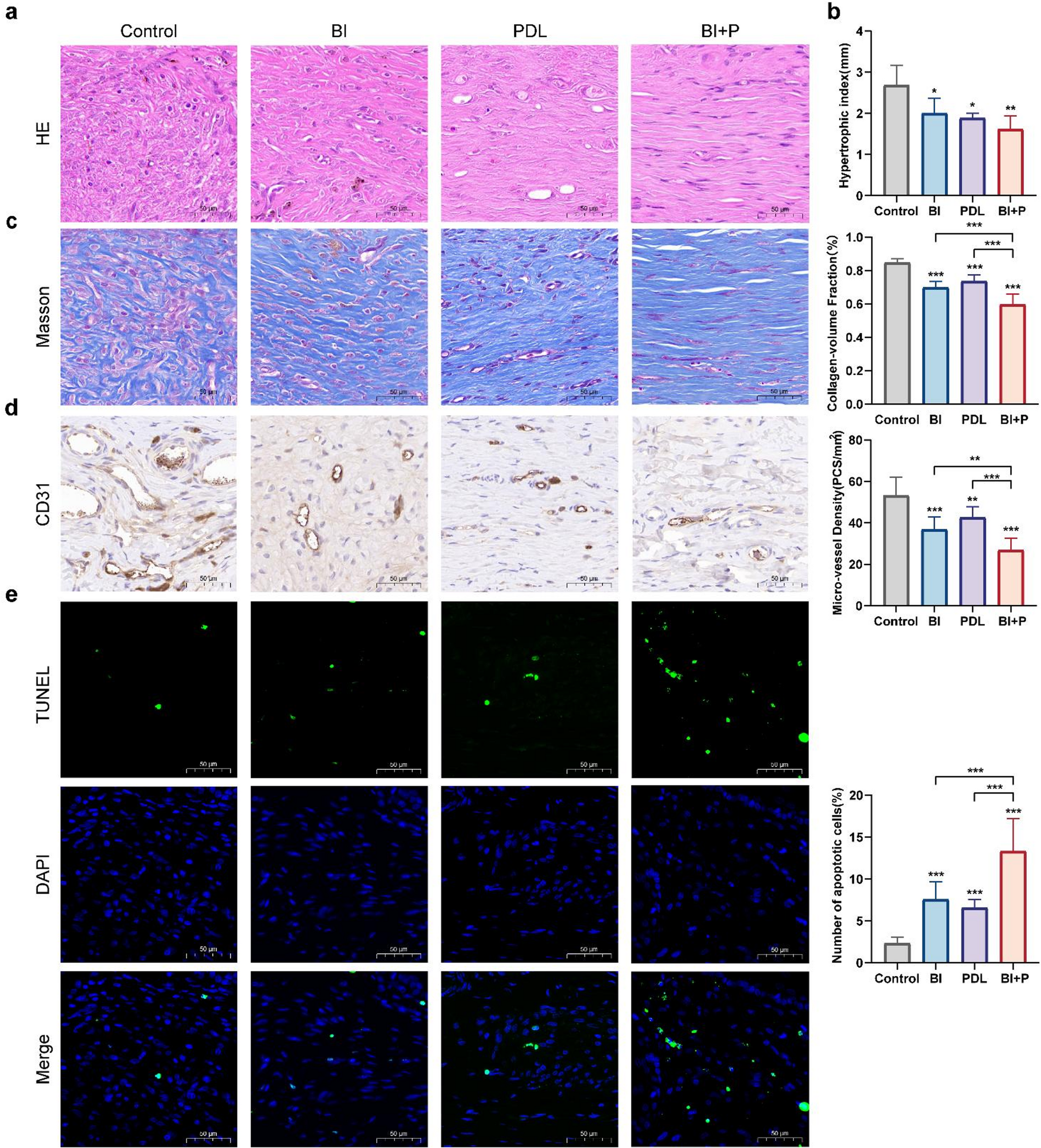
After establishment of the rabbit ear scar model and following two treatment sessions, HE staining revealed a marked reduction in inflammatory cells and a more orderly arrangement of fibroblasts in the BI, PDL, and BI + P groups compared with the control group (Fig. 1a). Scar height improvement was evaluated by measuring the HI. The BI + P group (1.61 ± 0.33) exhibited a lower HI than the BI group (2.00 ± 0.36) and the PDL group (1.88 ± 0.12). All three treatment groups showed significant differences compared with the control group (2.68 ± 0.48) (Fig. 1b).
Masson staining demonstrated dense and disorganized collagen in the control group. After treatment, collagen content decreased significantly in the BI (0.70 ± 0.03), PDL (0.74 ± 0.04), and BI + P (0.60 ± 0.06) groups compared with the control group (0.85 ± 0.02). The greatest reduction was observed in the BI + P group, which differed significantly from the two single-treatment groups. Collagen fibers also became more regularly arranged, with the most pronounced improvement in the BI + P group (Fig. 1c). CD31, used as an endothelial cell marker to assess microvascular changes in this study, showed that the BI + P group (26.91 ± 5.76) had the fewest micro-vessels and differed significantly from the BI (36.84 ± 6.01) and PDL (42.55 ± 5.20) groups (Fig. 1d). All three treatment groups also differed significantly from the control group (53.07 ± 8.87).
Apoptosis was assessed by TUNEL staining. After treatment, all three treatment groups exhibited increased apoptosis compared with the control group (2.32 ± 0.72%). The BI + P group (13.30 ± 3.40%) demonstrated significantly higher apoptosis than both the BI group (7.54 ± 2.12%) and the PDL group (6.56 ± 1.01%) (Fig. 1e).
*图注:图 1. Combined treatment with compound betamethasone injection and 595 nm PDL yielded superior improvement of HS compared with monotherapy. a Hematoxylin–eosin staining of the control and treatment groups. (Scale bar: 50 μm). b Scar hypertrophy index of the control and each treatment group. c Masson’s trichrome staining showed collagen alterations in the control and treatment groups (Scale bar: 50 μm). d IHC detection of CD31 expression (Scale bar: 50 μm). e TUNEL assay for apoptosis (Scale bar: 50 μm). The data were presented as the mean ± SD of each group (n = 3) from three independent experiments. ns indicates no significant difference; *P < 0.05, **P < 0.01, **P < 0.001
To investigate the mechanism by which the combined treatment attenuates scars, TEM was used to examine cellular structure. Compared with the control group, all three treatment groups exhibited varying increases in autophagosomes, with the combined therapy group showing a notably greater number of autophagosomes than either monotherapy group (Fig. 2a). LC3 and p62 are key autophagy-related proteins commonly employed to assess cellular autophagic status. Therefore, IHC was performed to evaluate LC3B and p62 expression. LC3B expression increased in all three treatment groups relative to the control group (1315.67 ± 214.37). The integrated optical density (IOD) of LC3B in the BI + P group (3331.74 ± 398.99) was significantly higher than in the BI group (2173.62 ± 397.98), and the PDL group (2118.39 ± 343.65) (Fig. 2b). Conversely, p62 expression was suppressed in all three treatment groups compared with the control group (6117.12 ± 722.79) after treatment. The degree of p62 suppression in the BI + P group (2066.62 ± 400.90) was significantly greater than in the BI group (3898.26 ± 622.82) and the PDL group (3834.70 ± 569.37) (Fig. 2c).
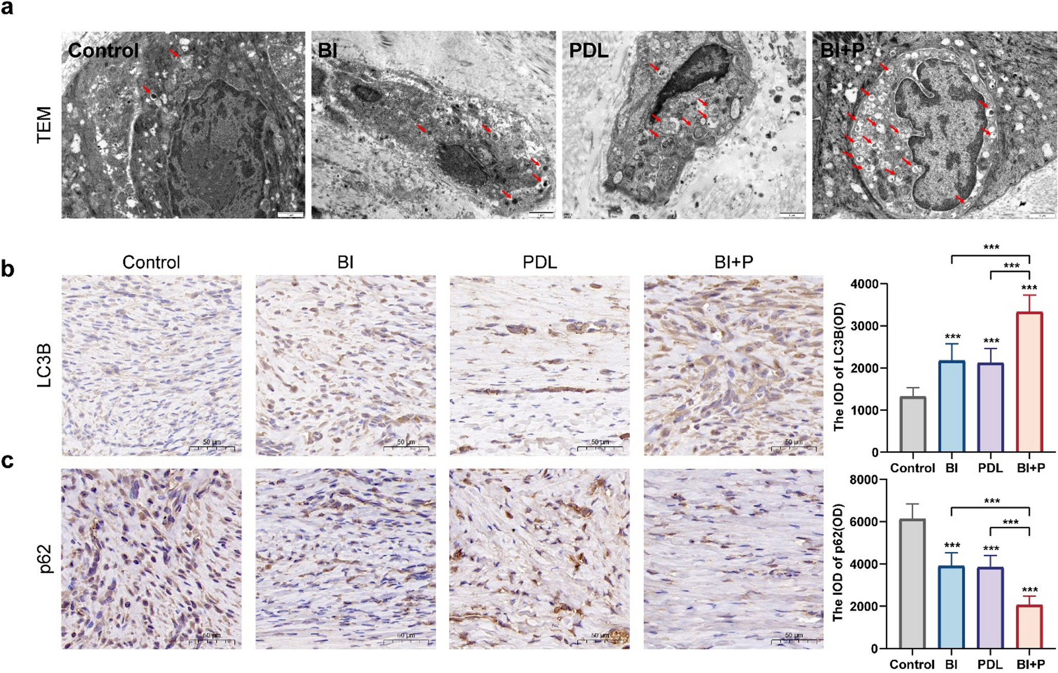
*图注:图 2. Compound betamethasone combined with 595 nm PDL markedly induced increased autophagy. a TEM detection of changes in autophagy levels in rabbit ear scars after treatment. Red arrows indicated autophagosomes (Scale bar: 1 μm). b IHC detection of LC3B expression (Scale bar: 50 μm). c IHC of p62 expression (Scale bar: 50 μm). The data were presented as the mean ± SD of each group (n = 3) from three independent experiments. ns indicated no significant difference; *P < 0.05, **P < 0.01, **P < 0.001
We conducted in vitro cell experiments to further investigate the mechanism by which the combined regimen treats HS. The compound betamethasone injection used in the animal experiments primarily contains betamethasone dipropionate and betamethasone sodium phosphate. Betamethasone sodium phosphate is rapidly absorbed, exerts a prompt effect and being cleared from tissues within 24 h. Whereas betamethasone dipropionate, after injection, forms a depot that is absorbed slowly and provides sustained activity, thereby controlling symptoms for an extended period. Therefore, betamethasone dipropionate was selected for this cell-based study.
To better model HS, primary cells were isolated from HS tissue obtained during surgical procedures and cultured. The resulting cells were characterized by immunofluorescence imaging using Vimentin and α-SMA as markers (Fig. 3a). Because the vertical distance from the PDL laser aperture to the cell monolayer was 1 cm, this affected the beam spot size and energy density. Based on this value, calculations and spot-size measurements yielded a spot diameter of 7.6 mm (Supplementary Fig. s1) and a fluence of 6.36 J/cm2 (Supplementary Fig. s2). Temperature before and after irradiation was measured with a thermocouple thermometer (YET-610 L, YOWEXA, China). The results showed that the liquid temperature did not deviate from baseline by more than 1 °C during recovery. In addition, a CCK-8 assay was performed immediately after irradiation to assess cell viability, and no significant difference in viability was observed before versus after irradiation (Supplementary Fig. s3).
Cell viability was assessed using the CCK-8 assay. First, this study determined that a final concentration of 0.4% v/v DMSO in the culture medium had no effect on cell viability (Supplementary Fig. s4). Next, betamethasone dipropionate was found to inhibit the viability of hypertrophic scar-derived fibroblasts in a dose-dependent manner, with a half-maximal inhibitory concentration (IC50) of 209 µM, which was used in subsequent experiments (Fig. 3b). ROS detection showed that BD at this concentration exhibited significantly lower levels compared with the control group (Supplementary Fig. s5a, b). Additionally, the BD group (53.06 ± 1.74%), the PDL group (47.19 ± 6.72%), and the BD + P group (15.05 ± 1.15%) all exhibited reduced cell viability, with the combination group showing the most pronounced inhibition (Fig. 3c).
The effects of each treatment group on collagen were assessed by Western blotting. Expression levels of type I and type III collagen were significantly lower in the BD group (0.59 ± 0.06; 0.75 ± 0.09), the PDL group (0.73 ± 0.06; 0.74 ± 0.13), and the BD + P group (0.38 ± 0.04; 0.56 ± 0.07) compared with the control group. Significant differences were also observed between the combined treatment group and each single-treatment group (Fig. 3d). These findings were corroborated by immunofluorescence (Fig. 3e).
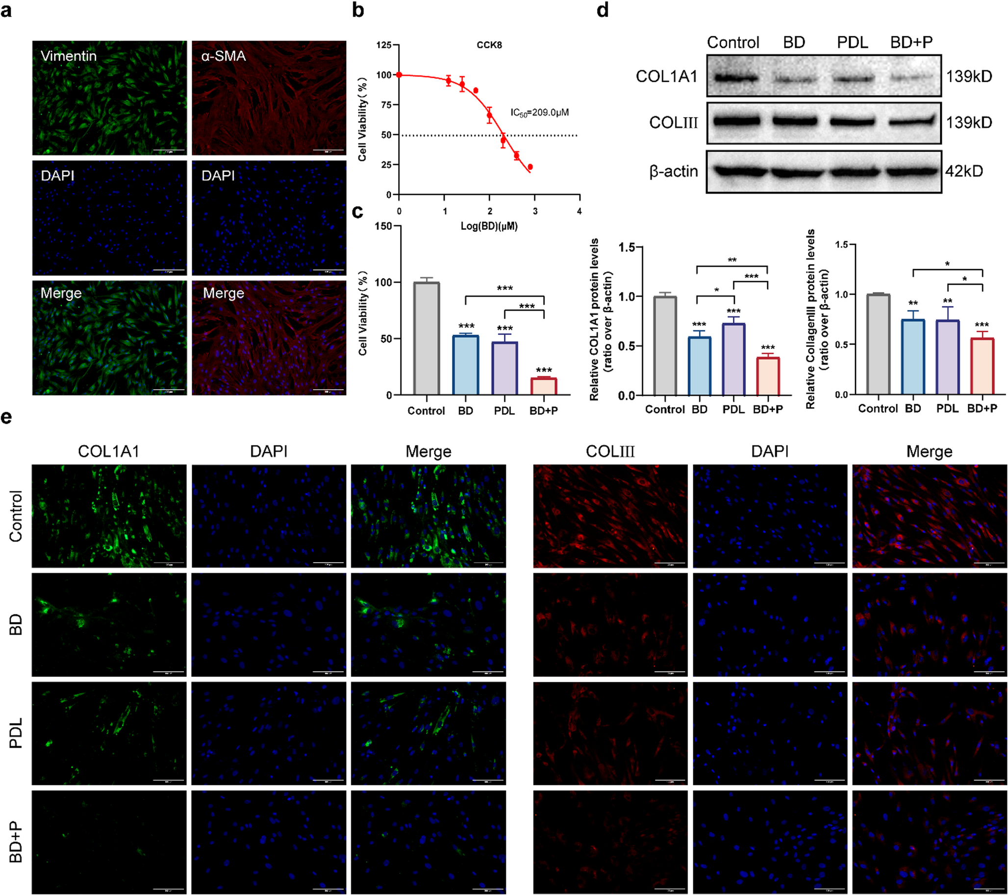
*图注:图 3. Betamethasone dipropionate combined with PDL suppressed the viability of hypertrophic scar myofibroblasts and collagen secretion. a Immunofluorescent identification of myofibroblasts isolated from scar tissue (Scale bar: 100 μm). b CCK-8 assay for IC50 determination. c CCK-8 assay of cell viability. d Western blot analysis of type I and type III collagen expression. e Immunofluorescence detection of type I and type III collagen expression (Scale bar: 100 μm). The data were presented as the mean ± SD of each group from three independent experiments. ns indicates no significant difference; *P < 0.05, **P < 0.01, **P < 0.001
Subsequently, Western blot was used to assess the ratio of LC3II to LC3I and the expression of p62 in myofibroblasts from the three treatment groups. The LC3II/LC3I ratios were elevated in the BD group (1.60 ± 0.22), the PDL group (1.43 ± 0.07) and the combination group (2.01 ± 0.28). The combined treatment produced the most pronounced increase and showed significant differences compared with the single treatment groups. Concurrently, a notable reduction in p62 expression was observed across all treated cohorts, including the BD (0.72 ± 0.07), PDL (0.64 ± 0.08), and the BD + P (0.32 ± 0.09) groups. And the BD + P group differed significantly from each single-treatment group (Fig. 4a). The MDC kit specifically labels autophagosomes, appearing as intracellular fluorescent puncta. Detection revealed a significant increase in fluorescent puncta in the combined treatment group (Fig. 4b). TEM showed an increased number of autophagosomes in all three experimental groups compared with the control group. The combined group exhibited the highest number. Western blot, MDC staining, and TEM collectively demonstrated that the combined intervention indeed enhanced autophagosome formation in myofibroblasts (Fig. 4c).
However, the increased number of autophagosomes may also result from downstream blockade of the autophagic process, namely impaired formation of autolysosomes. Because the above Western blot results showed an inverse correlation between LC3Ⅱ/LC3Ⅰ and p62 expression, we preliminarily inferred that the combination treatment significantly may enhanced autophagic flux in myofibroblasts. To further validate this finding, chloroquine (CQ) was applied as an autophagy inhibitor on top of each treatment group. The results showed that the trends of LC3Ⅱ/LC3Ⅰ and p62 changes in the BD, PDL and BD + PDL groups relative to control group were consistent with those described above. Administration of CQ (HY-17589 A; MCE) increased LC3Ⅱ/LC3Ⅰ levels in all treatment groups, with the greatest increase observed in the combination group. These differences were statistically significant. Under CQ treatment, elevated p62 expression was observed in all experimental cohorts. A notable difference was measured between the BD + P group and the BD + P+CQ group. These findings are consistent with the hypothesis that BD, PDL, and BD + P treatments may increase autophagy and preserve autophagic flux (Fig. 4d).
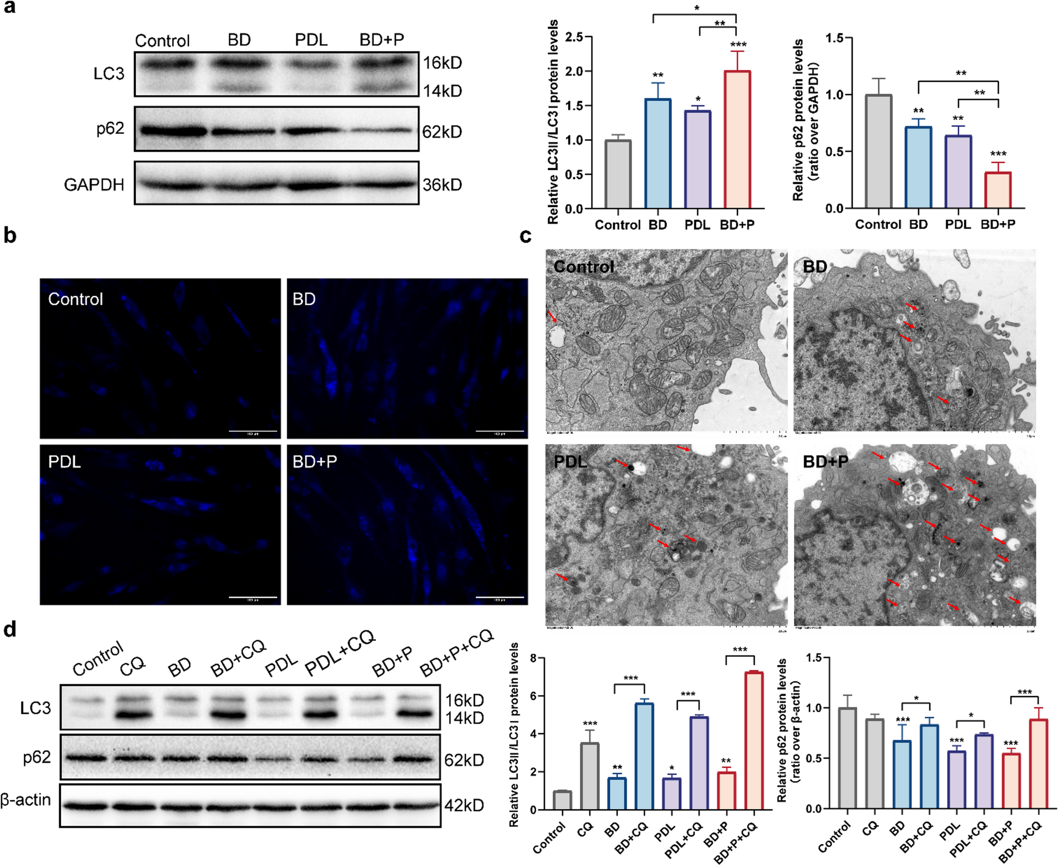
*图注:图 4. Betamethasone dipropionate combined with PDL enhances autophagic flux in hypertrophic scar myofibroblasts. a Western blot analysis of LC3Ⅱ/LC3Ⅰ and p62 expression. b MDC staining of autophagosomes (Scale bar: 100 μm). c TEM observation of autophagosomes. Red arrows indicated autophagosomes (Scale bar: 2 μm). d Western blot analysis of LC3Ⅱ/LC3Ⅰ and p62 expression following chloroquine treatment. The data were presented as the mean ± SD of each group from three independent experiments. ns indicates no significant difference; *P < 0.05, **P < 0.01, **P < 0.001
To assess the role of autophagy in HS modulation, we examined collagen expression in each treatment group following chloroquine administration. Western blot analysis revealed that COL1A1 and COLⅢ levels in the BD + P group were markedly reduced compared with the two single-treatment groups and the control. After the addition of CQ, the expression levels of COL1A1 and COLⅢ in the BD + P+CQ group were significantly elevated compared to the BD + P group. These finding indicate that autophagy inhibition may partially attenuate the suppressive effect of the combined treatment on collagen synthesis by myofibroblasts (Fig. 5a). Therefore, the combined therapy group may have exerted its therapeutic effect on HS by inducing excessive autophagy within cells. Immunofluorescence analysis corroborated these findings (Fig. 5b).
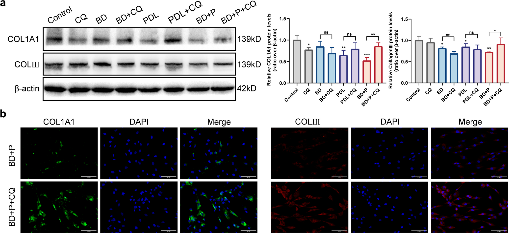
*图注:图 5. Adding chloroquine to each group revealed that inhibiting cellular autophagy reversed the combined treatment’s suppression of collagen production in myofibroblasts. a Western blot analysis of type I and type III collagen expression following chloroquine treatment. b Immunofluorescence detection of type I and type III collagen expression (Scale bar: 100 μm). The data were presented as the mean ± SD of each group from three independent experiments. ns indicates no significant difference; *P < 0.05, **P < 0.01, **P < 0.001
HS is a fibrotic skin disorder characterized primarily by excessive proliferation of fibroblasts and abnormal deposition of ECM. Using a rabbit ear scar model, we showed that combined intralesional compound betamethasone injection and PDL produced greater improvement of HS than either treatment alone. In vitro, we suggest that betamethasone dipropionate in combination with PDL enhances inhibition of hypertrophic scar myofibroblast viability and reduces type I and III collagen deposition more than either monotherapy. This enhanced effect was associated with an increase in autophagic activity and maintenance of autophagic flux, as evidenced by elevated conversion of LC3 I to LC3 II, decreased p62 levels, increased autophagosome numbers, and synchronous accumulation of LC3 II and p62 after chloroquine treatment. Meanwhile, chloroquine-mediated blockade of autophagic flux partially attenuated the collagen‑lowering effect of the combination treatment. These data suggest that enhanced autophagy in myofibroblasts may contribute to the observed reduction in collagen deposition.
The combined application of intralesional corticosteroid injections and PDL therapy has emerged as a pivotal strategy in the management of HS, with the objective of enhancing clinical outcomes and reducing the risk of recurrence [27, 28]. The primary mechanisms involve the anti-inflammatory and antiproliferative effects of glucocorticoids [29, 30], along with the selective microvascular photothermolysis and collagen remodeling caused by PDL [31, 32]. In the present study, using a rabbit ear scar model, we suggested that the combined therapy produced greater amelioration of HS compared with either monotherapy. Histological analysis revealed that the combination group not only markedly improved fibroblast and collagen organization, reduced inflammation, and decreased microvessel density, but also significantly increased apoptosis within the scar. Moreover, our findings extend previous observations by showing that compound betamethasone injection, PDL, and their combination all increased autophagosome abundance in scar tissue, with the combined treatment producing the largest effect.
Autophagy is a multi-step, tightly regulated biological process. This continuous dynamic sequence is referred to as autophagic flux. The impairment at any stage of the flux leads to inhibited autophagic activity and consequent disruption of its physiological functions [11]. LC3 and p62 are important autophagy-related proteins that are widely used as markers for autophagy assessment [33]. LC3 exists in two forms: during autophagy induction, cytosolic LC3Ⅰ is conjugated to phosphatidylethanolamine (PE) on the autophagosomal membrane to form the membrane-bound LC3Ⅱ, and thus the LC3Ⅱ/LC3Ⅰ ratio is used to reflect changes in autophagic status [34]. P62 was the first identified selective autophagy receptor and functions as a bridge between LC3 and ubiquitinated substrates. It recognizes and binds ubiquitin-tagged cargos via its ubiquitin-binding domain and simultaneously binds specifically to LC3II on the autophagosomal membrane via its LC3-interacting region. Thus, p62 is a key protein targeted for degradation by autolysosomes during the late stages of autophagy [35]. Autophagy has increasingly been recognized as a key regulator of tissue remodeling in both normal tissue repair and fibrotic processes [36]. HS is fundamentally a cutaneous fibrotic disorder, and regulation of autophagic activity is emerging as a novel focus of research. Yunxian Dong et al. found that compared with normal skin fibroblasts, hypertrophic scar fibroblasts exhibit impaired autophagic flux. Melatonin treatment enhances the autophagic process by acting through the MT2 receptor to suppress the PI3K/Akt/mTOR signaling axis, thus playing a role in treating HS [37]. Qing Zhang et al. found that shikonin improves HS by enhancing autophagy [38]. These findings collectively indicate that activation of autophagy is a potential therapeutic target for reducing scar fibrosis. Most existing studies have focused on hypertrophic scar fibroblasts. Myofibroblasts, as activated fibroblasts, exhibit markedly greater contractile activity and ECM production capacity [39–41]. In this study, we innovatively centered the cellular validation on hypertrophic scar myofibroblasts. The results indicated that combined treatment of betamethasone dipropionate and PDL could enhance the induction of autophagy activation in myofibroblasts. Pharmacological inhibition of autophagic flux using chloroquine partially attenuated the reduction in intracellular type I and type III collagens, suggesting that this enhanced autophagic activity may contribute to collagen degradation. This finding located the potential therapeutic strategy of autophagy from general fibroblasts to myofibroblasts with more pathological significance, and suggested that enhanced autophagy in hypertrophic scar myofibroblasts may contribute to the collagen degradation observed under combination therapy.
Although this study verified through in vivo and in vitro experiments that the effect of the combination group was superior to that of the single treatment group, providing a theoretical basis for better clinical treatment of HS. Meanwhile, the exploration of the mechanism suggested that increasing the autophagy of myofibroblasts might be a potential therapeutic approach for improving HS. Nonetheless, limitations remain. First, a major limitation of this study is the small sample size of the animal experiments. Due to animal death and sample loss, only three rabbits were ultimately included in the four treatment groups. This sample size was insufficient to conduct a rigorous comparative analysis of the four groups and multiple histological indicators. Although statistically significant differences were observed, these findings need to be further confirmed with sufficient sample sizes in the future. Second, in animal experiments we observed that the three treatment groups produced significant differences at the microvascular level compared with the control group. The in vivo mechanism of PDL fundamentally targets the vasculature: laser energy is selectively absorbed by oxyhemoglobin within scar microvessels, inducing selective photothermal lysis and thereby triggering tissue remodeling. However, in this study, PDL was used to irradiate cells covered with PBS as an in vitro model. Even with energy density calculations and real-time cell viability monitoring, this in vitro system still possesses inherent limitations. The in vitro PDL model cannot reproduce hemoglobin-mediated selective photothermolysis, vascular occlusion, heat diffusion, inflammatory responses, or tissue scattering. Cell-level PDL findings should therefore be presented as supportive mechanistic observations rather than direct simulations of clinical PDL treatment. The mechanisms revealed here at the cellular level should be interpreted with caution and regarded as hypothetical components of a combination therapy mechanism, rather than as a comprehensive explanation for the enhanced in vivo efficacy. Future studies should validate these mechanisms in model systems that more closely approximate the clinical scar-laser treatment environment, for example by constructing three-dimensional culture systems that include vascular structures or by using ex vivo skin organ culture models. Third, the animal experiments employed a compound betamethasone injection containing both the rapid-acting betamethasone sodium phosphate and the sustained-release betamethasone dipropionate to simulate clinical treatment. Considering chronic course of HS and the aim of elucidating mechanisms in cell experiments, only betamethasone dipropionate, the main active ingredient with a long - acting effect, was selected. Although this design focused on the core pharmacological effects, it did not account for potential pharmacokinetic synergy or complementary interactions between the two components in vivo. Future studies should evaluate the direct effects of the complete compound formulation on myofibroblasts to generate laboratory evidence that more closely aligns with clinical conditions. Simultaneously, the 209 µM BD concentration used in these experiments falls within the micromolar range. To exclude nonspecific cellular stress responses that can be induced by high drug exposure, intracellular reactive oxygen species (ROS) levels were measured. The results showed that ROS fluorescence intensity in the BD-treated group was significantly lower than in the control group, suggesting that at this concentration BD may exert classical anti-inflammatory/antioxidant effects mediated by glucocorticoid receptor (GR) activation. However, ROS data alone are insufficient to definitively ascribe the observed effects entirely to GR mediation. Furthermore, the in vitro cell culture system employed in this study cannot directly recapitulate the in vivo sustained-release pharmacokinetic behavior of betamethasone dipropionate. Consequently, the in vitro findings should not be directly used as a model for clinically translatable dosing. Further validation is required, including experiments using GR antagonists, metabolite profiling, and release systems that better mimic physiological conditions. Fourth, this study arrived at a preliminary conclusion that combined treatment may induce collagen degradation by enhancing autophagy in scar myofibroblasts, but the upstream signaling pathways remain undefined. Previous research has shown that glucocorticoids can upregulate Regulated in Development and DNA Damage Response 1 (REDD1), and REDD1 is an inhibitor of the mechanistic target of rapamycin complex 1 (mTORC1) [42]. PDL acting on cultured keloid fibroblasts has been reported to augment MAPK signaling cascade, with a marked increase in p38 phosphorylation [43]. Similarly, p38 is also a negative regulator of mTORC1. Based on the above, we propose a reasonable hypothesis: these two treatment modalities may converge on and jointly suppress mTORC1, the central negative regulator of autophagy, through complementary or cooperative actions. This hypothesis should be validated in future studies using approaches such as specific pathway inhibitors and gene knockdown. Given the known involvement of AMPK, ROS, and endoplasmic reticulum stress in autophagy regulation, future investigations may also explore mechanisms involving these pathways. Fifth, this study primarily assessed autophagic flux by using chloroquine to inhibit autophagosome–lysosome fusion. Although this pharmacological approach is a conventional strategy for evaluating autophagic flux, it remains an indirect inference at the pharmacological level. Chloroquine exerts broad effects on lysosomes rather than specifically inhibiting autophagy alone, so off-target effects or parallel mechanisms cannot be entirely excluded. In future studies, incorporation of the tandem fluorescent mRFP‑GFP‑LC3 reporter is required to precisely quantify dynamic changes in autophagic flux. Concurrently, genetic loss‑of‑function or rescue experiments targeting autophagy‑essential genes, together with direct measurement of collagen turnover, will be necessary to define the specific contribution and specificity of autophagy in the observed reduction of collagen. Furthermore, this study observed a decrease in collagen in the three treatment groups compared with the control group. The reduction of collagen needs to consider a variety of possibilities that are not mutually exclusive, such as the loss of active myofibroblasts, apoptosis-related degradation, and overall cell damage. In the combined treatment group of this study, a significant increase in the number of TUNEL-positive cells in scar tissue was observed. Meanwhile, through cell experiments, it was found that the ratio of autophagy markers LC3-II/LC3-I increased and the expression of p62 was down-regulated. These two results suggest that both apoptosis and autophagy were activated. Although our experiments using chloroquine indicate that autophagy inhibition partially restores collagen levels in the combination treatment group, the reduction in collagen cannot be solely attributed to increased autophagic degradation. It may also reflect decreased collagen secretion resulting from fibroblast apoptosis. Likewise, existing evidence indicates that autophagy and apoptosis are not isolated processes but are tightly coupled through complex signaling networks [44]. Under cellular stress, upregulation of autophagy can maintain homeostasis and inhibit apoptosis by engulfing damaged mitochondria and degrading pro-apoptotic proteins. However, some autophagy proteins can directly participate in apoptosis induction (such as Atg5 and Atg12). At the same time, anti-apoptotic and cell survival factors can be degraded during autophagy. Therefore, in cases where autophagy levels are overly activated or the apoptotic mechanism is impaired, autophagy also has the function of promoting apoptosis [45]. In light of this and in connection with our findings, the reduction of collagen observed in the combination treatment group may not be solely attributable to increased collagen degradation resulting from elevated autophagy. It may also include decreased collagen secretion due to fibroblast apoptosis. Future studies should perform multi-timepoint assessments of autophagy and apoptosis markers, combined with measurements of collagen synthesis rates, quantification of procollagen secretion, and assays of matrix metalloproteinase activity, to more fully disentangle the relative contributions of autophagy and apoptosis to collagen metabolism in scars under combination therapy and thereby provide a more precise theoretical basis.
In conclusion, our study suggests that compound betamethasone combined with 595 nm PDL significantly improve hypertrophic scars and that this effect is associated with enhanced autophagic activity and collagen degradation. These findings provide mechanistic insight into the enhanced effects of this widely used clinical regimen and highlight autophagy in myofibroblasts as a potential therapeutic target for optimizing hypertrophic scar treatment.
Below is the link to the electronic supplementary material.
Supplementary Material 1 (PDF 527 KB)
Supplementary Material 2 (PDF 291 KB)
声明:中文精读 · 仅供学术参考。内容来自公开文献检索,不代表本人观点,不构成诊疗建议。 医疗美容需在正规医疗机构由执业医师实施。