微创乳突悬吊(MIMS)颈部无痕提升
2026年8月5日31 min read
Aesthetic Surgery Journal. Open Forum 2026;8:ojag065
R Brannon Claytor; Patricia M Fuentes; Grace Tolan; Lauren LoweAesthetic Surgery Journal. Open Forum. 2026 Apr 11; 8: ojag065 | 开放获取 CC BY 4.0DOI: 10.1093/asjof/ojag065 | PMCID: PMC13418187摘要(中文精读) 颈部常是最早显现衰老迹象的面颈部区域,表现为颈颏角(CMA)变钝、下颌线模糊、颈阔肌条索、颏下脂肪堆积与皮肤冗余。本文目的是介绍一种微创乳突悬吊(MIMS)颈部提升技术,将吸脂与激光热作用同"双缝线系统"结合:一条永久缝线(4-0 Ethibond)锚定于乳突筋膜,一条可吸收缝线(4-0 Monocryl)针对皮肤松弛。方法:回顾性病例系列,纳入 2020 年 12 月至 2024 年 8 月由术者完成的全部 MIMS 病例。局麻肿胀麻醉下,先以动力辅助吸脂(preplatysmal 平面)去除颏下脂肪,再以 SmartLipo 激光于皮下/真皮下面热作用紧肤,最后置入双缝线并加止血网缝线。结果:共 73 例,平均 48.8 岁(22–82),平均 BMI 25.4。手术时间平均 150.6 分钟。最常见并发症为血清肿 3 例(4.1%,1–3 cc,门诊抽吸);1 例(1.4%)右下颌缘神经神经失用,术后第 5 天出现、2 周内自发恢复。13 例(16.9%)出现皮肤褶皱,经微针(±射频)处理,平均 24.75 天消退。无血肿、无皮肤坏死。1 年及 2 年随访见颈颏角与下颌线改善(图 4–6)。结论:吸脂 + 激光热作用 + 乳突悬吊三联法,针对早期颈皮肤松弛与颏下脂肪堆积,为开放颈成形术提供了一种微创替代方案。
颈部是最早显现衰老的面颈部特征,表现为颈颏角变钝、下颌线消失、颈阔肌条索、颏下脂肪堆积与皮肤冗余。文献中理想的颈部美学标准一直是锐利的颈颏角(90°–120°)与下颌角(120°–140°)。颈部年轻化技术从浅表的皮肤收紧,演进到针对颈阔肌、颏下与颈阔肌下脂肪、颌下腺的深层结构性处理。
Feldman 1988 年提出的"紧身胸衣式颈阔肌成形"开创了先河;Giampapa 与 DiBernardo 发展了缝线悬吊颈提升,1995 年 Giampapa 进一步用"乳突-乳突互锁悬吊缝线" refinement。Connell、Guerrero-Santos、Baker 等贡献了不同的瓣与锚定方式,多与面部提升联合,但代价是剥离范围大、恢复期长、并发症风险更高。
近年患者需求转向恢复短、可在诊室完成、创伤更小的术式。Mueller 的经皮颈阔肌成形用 LED 引导系统沿颈阔肌置入永久缝线矩阵,锚定于下颌缘保留韧带——避免大切口,但效果可能较温和、持久性有限。能量紧肤与注射治疗持久性则参差不一。本文目标即介绍 MIMS 技术:吸脂 + 激光热作用 + 双缝线系统(永久缝线锚定已剥离的乳突筋膜 + 浅层可吸收缝线针对皮肤松弛)。
回顾性病例系列,纳入 2020.12–2024.8 由主刀完成的全部 MIMS 颈部提升患者。获 Main Line Health IRB 豁免(E-24-5420,2025-09-16)。患者均知情同意,多人签署媒体授权用于照片发表。用 SPSS v30.0 做人口学描述性分析。
主刀在 QUAD-A 认证外科机构的局麻肿胀麻醉下完成。颈部以约 250 cc 含利多卡因与肾上腺素的生理盐水肿胀。顺序是: 先吸脂与激光热作用,后缝线悬吊 。
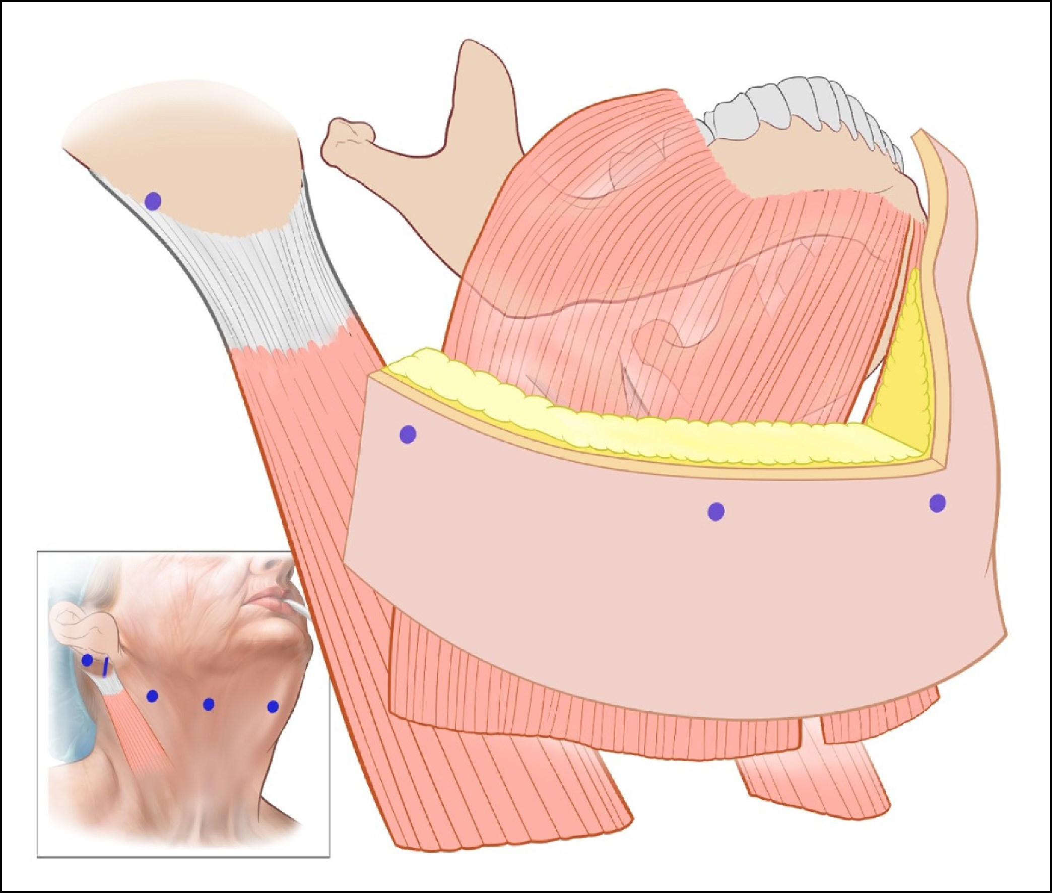
图 1 穿刺位点
图 1. 穿刺位点:舌骨体上 1 cm 中央、颌下腺正上方、下颌体中部、下颌角下方。图 2 乳突悬吊隧道制作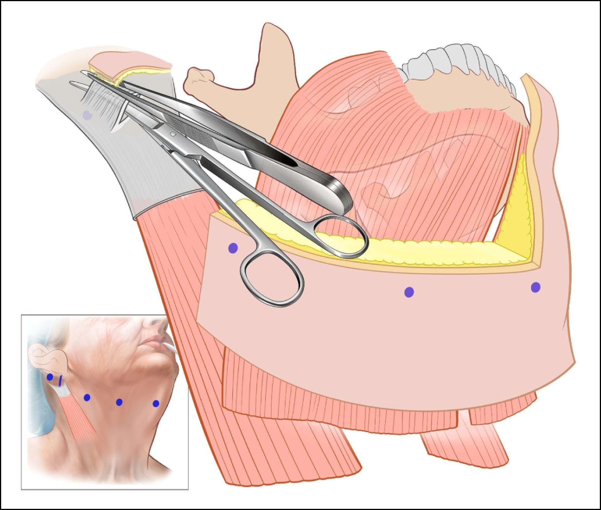
图 2. 制作乳突悬吊:乳突骨上方做斜切口;皮肤与乳突筋膜间做浅隧道;筋膜深面另做一袋穴。图 3 Ethibond 缝线走行(A/B/C)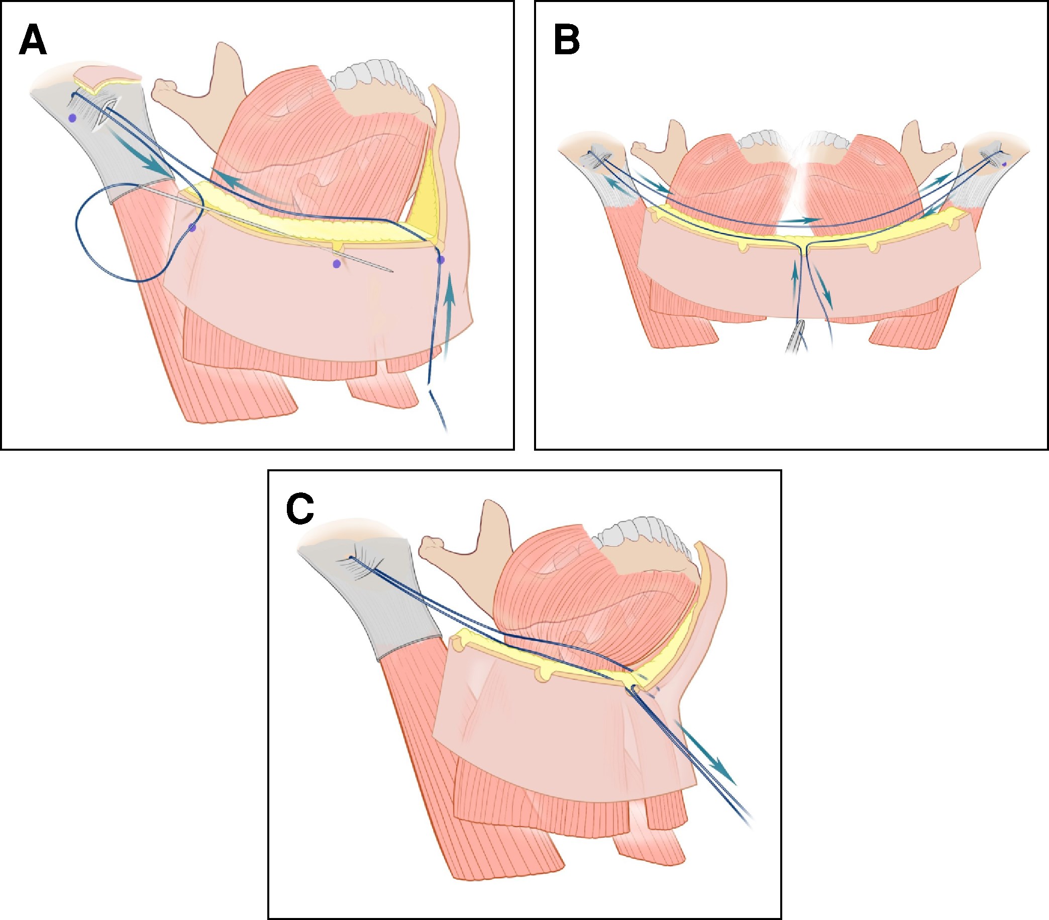
图 3. (A) 4-0 Ethibond 于颈中线部署,经真皮深面穿越至乳突筋膜;(B) 经筋膜深面隧道返回浅隧道,完全包绕乳突骨;(C) 经皮肤穿出,下行至颈中线达对侧,同法绕对侧乳突骨。
2025 年 8 月于 Miami Anatomical Research Center 用新鲜尸体验证 MIMS 的效能与抗拉强度:4-0 Ethibond 自颈中线(舌骨上)起,经真皮深面隧道环包乳突筋膜并牢固锚定形成悬吊;4-0 Monocryl 置于表皮下方。两缝线依次紧扎。
共 73 例接受 MIMS 颈部提升(表 1)。典型入选者表现为颏下脂肪过多与轻度颈皮肤冗余。术后结果汇总于表 2。
| 特征 | 数值 |
|---|---|
| 患者总数,n | 73 a |
| 年龄(岁),均值 ± SD(范围) | 48.78 ± 12.89(22–82) |
| 性别,n 女 / 男 | 67 / 6 |
| BMI(kg/m²),均值 ± SD(范围) | 25.44 ± 3.77(19–38) |
| 手术时间(分钟),均值 ± SD(范围) | 150.57 ± 74.78(57–415) |
| 肿胀液量(mL),均值 ± SD(范围) | 293.89 ± 134.17(40–560) |
| 局麻药量(mL),均值 ± SD(范围) | 33.48 ± 10.94(15–60) |
| 联合其他操作 | 62(80.5%) |
表 1 人口学与手术特征。SD = 标准差。a指所有接受 MIMS 技术的患者。最常见并发症为血清肿 3 例(均门诊抽吸,无后遗症):1 例术后第 8 天 3 mL、1 例第 1 天 2 mL、1 例第 11 天 1 mL。1 例术后第 5 天出现右下颌缘神经神经失用,2 周内自发恢复,无残留缺陷。
| 特征 | 数值 |
|---|---|
| 皮肤过度褶皱,n (%) | 13(16.9%) |
| 微针治疗 a | 6(8.2%) |
| 微针 + 射频 a | 3(4.1%) |
| 褶皱消退时间(天),均值 ± SD(范围) | 24.75 ± 8.79(16–42) |
| 总并发症,n (%) | 4(5.4%) |
| 血清肿 | 3(4.1%) |
| 神经失用 | 1(1.4%) |
| 血肿 | 0 |
| 皮肤坏死 | 0 |
表 2 术后结果。a指皮肤过度褶皱并接受微针(±射频)补充治疗的患者。13 例(16.9%)因皮肤褶皱接受补充治疗(6 例微针、3 例微针+射频),褶皱平均 24.75 天消退。1 年与 2 年随访的临床效果见图 4–6。
图 4 45 岁女性,术后 1 年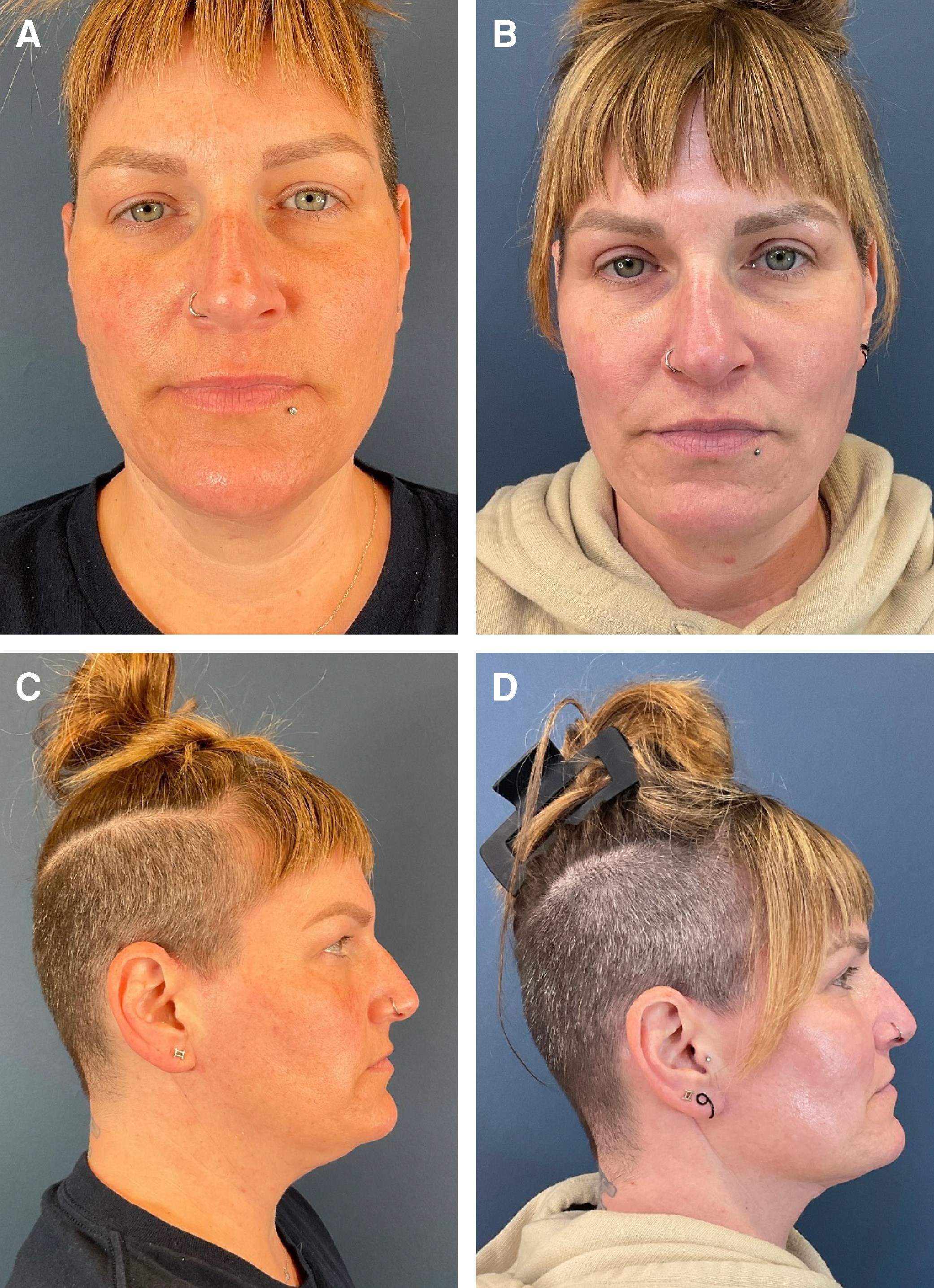
图 4. 45 岁女性,MIMS 术后 1 年。(A、C) 术前;(B、D) 1 年随访。图 5 67 岁女性,术后 1 年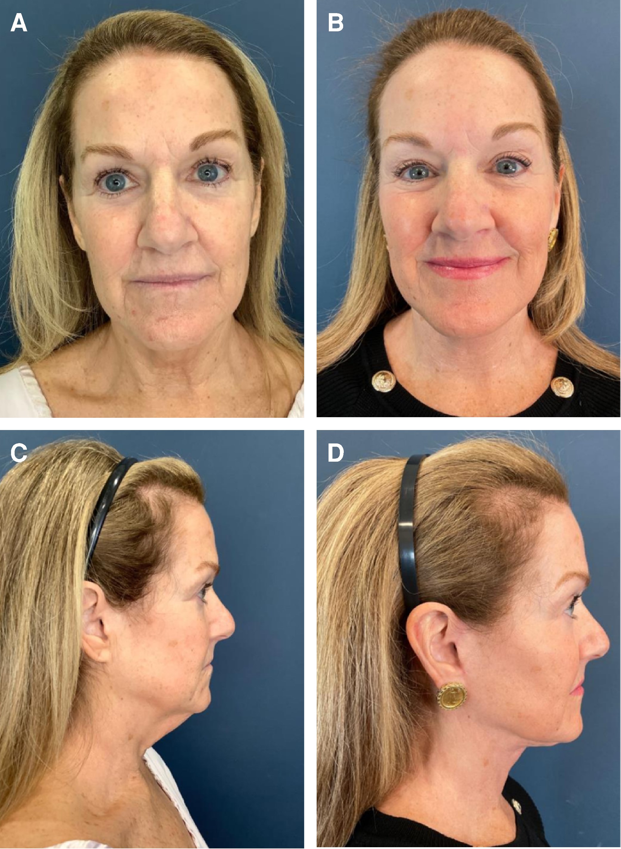
图 5. 67 岁女性,MIMS 术后 1 年。(A、C) 术前;(B、D) 1 年随访。图 6 40 岁女性,术后 2 年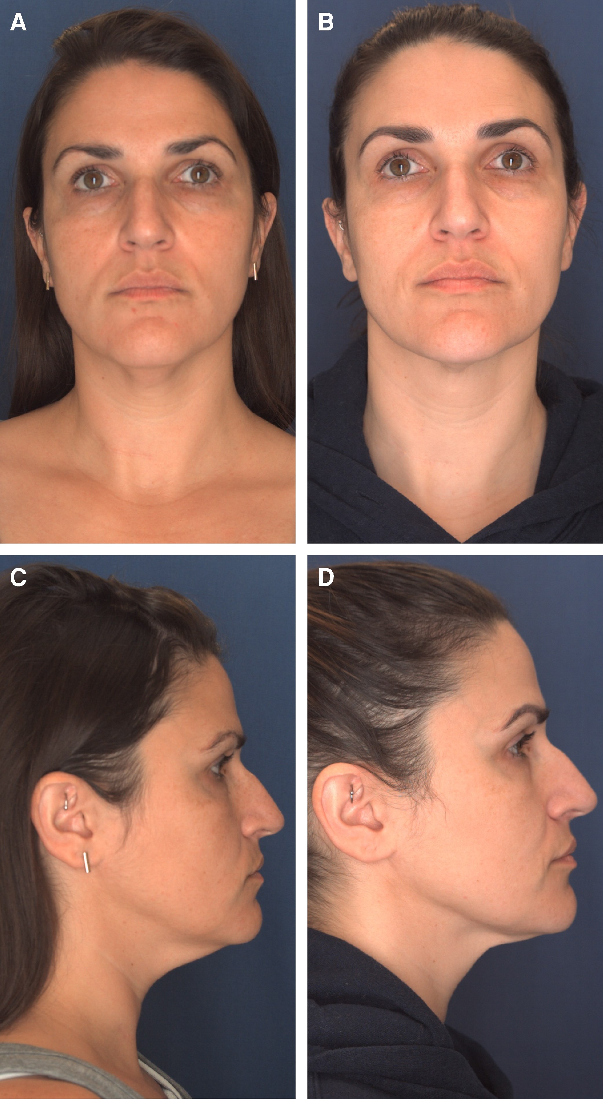
图 6. 40 岁女性,MIMS 术后 2 年。(A、C) 术前;(B、D) 2 年随访。
开放颈阔肌成形仍是中重度颈部衰老(颈颏角/下颌角变钝、动态颈阔肌条索、皮肤弹性差)的金标准。但许多表现为软组织萎缩轻、颏下饱满、颈阔肌条索、颌下腺下垂的患者,并不需要广泛剥离即可获得年轻化的颈颏角与下颌角。随着微创偏好上升,MIMS 适用于 早期至轻度 颈皮肤松弛 + 颏下脂肪过多的患者。
与既往技术的区别 :Giampapa 单条互锁缝线悬吊仅于皮下平面制造支持韧带;MIMS 的强乳突筋膜锚定提供了持久可靠的牵引点,可将提升力传导越过颌下腺与下颌下区。Mueller 的经皮颈阔肌成形用光导系统;Kaplan 等则经中央颈颏切口单切口完成、免耳后入路。MIMS 的核心贡献是 双缝线模式 :永久编织缝线(Ethibond,软组织内触感 softer、缝线处更不明显)置于深层依乳突筋膜强锚定塑形;可吸收 Monocryl 置于真皮深层,其轻微真皮反应(轻度炎症、单核细胞浸润、少量纤维蛋白合成)使松弛皮肤暂时重分布至颈阔肌/筋膜层,可能促进纤维蛋白介导的皮肤-新轮廓粘连;随缝线溶解,不规则皮褶放松,留下平滑、贴附新轮廓的软组织,改善颈颏角。
为什么先吸脂+热作用 :单纯颈颏吸脂适合皮肤弹性好、颈阔肌条索轻者,但仅针对 preplatysmal 脂肪、无显著紧肤。联合激光热作用诱导脂肪溶解、真皮温度升至 60–70°C 引发胶原收缩与新胶原生成,临床可见紧肤。充分吸脂既清除多余脂肪,又助建立皮下隧道以利缝线精准置入。
止血网 :术后维持 4 天,以绗缝样压迫组织、减少死腔、促进止血。Pellini 等在 480 例面部除皱中报告止血网使血肿率从 5.42% 降至 0;本研究亦无血肿或皮肤坏死。其对胶原合成的直接证据仍有限,是未来方向。
微针补充 :16.9% 患者术后皮肤褶皱,经微针(±射频)处理,通过真皮微损伤/热作用促进真皮重塑与新胶原,平均约 25 天消退。
局限性 :单医生、小样本回顾性系列,外推性有限;因回顾性设计不能得出因果结论。需未来多中心前瞻性研究验证。
吸脂 + 激光热作用 + 乳突悬吊三联法,有效针对早期颈皮肤松弛迹象,综合处理多余脂肪、皮肤松弛与结构支撑,为合适患者的开放颈成形术提供了微创替代方案。
英文原文全文(PMC XML,供溯源核对)The neck is often the first facial feature to show signs of aging, with blunting of the cervicomental angle (CMA), loss of a defined jawline, platysmal banding, submental fat accumulation, and redundant skin.1 These changes can significantly affect overall facial aesthetics, and their correction has long been the focus of neck rejuvenation surgery. From Ellenbogen et al to Halani et al, both described the ideal neck as a sharp CMA of 90 to 120° and a gonial angle of 120 to 140°.2,3 These are the current gold standard benchmarks for neck and jawline aesthetics.
Historically, rejuvenation techniques evolved from superficial skin tightening to deeper, structural procedures targeting the platysma, submental and subplatysmal fat, and submandibular glands.4,5 Feldman's corset platysmaplasty, introduced in 1988, combined midline and lateral platysmal plication with resection of submental fat, anterior digastric muscles, and submandibular glands to restore cervical definition.6 Shortly thereafter, Giampapa and DiBernardo developed the suture-suspension neck lift, later refined by Giampapa in 1995 with an interlocking mastoid-to-mastoid suspension suture. Other contributions, such as Connell's platysma flap anchored to the mastoid fascia, Guerrero-Santos's 2-flap platysma sling, and Baker's reinforced mastoid anchoring, further advanced the field, often in combination with facelift techniques to achieve durable results—though at the expense of greater dissection, longer recovery, and higher complication risks.4,7,8
In recent years, patient demand has shifted toward less invasive procedures with shorter recovery and in-office feasibility, leading to the development of a wide range of nonsurgical alternatives to deliver improved jawline definition.9-11 However, the unique anatomy and dynamic forces of the neck, particularly mimetic activity and fat distribution, limit the effectiveness of approaches that rely solely on skin traction or superficial support.12
There are options for minimally invasive techniques that can produce sharper jawlines. One such technique is the percutaneous platysmaplasty, introduced by Mueller, which uses a transcutaneous light-emitting diode (LED)-guided system to pass a permanent suture matrix along the platysma, anchored to neck-retaining ligaments at the mandibular border.13 This approach avoids large incisions and provides cervical support, but results may be modest and less durable, with recurrent laxity possible over time or with mandibular motion.14 Other options include energy-based skin tightening and injectable treatments, however, the longevity of these treatments are variable.15,16
The objective of this paper is to introduce the minimally invasive mastoid sling (MIMS) neck lift technique that combines liposuction and laser thermal energy with a dual-suture system to anchor a permanent suture to the dissected mastoid fascia and a second superficial absorbable suture that targets skin laxity.
This retrospective case series included all patients who underwent the minimally invasive mastoid sling (MIMS) neck lift performed by the primary surgeon between December 2020 and August 2024. IRB exemption (E-24-5420) by the Main Line Health was granted on September 16, 2025. The patients included provided informed consent, and several patients signed media release forms for their photographs to be used. A descriptive analysis of patient demographics was performed using SPSS v30.0 (IBM, Armonk, NY).
Video 1, available online at www.asjopenforum.com, summarizes all surgical steps. The primary surgeon performs the minimally invasive mastoid sling neck lift under tumescent local anesthesia at a Class A American Association for Accreditation of Ambulatory Surgery Facilities (AAAA SF) (QUAD-A)-certified surgical facility. The neck is tumesced with approximately 250 cc solution of normal saline infused with lidocaine and epinephrine. First, submental fat was removed with power-assisted liposuction in the preplatysmal plane. Followed by laser thermal heating using the SmartLipo (Cynosure, Westford, MA) performed into the subdermis and subcutaneous layers delivering thermal energy to the soft tissue. Liposuction and laser thermal heating are completed prior to suture suspension.
Anticipated puncture sites are made in these areas: (1) centrally 1 cm superior to the body of the hyoid bone, lateral to this, (2) directly over the submandibular gland, (3) midportion of the body of the mandible, and (4) inferior to the angle of the mandible (Figure 1).
[FIG] ojag065-F1 | Figure 1. | Puncture sites are created centrally 1 cm superior to the body of the hyoid bone, directly over the submandibular gland, midportion of the body of the mandible, and inferior to the angle of the mandible.
In the postauricular region, directly over the mastoid process, an oblique 1 cm incision is made, extending through the skin and the mastoid fascia, superficial to the periosteum. A superiorly oblique tunnel, measuring 1 cm by 2 cm, is created to form the mastoid fascial tunnel, with communication made through the skin at the distal aspect of the tunnel. A separate subcutaneous tunnel is created directly overlying the mastoid fascia tunnel. This is also connected through the skin.
With a 4-0 Ethibond (Ethicon, Inc., Raritan, NJ), this allows the suture to be placed deep to the mastoid fascia along the mastoid bone and then brought through the skin and reintroduced into the subcutaneous tunnel. This creates a cerclage grasping attachment to the mastoid fascia, providing a strong anchor that serves as the mainstay of this innovative technique.
The suture is percutaneously passed through subcutaneous tunnels across the anterior neck to the contralateral side and similarly passed around the mastoid fascia sling and then brought back centrally in the neck and temporarily secured with a hemostat (Figures 2, 3).
[FIG] ojag065-F2 | Figure 2. | To develop the mastoid sling, an oblique incision is made directly overlying the mastoid bone. A superficial tunnel is made between the skin and mastoid fascia. A second pocket is made deep to the mastoid fascia.
[FIG] ojag065-F3 | Figure 3. | (A) A 4-0 Ethibond suture is deployed at the neck midline. The suture is threaded deep to the dermis and traversed from the neck midline to the mastoid fascia. (B) The suture is passed through the tunnel deep to the mastoid fascia and then back through the superficial tunnel to fully envelope the mastoid bone. (C) The suture is delivered through the skin, passed inferiorly to the neck midline to reach the contralateral side, and similarly passed around the mastoid bone.
A second suture, which is a 4-0 Monocryl (Ethicon, Inc., Raritan, NJ), is introduced through a separate puncture site directly over the submandibular gland on the left side. This suture is passed through a similar path as the 4-0 Ethibond; however, the 4-0 Monocryl is placed into the deep dermis, whereas the 4-0 Ethibond is placed deep to the dermis. It is similarly passed around the mastoid sling on each side and brought out through a percutaneous hole directly over the left submandibular gland.
The operating room table is adjusted to flex the patient's neck to a 30° angle. The 4-0 Ethibond is tied with sufficient force to hold the suture in place while manipulating the tissue to ensure no puckering of the skin is seen. Then, the 4-0 Monocryl suture is tied tightly to create significant skin puckering.
Lastly, a subsequent hemostatic net suture is placed from the posterior region across the jawline and the hyoid bone to the contralateral side. The puncture sutures are covered by the hemostatic net suture. No drains are placed. Sutures are left in place for 3 days. Patients remain wrapped for 1 week.
In August 2025, a fresh cadaver dissection was conducted at the anatomy lab of the Miami Anatomical Research Center (MARC) to evaluate the efficacy and tensile strength of the MIMS neck lift technique. A 4-0 Ethibond permanent suture was started at the neck midline, superior to the hyoid bone. The suture was threaded deep under the dermis to create a tunnel around the mastoid fascia in the retroauricular region. The suture was passed under the mastoid tunnel, through the skin and back, securely anchoring it around the mastoid fascia to create a sling. Then, a 4-0 Monocryl suture was placed superficially just below the epidermis. The Ethibond, followed by the Monocryl suture, was tied tightly (Video 2, available online at https://doi.org/10.1093/asjof/ojag065).
A total of 73 patients underwent the MIMS neck lift. The mean age was 48.7 years (range: 22-82), and the mean BMI was 25.44 kg/m2 (range: 19-38). Patients selected for this procedure typically presented with excess submental fat and modest excess neck skin (Table 1).
[TABLE] ojag065-T1
Table 2 summarizes all postoperative outcomes. In this cohort, the most common complication was seroma which developed in 3 patients, all of whom were managed with in-office aspiration, without any sequelae. Seroma volumes ranged from 1 to 3 cc and occurred within days of surgery: Patient 1 developed a 3 mL seroma on postoperative Day 8; Patient 2 developed a 2 mL seroma on postoperative Day 1; and Patient 3 developed a 1 mL seroma on postoperative Day 11. One patient experienced neuropraxia of the right marginal mandibular nerve that presented on postoperative Day 5 and resolved spontaneously within 2 weeks without residual deficit.
[TABLE] ojag065-T2
A total of 13 patients (16.9%) underwent additional treatments for excessive skin puckering, of whom 6 (8.2%) were treated with microneedling alone and 3 (4.1%) with microneedling plus radiofrequency. Overall, the mean time to resolution of skin puckering was 24.75 days (range: 16-42). Clinical outcomes at 1- and 2-year follow-ups are demonstrated in Figures 4 to 6. The dynamic neck video is available in Video 3, accessible online at www.asjopenforum.com.
[FIG] ojag065-F4 | Figure 4. | A 45-year-old female patient 1 year after the minimally invasive mastoid sling (MIMS) technique. (A and C) Preoperative. (B and D) One-year follow-up.
[FIG] ojag065-F5 | Figure 5. | A 67-year-old female patient 1 year after the minimally invasive mastoid sling (MIMS) technique. (A and C) Preoperative. (B and D) One-year follow-up.
[FIG] ojag065-F6 | Figure 6. | A 40-year-old female patient 1 year after the minimally invasive mastoid sling (MIMS) technique. (A and C) Preoperative. (B and D) Two-year follow-up.
With the growing interest in minimally invasive procedures that provide durable and effective aesthetic results, surgeons have sought to address the cosmetically vexing problem of the aging, sagging neck through less invasive approaches. Therefore, this paper describes the MIMS neck lift, a modified dual-suture suspension technique that combines suture suspension with liposuction and thermal injury designed to restore cervical definition and youthfulness.
Open platysmaplasty remains the gold standard treatment for cervical rejuvenation in patients with advanced neck aging, including blunting of the cervicomental and gonial angles, dynamic platysmal banding, and poor skin elasticity.1,3,17 However, many patients who present with less severe soft tissue atrophy, submental fullness, platysmal muscle banding, and submandibular gland ptosis do not require the extensive dissection of a traditional open neck lift technique to achieve a youthful and defined cervicomental and gonial angles.10,12 With the growing preference for minimally invasive treatments, patients desire techniques that offer decreased downtime while achieving highly desirable outcomes.18,19 This shift in the cosmetic market for minimally invasive rejuvenation has motivated surgeons to develop minimally invasive or nonsurgical techniques that provide effective and durable results.20 Patient selection remains critical when deciding between a minimally invasive and an open neck lift approach based on the degree of laxity, jowling, and banding, guiding the surgeon in determining when minimally invasive approaches may be sufficient to achieve aesthetic outcomes.21,22 The MIMS neck lift is most suitable for patients with early-to-minimal neck skin laxity and excess submental fat, who do not require extensive dissection of the neck to achieve the desired aesthetic outcome.
Giampapa introduced the interlocking suture suspension for minimally invasive neck lifting, which deploys a single, long, permanent interlocking suture placed in the subcutaneous plane to create a supportive ligament under the mandible.1,10 The difference between Giampapa's technique and the MIMS is the strong mastoid fascia anchor, which provides a durable and reliable traction point to transmit elevating forces over the submandibular glands and underneath the jaw. The secondary absorbable suture as well as the hemostatic net suture adds to the neck contouring and sculpting with tissue manipulation that maximizes lifted and repositioned tissue to adhere to the new contour through temporary suture adhesion. Moreover, Mueller's percutaneous platysmaplasty uses a light-guided system that introduces a continuous suture matrix along the platysma and anchors it to cervical retaining ligaments, creating a trampoline-like support framework.13 More recently, Kaplan et al described a single-incision approach performed entirely through a central cervicomental incision, thereby eliminating the need for postauricular access while achieving a minimally invasive neck lift.11 These innovations reflect a clear trend toward minimally invasive procedures that minimize scarring and reduce downtime and complication risk while still providing meaningful rejuvenation of the cervical contour.
The primary contribution of the MIMS technique is the dual-suture modality of the permanent braided suture placed in a deep layer to contour the neck and jawline based on a strong anchor support of the mastoid fascia and centrally tied knot. Braided sutures have inherently soft tissue characteristics, which make them less palpable and less noticeable at the suture line. The second is the deployment of a 4-0 Monocryl suture in the deep dermis. Monocryl, also known as poliglecaprone 25 suture, causes a mild dermal reaction during would healing, which has been characterized as minimal inflammation with infiltration of mononuclear cells and mild fibrin synthesis.23,24 Employing this absorbable second suture temporarily redistributes the loose skin to the platysmal and fascial layers, which may promote fibrin-mediated adhesion of the skin to the new neck contour. As these sutures dissolve, the undulating and irregular skin folds relax, leaving a soft, smooth contour that is adherent to the new neck contour, therefore achieving an improved CMA.
Building upon these principles, the MIMS neck lift offers an alternative approach by also combining soft-tissue debulking with power-assisted liposuction and thermal energy skin tightening with the permanent suspension. The technique employs both a permanent and an absorbable suture through subdermal tunnels. A critical initial step is thorough power-assisted liposuction of the preplatysmal plane, which not only evacuates excess adipose tissue but also assists in creating subdermal channels that facilitate more precise suture placement.
Isolated cervicomental liposuction is a well-established option for patients with favorable skin elasticity and minimal platysmal banding; however, when used alone, it primarily targets preplatysmal fat without significant skin-tightening benefits.25 Combining liposuction with thermal energy induces lipolysis to liquefy adipocytes, targeting preplatysmal fat removal while also delivering a skin-tightening effect through neocollagenesis and adipolysis.26 This combination treatment promotes collagen production and simultaneously cauterizes small blood vessels, which decreases bleeding and swelling. Badin et al and Ichikawa et al showed in their histological studies that laser lipolysis can rupture fibrous septa, freeing retracted skin and remodel dermal collagen, resulting in clinical skin tightening. Studies have shown that inducing dermal temperature between 60 and 70°C is associated with collagen contraction and neocollagenesis.27,28 By integrating power-assisted liposuction and laser thermal energy, the MIMS technique reduces preplatysmal fat and creates a subdermal plane through which thermal energy can be delivered directly to the dermis, inducing neocollagenesis that results in clinically visible skin tightening. The importance of comprehensive power-assisted liposuction to evacuate the fat from the preplatysmal plane as well as allowing for heat energy devices to aid in the shrinking of loose dermal elements is an essential step for preparing the neck for suture deployment.
In this cohort, the hemostatic net was maintained for 4 days postoperatively to solidify these new skin-to-platysma connections through a quilting pattern. By evenly compressing the tissues captured within the net, these sutures also help minimize dead space and promote hemostasis. Pellini et al reported, in a retrospective cohort study of 480 cervicofacial rhytidectomy patients, a reduction in hematoma formation from 5.42% to 0% after implementing a hemostatic net suture.29 Moreover, previous studies have demonstrated that the hemostatic net does not compromise skin blood supply and perfusion.30,31 This finding is corroborated by our study in which no hematomas or skin necrosis occurred. Beyond hemostasis, suture techniques have been found to influence collagen deposition and wound healing.32 Ismail et al sought to demonstrate if a hemostatic net suture induced collagen remodeling or synthesis using a rat model; however, at 20 days, the findings did not detect any significant differences in neocollagenesis, although the authors acknowledged that the small sample size may have limited their ability to detect subtle effects.33 Although direct evidence regarding the hemostatic net's impact on collagen synthesis and extracellular matrix remodeling remains limited, this represents an important area for future investigation, particularly in the context of minimally invasive neck rejuvenation techniques.
Microneedling with and without radiofrequency (RF) has been found to be effective in reducing skin folds and puckering. In this study, patients who had skin puckering underwent additional microneedling with or without RF to target these irregularities. A total of 13 patients in this cohort underwent additional treatment for skin puckering. Microneedling alone stimulates dermal remodeling and neocollagenesis by introducing micropunctures into the dermis to improve skin texture and irregularities.34,35 Moreover, microneedling with RF combines mechanical injury with thermal energy to augment neocollagenesis, elastogenesis, and dermal extracellular matrix remodeling, providing additional skin tightening.36,37 Hwang et al found that microneedling with RF increased the number of nonsenescent fibroblasts in the dermis, thus increasing collagen and elastin synthesis.38 These findings suggest that microneedling with or without RF can be used as a postprocedure adjunct to address any residual skin irregularities and optimize minimally invasive neck lifting results.
This study does have limitations worth discussing. It is a single-surgeon series with a small sample size, which limits the generalizability of our findings. Due to its retrospective design, causative conclusions cannot be drawn from the results of this study. Future multicenter, prospective studies are needed to validate the efficacy and reproducibility of the MIMS technique.
By combining liposuction, laser thermal energy, and the mastoid sling, this trifecta approach effectively targets early signs of cervical skin laxity. Together, these modalities address excess fat, skin laxity, and structural support, offering a minimally invasive alternative to open platysmaplasty for appropriately selected patients.
声明:中文精读 · 仅供学术参考。内容来自公开文献检索,不代表本人观点,不构成诊疗建议。 医疗美容需在正规医疗机构由执业医师实施。
基于相同主题推荐 · 继续深挖
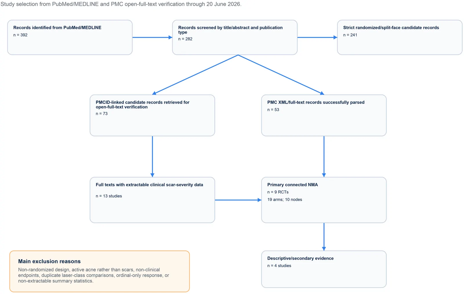
PROSPERO注册的系统综述,纳入9项随机/分脸随机试验、19个治疗臂,构成稀疏连接网络并采用频率学随机效应网络荟萃分析。
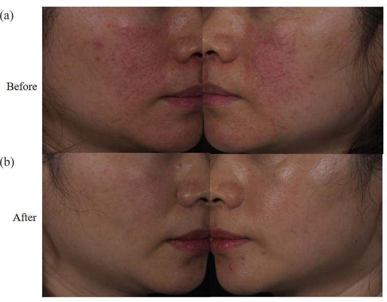
回顾性纳入63例红斑毛细血管扩张型玫瑰痤疮(ETR)中国患者,均采用555–950 nm与530–750 nm双滤光片窄谱IPL治疗。
真实世界回顾性队列纳入50例临床诊断玫瑰痤疮成人,80%为女性、中位年龄33岁,多数(76%)为Fitzpatrick III型肤色。