维生素CE阿魏酸精华分脸双盲临床试验
2026年9月11日28 min read
Clinical, Cosmetic and Investigational Dermatology
结论与实践要点
循证层面:本研究为随机双盲分脸自身对照试验(倾向 II 级证据),方法学质量较高;在 12 周内,微针联合维生素 C/E/阿魏酸精华在光老化多项指标上均显著优于单用微针(p<0.001),证据支持该联合方案。
局限与注意:样本量小(31 例)、随访仅 12 周、为单中心韩国 III–IV 型肤质人群,外推至其他肤色 / 族群需谨慎;分脸设计主要比较局部效应,未评估全身性或长期安全性;虽多为轻度一过性反应,仍有 2 例对微针金属组件接触性皮炎。
落地提示:可将“微针 + 维生素 C/E/阿魏酸抗氧化精华”作为面部光老化(细纹、色素、弹性下降)的联合抗衰方案,术后即刻使用有助于活性成分透皮;操作中须询问金属过敏史并做斑贴 / 过敏评估,以降低接触性皮炎风险。
PurposeMicroneedling achieves skin rejuvenation through controlled microinjury, while topical antioxidants may enhance its outcomes by neutralizing reactive oxygen species and promoting tissue repair. This study evaluated whether a topical antioxidant formulation containing vitamins C, E, and ferric acid enhances facial rejuvenation outcomes when combined with microneedling.
Patients and MethodsA 12-week, prospective, randomized, double-blinded, split-face trial was conducted in 31 adults with signs of facial aging. After subjects receiving microneedling treatments on both sides of the face, one side was randomly assigned to receive application of antioxidant serum while the other receiving placebo serum. Microneedling was performed at baseline and Week 4. Clinical efficacy throughout 12 weeks at four-week interval was assessed using the modified Griffith’s scale, hemi-MASI, Global Aesthetic Improvement Scale (GAIS), and non-invasive skin biophysical measurements.
ResultsAt Week 12, the Microneedle + Antioxidant side demonstrated significantly greater improvements in modified Griffith’s scale scores (23.9% vs 6.8%, p<0.001), hemi-MASI scores (31.2% vs 5.1%, p<0.001), and skin elasticity (39.0% vs 6.8%, p<0.001) compared to Microneedle + Placebo. GAIS assessment revealed 89.3% of antioxidant-treated sides achieved marked/near-total improvement versus 7.1% of placebo-treated sides. The antioxidant-treated sides also demonstrated superior improvements in skin elasticity (39.0% vs 6.8%, p<0.001) and melanin index (21.3% vs 3.4%, p<0.001). Only mild, transient side effects were reported.
ConclusionCombining topical antioxidant serum with microneedling provides superior outcomes for facial rejuvenation than microneedling alone. This combination protocol significantly improves clinical signs of photoaging, including skin elasticity, rhytides, and dyspigmentation, suggesting an optimized treatment protocol with minimal recovery time.
Keywords: microneedling、antioxidants、photoaging、vitamin C、vitamin E、ferulic acid
Microneedling is a minimally invasive procedure that creates controlled micro-injuries in the skin, leading to noticeable improvements in signs of aging. Studies have shown that it works by stimulating organized collagen production, promoting elastin synthesis, and thickening the epidermis. 1 With its accessibility and minimal downtime, microneedling has a variety of additional applications, including the treatment of acne scars, melasma, and enhancing the transdermal delivery of topical agents. 2 Post-procedure topical treatments have been shown to have a synergistic effect with microneedling. 3–6 For instance, several studies have demonstrated that microneedling enhances the absorption of vitamin C, tranexamic acid, platelet-rich plasma, and other substances, providing added benefits for improving melasma. 7–10
When it comes to skin aging, one of the most significant environmental factors that accelerate the process is the presence of reactive oxygen species (ROS). Antioxidants are key compounds in skincare formulations that help target and neutralize ROS. 11 Topical antioxidants, such as vitamin C, vitamin E, ferulic acid, and resveratrol, are frequently recommended after non-invasive energy-based device (NI-EBD) treatments, particularly those that enhance the penetration and efficacy of topical agents. 12 Ferulic acid is not only a powerful antioxidant, forming stable phenoxyl radicals when interacting with free radicals, but also stabilizes Vitamin C and E when combined to provide synergic effects of antioxidant. 4 A single-blinded, prospective, randomized split-face trial of 18 patients found that combining 15% vitamin C, 1% vitamin E, and 0.5% ferulic acid serum with Q-switched 1064-nm Nd:YAG laser treatment resulted in a safe and significantly greater reduction in the melanin index compared to laser treatment alone. 13 In another evaluator-blinded, split-face, randomized study of 49 patients, application of two different antioxidant formulations (15% vitamin C, 1% vitamin E, and 0.5% ferulic acid; 1% resveratrol, 0.5% baicalin, and 1% vitamin E) after using a home fractional non-ablative 1440-nm diode laser device, led to significant improvements in skin appearance, texture, tone uniformity, and overall facial wrinkles. 14 Antioxidants also help minimize complications by addressing the ROS produced during the treatment process itself. A randomized, investigator-blinded, split-face, controlled trial involving 51 patients demonstrated the efficacy of antioxidant serum (15% vitamin C, 1% vitamin E, and 0.5% ferulic acid) combined with resveratrol BE serum in reducing erythema and hyperpigmentation, promoting wound healing, and maintaining skin hydration after ablative CO 2 laser treatment. 3
Given that microneedling enhances the transcutaneous delivery of topical agents, combining topical antioxidants with microneedling can significantly improve the signs of skin aging. A split-face comparative study involving 16 women demonstrated that eight weekly treatments of a 15% ferulic acid peel followed by microneedling were more effective than the ferulic acid peel alone in reducing photoaging, particularly improving skin elasticity. 4 More recently, a split-face study of 15 participants showed that combining microneedling with antioxidant serum with 15% vitamin C, 1% vitamin E, and 0.5% ferulic acid produced a synergistic effect, significantly improving pigmentary disorders. 5
Despite the growing body of evidence supporting this approach, there remains a lack of clinical studies that measure biophysical parameters of skin rejuvenation evaluating antioxidant use post-microneedling. This study aims to evaluate the clinical effectiveness of topical antioxidant serum containing vitamins C, E, and ferulic acid in addressing signs of photoaging, including skin elasticity, rhytides, erythema, and pigmentation, when applied topically after microneedling treatment. We hypothesized that post-microneedling application of a vitamin C, E, and ferulic acid serum would produce superior clinical and biophysical outcomes compared with placebo.
A 12-week, prospective, randomized, double-blinded, split-face trial was conducted to investigate the effectiveness of microneedling combined with a post-procedural antioxidant serum (containing vitamin C, vitamin E, and ferulic acid) for facial skin rejuvenation. A total of 31 Korean adults (Skin Type III and IV) aged 31–68 with clinically evident facial aging signs (including rhytides, dyspigmentation, and decreased skin elasticity) were recruited. Exclusion criteria encompassed a history of keloid, pregnancy, significant psychiatric illness, and any facial interventions (including chemical peeling, energy-based treatments, or cosmetic surgery) within the preceding six months.
Sample size was determined based on a priori power analysis. Based on previous studies with similar designs, expected GAIS scores were 3.5 ± 0.9 for the treatment group and 2.9 ± 0.7 for the control group. With α = 0.05, power = 0.80, and a two-tailed test, the required sample size was estimated at approximately 30 subjects per group. 15
Randomization was accomplished using computer-generated allocation sequences (Microsoft Excel 2019; Microsoft, Redmond, WA, USA). Individual randomization codes remained sealed in opaque envelopes until the completion of data analysis. The two sides of face were systematically randomized to receive an application of either the antioxidant serum or placebo following microneedling treatment. The study was approved by the Institutional Review Board of Yongin Severance Hospital (IRB No.: 9–2024-0051), and all participants provided written informed consent prior to enrollment. This study was conducted in accordance with the principles of the Declaration of Helsinki.
Treatment was administered using a microneedle device (GENIUS, Lutronic Co., Korea) fitted with a sterile disposable tip containing 49 (7×7) microneedles. All procedures were conducted by a single board-certified dermatologist (JHK). Prior to each session, topical anesthesia consisting of 2.5% lidocaine hydrochloride and 2.5% prilocaine cream mixture was applied under occlusion for 30 minutes. The microneedling procedure was performed at 1.2–1.5 mm penetration depths using a single-pass technique with minimal overlap (<20%) on both sides od face. Participants underwent two treatment sessions separated by a four-week interval.
Beginning on the day of the initial microneedling session, participants were directed to apply approximately 1 mL (3–4 drops) of either the active antioxidant serum (CE Ferulic ® , SkinCeuticals Inc., containing 15% vitamin C, 1% vitamin E, and 0.5% ferulic acid) or the visually identical placebo vehicle (devoid of any active antioxidant components) to their respective randomized facial sides immediately post microneedling followed by once daily at a specified time.
To minimize cross-contamination, subjects were instructed to apply products carefully to each respective side without crossing the midline. Additionally, oral antioxidant supplements and nutritional supplements were prohibited throughout the study period to eliminate potential systemic confounding effects. Also, all subjects were instructed to use a moisturizer and physical sunscreen on both sides of face throughout the study. Adherence to the treatment protocol was verified through patient-completed diaries and regular monitoring of product consumption. The investigation spanned 12 weeks total, with four clinical evaluations: baseline (Week 0, coinciding with first microneedling treatment), Week 4 (second microneedling treatment), Week 8, and Week 12.
The primary efficacy parameters were independently evaluated during each follow-up visit by two experienced board-certified dermatologists (JMK and SWH) who remained blinded to treatment allocation. Assessments utilized standardized digital photographs presented in random, non-sequential order. Treatment outcomes were measured using three validated clinical scales. First, the modified Griffith’s scale was employed to evaluate overall photodamage, coarse and fine lines and rhytides, evenness of skin tone, and tactile roughness. 16 Second, hemi-MASI (Melasma Area and Severity Index) scores were calculated to quantify melasma severity on each side of face. 17 Lastly, the Global Aesthetic Improvement Scale (GAIS) was applied to assess overall clinical improvement (Grade 1 = worsening; Grade 2 = minimal improvement/no change [0–25%]; Grade 3 = moderate improvement [26–50%]; Grade 4 = marked improvement [51–75%]; Grade 5 = near-complete improvement [>75%]). Each evaluator assessed parameters independently, with mean values utilized for subsequent analysis.
Objective skin biophysical parameters were obtained using non-invasive instrumentation to complement physician evaluations. The Mexameter® MX18 (Courage Khazaka, Köln, Germany) quantified cutaneous erythema and pigmentation via erythema and melanin indices. Skin elasticity was measured using the Elastometer® EM 25 (Courage Khazaka), while skin hydration was assessed with the Corneometer® CM 825 (Courage Khazaka). All measurements were performed at baseline (Week 0) and during subsequent visits at Week 4, 8, and 12. All biophysical measurements were conducted under standardized environmental conditions at a controlled temperature of 20–24°C and relative humidity of 45–55%. Participants acclimated to the climate-controlled room for at least 20 minutes before each examination. To minimize confounding effects on biophysical measurements, all subjects were instructed to refrain from applying any topical products, including the study serum, placebo, and moisturizers, for at least 8 hours prior to each assessment. Measurements were taken in triplicate from predetermined, consistent locations on both cheeks at each timepoint, with mean values used for statistical analysis.
Safety evaluations were conducted at each study visit through comprehensive clinical examination and visual assessment. Any treatment-related adverse reactions (including erythema, edema, pigmentary alterations, punctate bleeding, ecchymosis, and scar formation) were systematically recorded based on subjects’ reports and clinicians’ observations.
Results are presented as either frequencies (percentages) or means ± standard deviations, as appropriate for the variable type. The principal statistical evaluation employed Linear Mixed Models (LMM), which effectively account for inter-subject variability and fluctuations in repeated measurements. LMM methodology inherently manages missing observations without necessitating explicit data imputation techniques. In this study, three participants dropped out after Week 4, creating missing data in subsequent follow-up; the LMM framework appropriately handled these missing values. As a complementary approach, the Last Observation Carried Forward (LOCF) method was applied to impute absent data points and performed Repeated Measures ANOVA (RM-ANOVA) on the completed dataset.
Post-hoc analyses utilizing Student’s t-tests with Bonferroni adjustments were performed to compare the parameters at each discrete time point between treatment groups. When analyzing ordered categorical variables arranged in 2×N tables, linear-by-linear association tests were implemented. Statistical significance was established at p<0.05. All data analyses were executed using Python version 3.9.7 and R version 4.1.3.
A total of 31 participants (28 females [90.3%], mean age 49.5 ± 10.4 years, range 31–64 years) were recruited between May and August 2024. The modified Griffith’s scale at baseline showed a mean score of 26.7 ± 4.72 points, with scores ranging from 18 to 35 points. A total of 28 (90.3%) individuals completed the 12-week study, while three discontinued after the Week 4 evaluation (two developed allergic contact dermatitis from microneedling device, one due to photoallergic dermatitis due to sunscreen). Analysis of skin aging parameters and safety outcomes utilized the full analysis set, which included all enrolled subjects, including those who withdrew from the study. Conversely, GAIS evaluations conducted after Week 8 were analyzed using the per-protocol approach, including only subjects who completed the study.
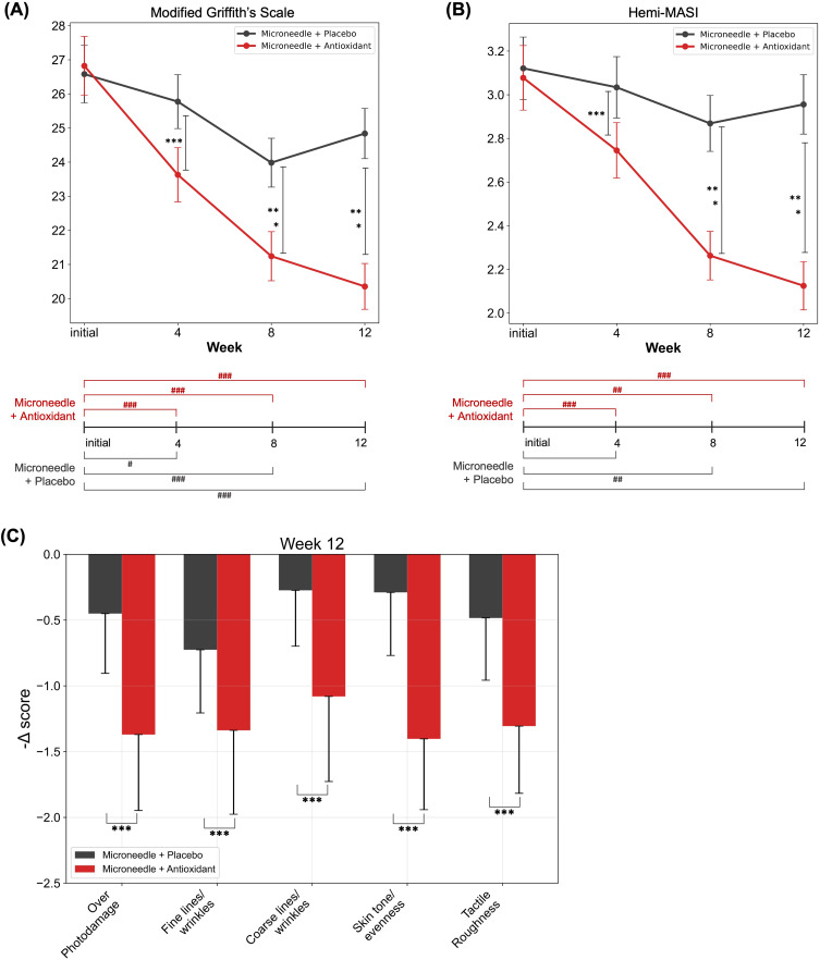
Table 1 and Figure 1 present the measurements of skin aging parameters evaluated throughout the study period. Linear mixed model (LMM) analysis demonstrated significant group-by-time interactions for both the modified Griffith’s scale (p<0.001) and hemi-MASI scores (p<0.001). In the Microneedle + Antioxidant group, the modified Griffith’s scale scores decreased from 26.8 ± 4.82 at baseline to 20.4 ± 3.74 at Week 12, representing a 23.9% improvement ( Figure 1A , p<0.001). Similarly, hemi-MASI scores in this group decreased from 3.08 ± 0.82 to 2.12 ± 0.62, showing a 31.2% reduction ( Figure 1B , p<0.001).
Table 1 Comparison of Parameters of Skin Aging During the Study Period
| Parameters | Baseline | Week 4 | Week 8 | Week 12 | P -Value |
|---|---|---|---|---|---|
| LMM | RM-ANOVA | ||||
| Modified Griffith’s scale | |||||
| Microneedle + Antioxidant | 26.8 ± 4.82 (23.3–30.8) | 23.6 ± 4.43 (19.8–27.0) | 21.2 ± 4.01 (18.0–24.0) | 20.4 ± 3.74 (17.5–23.8) | <0.001 |
| Microneedle + Placebo | 26.6 ± 4.70 (22.8–30.0) | 25.8 ± 4.40 (21.8–29.0) | 24.0 ± 3.99 (20.8–26.8) | 24.8 ± 4.11 (21.5–28.3) | |
| P -value ‡ | 0.35 | <0.001 | <0.001 | <0.001 | |
| Hemi-MASI | |||||
| Microneedle + Antioxidant | 3.08 ± 0.82 (2.53–3.60) | 2.74 ± 0.71 (2.20–3.27) | 2.26 ± 0.62 (2.00–2.58) | 2.12 ± 0.62 (1.85–2.50) | <0.001 |
| Microneedle + Placebo | 3.12 ± 0.80 (2.60–3.55) | 3.03 ± 0.78 (2.50–3.50) | 2.87 ± 0.71 (2.45–3.24) | 2.96 ± 0.76 (2.47–3.33) | |
| P -value ‡ | 0.55 | <0.001 | <0.001 | <0.001 | |
| Skin elasticity (%) | |||||
| Microneedle + Antioxidant | 57.2 ± 9.25 (50.8–63.0) | 71.8 ± 9.00 (66.8–76.2) | 75.9 ± 6.99 (74.3–78.5) | 79.5 ± 6.14 (75.8–82.2) | <0.001 |
| Microneedle + Placebo | 56.2 ± 9.54 (48.7–62.2) | 65.3 ± 9.15 (59.7–71.7) | 66.5 ± 8.05 (61.2–71.2) | 66.0 ± 8.22 (61.0–70.7) | |
| P -value ‡ | 0.42 | <0.001 | <0.001 | <0.001 | |
| Skin hydration (AU) | |||||
| Microneedle + Antioxidant | 73.6 ± 7.77 (69.8–79.2) | 79.0 ± 6.49 (74.3–84.8) | 79.3 ± 8.16 (73.8–84.7) | 80.0 ± 9.29 (73.7–86.2) | 0.75 |
| Microneedle + Placebo | 74.0 ± 8.57 (68.5–81.5) | 77.6 ± 8.41 (74.2–85.7) | 79.7 ± 9.00 (73.9–87.5) | 81.0 ± 9.55 (75.4–88.1) | |
| P -value ‡ | 0.74 | 0.3 | 0.84 | 0.77 | |
| Melanin index (AU) | |||||
| Microneedle + Antioxidant | 99.3 ± 51.3 (68.5–117.0) | 85.3 ± 46.6 (59.2–91.2) | 83.3 ± 46.5 (57.0–92.7) | 78.1 ± 42.8 (50.0–93.3) | <0.001 |
| Microneedle + Placebo | 95.5 ± 49.9 (60.8–115.0) | 86.5 ± 45.1 (53.7–99.8) | 87.1 ± 43.7 (59.0–105.8) | 92.3 ± 45.5 (61.3–107.2) | |
| P -value ‡ | 0.22 | 0.70 | 0.46 | <0.001 | |
| Erythema index (AU) | |||||
| Microneedle + Antioxidant | 223 ± 78.6 (166.2–257.5) | 179 ± 68.3 (129.5–203.7) | 183 ± 79.0 (132.0–204.7) | 147 ± 65.9 (109.0–156.0) | 0.13 |
| Microneedle + Placebo | 221 ± 83.7 (170.7–238.7) | 184 ± 71.2 (142.8–212.3) | 176 ± 72.9 (134.8–193.0) | 162 ± 66.7 (128.5–175.5) | |
| P -value ‡ | 0.84 | 0.50 | 0.38 | 0.02 |
Post-hoc analysis revealed that the Microneedle + Antioxidant group demonstrated significantly greater improvements compared to the Microneedle + Placebo group as early as Week 4 and the improvement continued until the end of the study period for both modified Griffith’s scale (p<0.001 for Week 4, 8, and 12) and hemi-MASI scores (p<0.001 for Week 4, 8, and 12). Notably, when evaluating the individual components of the modified Griffith’s scale at Week 12 ( Figure 1C and Supplementary Figure S1), the Microneedle + Antioxidant group showed significantly greater improvements across nearly all parameters assessed, including photodamage, fine/coarse rhytides, and evenness of skin tone (p<0.05 to p<0.001 for all comparisons). Inter-rater reliability between the two independent physicians was assessed using the Intraclass Correlation Coefficient (ICC). The ICC values for the modified Griffith’s scale and hemi-MASI assessments were sufficiently high (>0.95), indicating excellent agreement between evaluators (Supplementary Table S1).
The GAIS evaluation independently conducted by two blinded dermatologists revealed significantly superior outcomes in the Microneedle + Antioxidant group compared to the Microneedle + Placebo group at both assessment timepoints ( Figure 2 , p<0.001 for Week 8 and Week 12). At Week 8, 71.4% of facial sides treated with Microneedle + Antioxidant demonstrated marked/near-total improvement (Grade 4 and 5), in comparison with only 14.3% for Microneedle + Placebo-treated sides. This disparity became even more evident at the final 12-week assessment, with 89.3% of Microneedle + Antioxidant-treated sides exhibiting marked or near-total improvement, while 7.1% of Microneedle + Placebo-treated sides reached similar outcomes. Importantly, no participants reported minimal improvement (Grade 2) for Microneedle + Antioxidant-treated sides at either assessment time point, in contrast to the Microneedle + Placebo group, where 35.7% of treated sides showed only minimal improvement at Week 12.
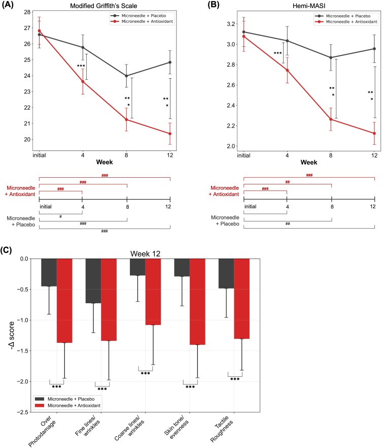
Figure 1 Time Course of Improvement in Facial Aging Parameters over 12 Weeks of Treatment: (A) Modified Griffith’s Scale Scores, (B) Hemi-MASI (Melasma Area and Severity Index) Scores, (C) Sub-analysis of the Modified Griffith’s Scale at Week 12. Data are presented as mean ± standard deviation (interquartile range; 25th–75th percentile). In (A) and (B), upper graphs show the temporal changes in clinical parameters, while lower bars indicate statistical difference of corresponding scores between baseline and relevant time points. *p<0.05, **p<0.01, ***p<0.001 for placebo-vs-antioxidant group comparisons at each timepoint with Bonferroni correction, and #p<0.05, ##p<0.01, ###p<0.001 for within-group comparisons versus baseline values.12 周上面部老化参数(改良 Griffith、hemi-MASI)随时间的改善曲线,抗氧化侧改善幅度更大。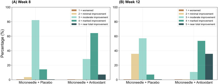
Figure 2 Distribution of Global Aesthetic Improvement Scale (GAIS) Scores at (A) Week 8 and (B) Week 12. Data are presented as percentage of participants in each improvement category for both Microneedle + Placebo (left of (A and B) and Microneedle + Antioxidant (right of (A and B) treated sides of face.第 8 与 12 周 GAIS 评分分布,抗氧化侧明显 / 近完全改善占比更高。
Table 1 and Figure 3 illustrate the changes in skin biophysical parameters throughout the study period. LMM analysis revealed significant group-by-time interactions for skin elasticity (p<0.001) and melanin index (p<0.001). In the Microneedle + Antioxidant group, skin elasticity increased substantially from 57.2 ± 9.25% at baseline to 79.5 ± 6.14% at Week 12, representing a 39.0% improvement ( Figure 3A , p<0.001). Post-hoc analysis demonstrated that the Microneedle + Antioxidant group exhibited significantly greater enhancement in skin elasticity compared to the Microneedle + Placebo group from as early as Week 4 with persistent superiority until the end of study period (p<0.001 for Week 4, 8, and 12).
For pigmentation parameters, the Microneedle + Antioxidant group showed a progressive reduction in melanin index from 99.3 ± 51.3 AU at baseline to 78.1 ± 42.8 AU at Week 12, representing a 21.3% decrease ( Figure 3B , p<0.001). Only at Week 12, the difference between groups was statistically significant (p<0.001). Additionally, the Microneedle + Antioxidant group demonstrated a greater reduction in erythema index (34.1% decrease) compared to the Microneedle + Placebo group (26.7% decrease) with statistical significance between groups (p=0.02, Figure 3C ), but the difference was not noticed until Week 12, similar to melanin index reduction.
LMM analysis showed no significant group-by-time interactions for skin hydration, with both groups exhibiting similar improvements at Week 12 ( Figure 3D ). Supporting analysis using repeated measures ANOVA confirmed these findings, validating the consistency of the study results ( Table 1 ). Additionally, representative results of individual participants’ skin aging parameters and clinical images are illustrated in Supplementary Figure S2.
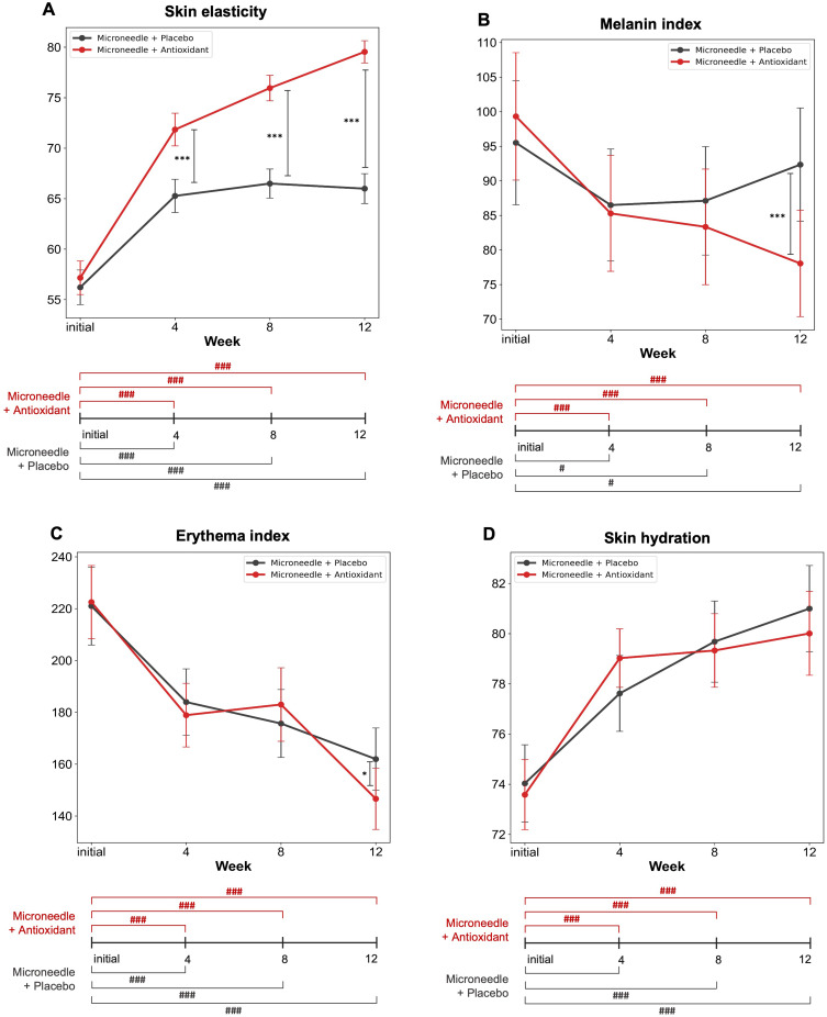
Figure 3 Time Course of Improvement in Skin Biophysical Parameters over 12 Weeks of Treatment: (A) Skin Elasticity, (B) Melanin Index, (C) Erythema Index, and (D) Skin Hydration. Data are presented as mean ± standard error. Upper graphs show the changes in parameters over time, while lower bars indicate statistical difference of corresponding scores between baseline and relevant time points. *p<0.05, ***p<0.001 for placebo-vs-antioxidant group comparisons at each timepoint with Bonferroni correction, and #p<0.05, ###p<0.001 for within-group comparisons versus baseline values.皮肤生理功能参数(弹性、黑素、红斑、水合)随时间变化,抗氧化侧弹性与色素改善更显著。
The treatment procedures and test products were generally well-tolerated, with most participants experiencing only mild and transient reactions such as erythema, stinging, or pruritus that resolved spontaneously within minutes after application. However, two participants developed eczematous papular eruptions on both sides of their face at the microneedle contact points several days following their first treatment session. The clinical presentation was suggestive of allergic contact dermatitis (ACD) to the metal components of the microneedle device, which was subsequently confirmed with positive patch test reactions to nickel and cobalt in both patients. While their dermatitis resolved within a few days following topical corticosteroid therapy, both withdrew from the study and did not undergo the second scheduled treatment session.
This study demonstrated the combining microneedling with a topical antioxidant formulation containing vitamins C, E, and ferulic acid can effectively enhance clinical outcomes in facial rejuvenation over a 12-week course with minimal side effects. It was clinically supported by the significant improvements in modified Griffith’s scale scores, hemi-MASI scores, and skin elasticity as early as four weeks with even more pronounced improvement at Week 12. The 39.0% improvement at Week 12 in skin elasticity is clinically translated into improved firmness, reduction in fine lines, and smoother texture; 21.3% reduction in melanin index means visible brightening and reduction in dark spot intensity leading to more even skin tone. This study highlights the importance of combination protocol of non-invasive device with topical antioxidant to achieve synergic outcome of facial rejuvenation.
Non-invasive device-based procedures for skin rejuvenation are gaining increasing popularity worldwide. While post-treatment care is crucial, there is limited clinical evidence regarding integrated skincare routines following non-invasive cosmetic procedures, including microneedling. For these procedures, it was suggested that post-treatment skincare should prioritize alleviating skin dryness, reducing oxidative stress and inflammation, preventing post-inflammatory hyperpigmentation, promoting the repair of treated areas to shorten downtime, and enhancing the overall effects of the procedure. 11 , 12 Topical ingredients with both anti-inflammatory and antioxidant properties are essential in achieving these goals. 12
For post-microneedling skincare, traditional recommendations typically include the application of hyaluronic acid gel, topical corticosteroids (eg, 1% hydrocortisone), and nonallergenic moisturizing creams. 18 However, there is growing evidence supporting a variety of newer topical treatments after microneedling. The healing process following the micro-injuries caused by microneedling is vital for restoring skin integrity. A randomized, double-blinded, split-face trial involving 30 patients demonstrated that a ceramide-containing moisturizer post-microneedling can significantly improve skin barrier recovery, reduce trans-epidermal water loss, and enhance skin texture. 19 An antioxidant serum (15% vitamin C, 1% vitamin E, and 0.5% ferulic acid) has also shown to facilitate more rapid wound healing after fractional laser treatments, with evidence indicating increased beta fibroblast growth factor. 20 In addition, microneedling’s ability to enhance the penetration of active ingredients offers an opportunity for post-procedure treatments that can further amplify the effects of the procedure. For instance, platelet-rich plasma and 15% trichloroacetic acid, when applied topically after microneedling, have demonstrated improvements in skin texture, wrinkle depth, and pigmentary dyschromia. 6
In addition to the global improvement of skin rejuvenation, the most significant finding of this study is that the Microneedle + Antioxidant-treated side of the face showed substantial improvement in the objective measurement of skin elasticity than microneedling alone. Microneedling creates micro-injuries that stimulate the release of growth factors such as platelet-derived growth factor, epidermal growth factor (EGF), fibroblast growth factor (FGF), and transforming growth factors (TGF-alpha and TGF-beta). Studies have also shown that TGF-beta3, a key marker for preventing scarring, is upregulated after microneedling, followed by a downregulation of TGF-beta1 and TGF-beta2. 21 This initiates fibroblast proliferation and migration, followed by the formation of a fibronectin matrix. Neocollagenesis and neoelastogenesis occurs, with initial Collagen III deposition, which is later replaced by Collagen I along with increased elastin. 22 Despite this profound dermal healing process, microneedling results in minimal epidermal damage and carries a low risk of dyspigmentation. 1 Given that the primary goal of microneedling is to improve signs of skin aging, combining topical antioxidants with microneedling can further enhance skin elasticity and rejuvenate the skin, as demonstrated in this study with antioxidant serum. 12
Vitamin C, for instance, boosts the activity of transcription factors responsible for collagen synthesis, stabilizes procollagen mRNA, and prevents collagen breakdown by inhibiting matrix metalloproteinase I. 23 , 24 A study involving 17 patients who underwent four microneedling treatments with vitamin C serum (0.5 g L-ascorbic acid dissolved in 2.5 mL of strawberry hydrolysate, 10 days apart) found significant improvements in skin firmness, elasticity, hydration, and skin tone, further emphasizing the synergic effects of topical antioxidant with microneedling. 10 Combining topical vitamins C and E has also been shown to significantly improve signs of aging, including skin smoothing, brightening of the skin tone, and wrinkle reduction. 25 Ferulic acid, a powerful antioxidant, forms stable phenoxyl radicals when interacting with free radicals, effectively preventing the further cascade of radical formation. It also acts as a hydrogen donor, supplying atoms directly to the radicals. 4 In addition to its antioxidant properties, ferulic acid inhibits matrix metalloproteinases and promotes the production of tissue inhibitors of metalloproteinases, which likely contributed to the increased skin elasticity observed in this study. 26 , 27
The enhanced penetration provided by microneedling, coupled with the combination of these three antioxidant ingredients, explains the synergistic effects of antioxidant serum (15% vitamin C, 1% vitamin E, and 0.5% ferulic acid) observed in this study at a time point as early as four weeks. This combination resulted in objective superiority in improving in skin elasticity and pigmentation. The practical implication for dermatologic professionals is the need to reassess the role of post-microneedling skincare. Rather than relying solely on traditional recommendations of bland moisturizers, these findings highlight the value of incorporating targeted antioxidants to optimize clinical outcomes. Likewise, product developers should prioritize antioxidant-rich formulations when designing post-procedural skincare products.
The combination therapy was well-tolerated among the participants without major adverse effects. Erythema, stinging, or pruritus was minimal and resolves within a few minutes. Granulomatous reactions after microneedling are an exceedingly rare with only 15 patients reported in the literature. It was suggested to be driven by either a specific delayed-type hypersensitivity or a non-specific inflammatory response to a foreign body or local infection. 28 Although vitamin C (ascorbic acid or tetrahexyldecyl ascorbate) was implicated in 60% of these cases, there is currently insufficient evidence to suggest direct causation. 28 However, two cases of ACD to microneedles in this study suggests caution prior to treatment for patients with known ACD to metals.
This study has several limitations to consider. First, the relatively short follow-up period (12 weeks) may not fully capture long-term efficacy and durability of treatment effects, particularly for ongoing collagen remodeling processes. Second, as a single-center study conducted primarily in Asian participants with Fitzpatrick Skin Type III–IV, these findings may have limited generalizability to other ethnic groups and skin phototypes. Third, despite the standardized measurement protocols and split-face design, multiple confounding factors including seasonal weather variations and slight variations in measurement locations may have influenced the skin biophysical parameters assessed in this study. 29 Fourth, although all subjects were provided with a designated sunscreen and instructed to apply it before outdoor activities, UV exposure may have influenced pigmentation and vascular responses. However, as subject recruitment occurred between May and August, the period of highest UV intensity in Korea, any potential UV-related confounding would have been unfavorable to the observed improvements in Melanin Index and Erythema Index. Lastly, our study design did not include a control group receiving antioxidant serum without microneedling, which would be necessary to fully dissociate the individual contributions of each treatment modality. Future studies incorporating such a control group are warranted to further elucidate the synergistic effects of this combination therapy.
This study demonstrates that combining microneedling with topical antioxidant serum containing vitamins C, E, and ferulic acid significantly enhances facial rejuvenation outcomes compared to microneedling alone. This combination approach yielded superior improvements in multiple parameters of skin aging, including elasticity, rhytides, and pigmentation, with minimal recovery time. The synergistic effect likely stems from enhanced penetration of antioxidant ingredients through microchannels created by the procedure, coupled with their complementary mechanisms against photoaging. This treatment protocol represents an effective, minimally invasive approach for patients seeking facial rejuvenation with measurable clinical benefits. Further long-term studies involving diverse populations would be valuable to confirm the durability of these promising results.
声明:中文精读 · 仅供学术参考。内容来自公开文献检索,不代表本人观点,不构成诊疗建议。 医疗美容需在正规医疗机构由执业医师实施。
基于相同主题推荐 · 继续深挖
背景:微针通过可控微损伤促进皮肤年轻化;外用抗氧化成分或可增强其疗效,本研究评估含维生素 C、维生素 E 与阿魏酸的精华。
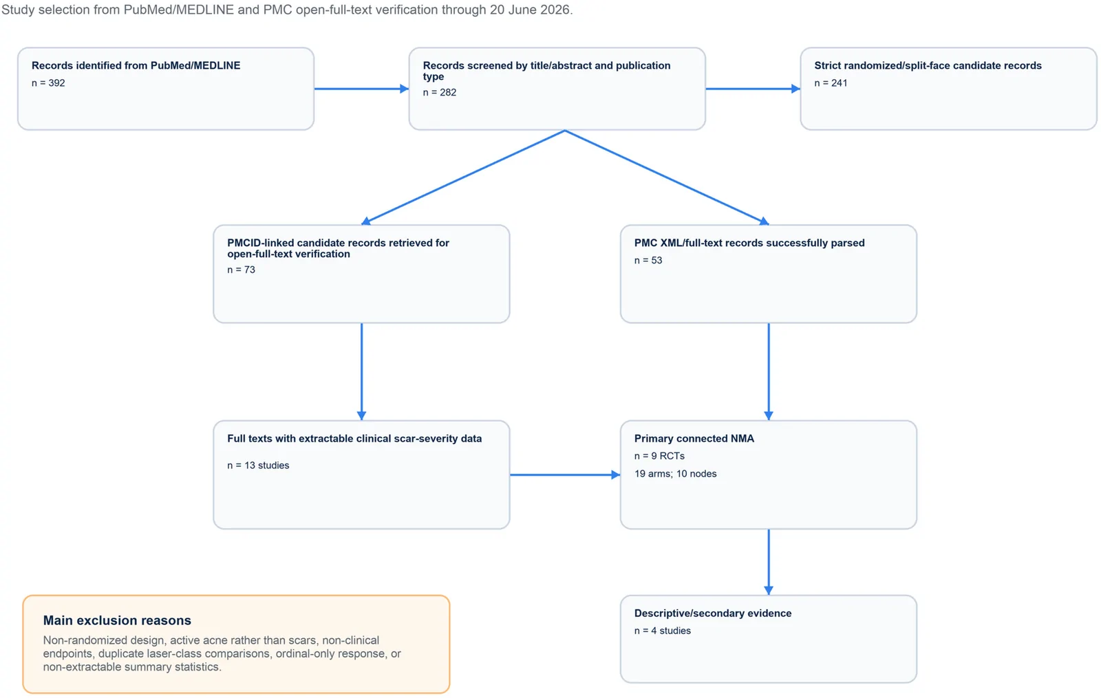
PROSPERO注册的系统综述,纳入9项随机/分脸随机试验、19个治疗臂,构成稀疏连接网络并采用频率学随机效应网络荟萃分析。
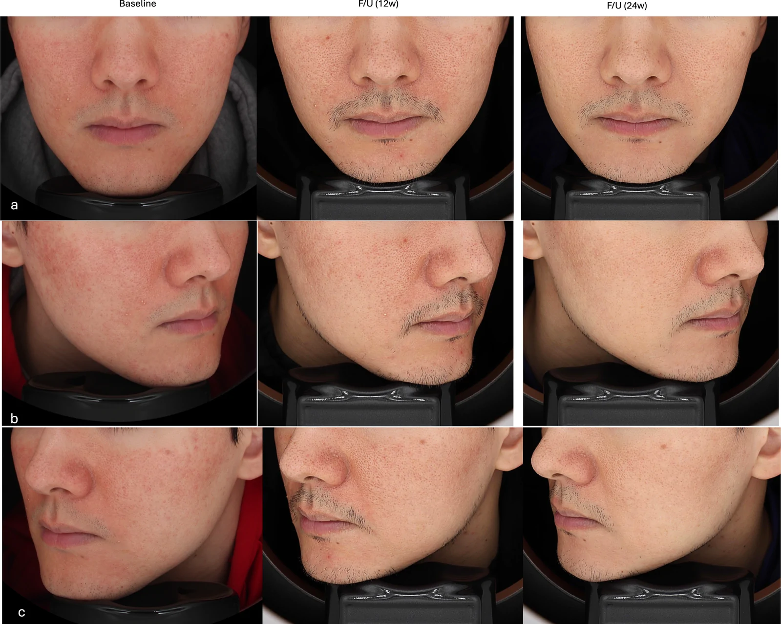
真实世界回顾性队列纳入50例临床诊断玫瑰痤疮成人,80%为女性、中位年龄33岁,多数(76%)为Fitzpatrick III型肤色。