倒刺线双平面提升矫正外眦下垂与颞部发际线
2026年9月19日24 min read
Plastic and Reconstructive Surgery Global Open
结论与实践要点
循证层面: 本研究为回顾性无对照队列(证据等级 V),样本量小(22 例亚洲女性)且单术者操作,结论的外推性有限;但其解剖学部分(筋膜层次、神经血管危险区)对安全布线具参考价值。
安全要点: 并发症率低且均为轻度,关键在于保持线材在 sub-STF 蜂窝平面穿行、倒刺段于上颞线以远腱膜下锚定,避免偏离平面或过于浅表,以规避面神经颞支与颞浅动脉相关风险;文中未出现切口感染、神经损伤或线材外露。
落地提示: 对轻中度颞部下垂/外眦下垂的亚洲求美者,双平面(sub-STF 通路 + 腱膜下锚定)颞部线雕可作为改善颞部遮盖与外眦位置的选项;临床须结合术前血管神经体表标记(如 Pitanguy 线)、规范进针层次,并向求美者说明 12 月时部分指标有回落、需更长期随访数据。
Background:The temporal region stabilizes the lateral canthus and brow. Age-related weakening of the superficial temporal fascia and galeal continuity contributes to temporal hooding and lateral canthal ptosis, but outcome data for 2-plane temporal thread lifting are limited.
Methods:Retrospective review of 22 Asian women treated with an I-type multidirectional barbed thread technique. A blunt cannula was advanced in the sub- superficial temporal fascia areolar plane and the barbed segment anchored beyond the superior temporal line in the subgaleal aponeurosis. Standardized photographs assessed lateral canthal tilt, lateral canthus vertical displacement, brow tail height, temporal hooding severity, global aesthetic improvement scale, and satisfaction at 6 and 12 months.
Results:Lateral canthal tilt increased from 2.1 ± 1.4 degrees to 4.3 ± 1.6 degrees at 6 months and 3.6 ± 1.5 degrees at 12 months. Lateral canthus vertical displacement improved by +1.8 ± 0.7 and +1.2 ± 0.6 mm; brow tail height by +2.4 ± 1.0 and +1.8 ± 0.9 mm. Temporal hooding improved from 2.6 ± 0.5 to 1.4 ± 0.5 and 1.6 ± 0.6. Global aesthetic improvement scale was “improved” or better in 90.9% at 6 months and 77.8% at 12 months, with high satisfaction. Adverse events were minor and transient.
Conclusions:In this retrospective uncontrolled cohort (evidence level V), subgaleal anchoring after subsuperficial temporal fascia passage was associated with measurable improvement maintained at 12 months. Prospective comparative studies are needed to define durability and generalizability.
The temporal region plays a central role in shaping periorbital and upper-face contours, as it houses a complex arrangement of fascial layers, neurovascular structures, and retaining ligaments that collectively determine lateral canthal position and brow support. 1 , 2 Age-related descent in this region is driven by progressive attenuation of the superficial temporal fascia (STF), relaxation of the temporal septa, and volume shifts within temporal fat compartments, all of which contribute to lateral canthal ptosis and temporal hollowing. 2 , 3
Degeneration of these fascial relationships alters the mechanical balance between the upper and midface, producing visible descent that cannot be corrected by skin manipulation alone. Prior studies have demonstrated that gravitational descent and weakening of the temporoparietal fascia–galea continuum reduce the structural tension that normally stabilizes the lateral brow and canthus. 4 , 5 Thus, treatments targeting fascial planes—rather than cutaneous repositioning—are required for durable correction.
Given that the temporal region contains critical structures such as the superficial temporal artery, zygomatico-orbital artery, sentinel vein, and the temporal branch of the facial nerve, minimally invasive rejuvenation demands precise identification of safe anatomical planes. 6 – 8 The STF, continuous inferiorly with the superficial musculoaponeurotic system and superiorly with the frontalis–galea complex, provides a predictable and accessible plane for controlled lifting, making it a key reference layer for thread-based interventions. 9
Recent advances in thread design, including multidirectional barbed constructs, and improved fascial mapping through cadaveric and imaging studies have enabled more predictable outcomes when procedural vectors are aligned with verified anatomic planes such as the STF, deep temporal fascia (DTF), and adjacent danger zones. 3 , 10 These developments have expanded the role of thread lifting in addressing lateral canthal ptosis and temporal descent while maintaining procedural safety.
However, despite these anatomical and technical developments, clinical validation of temporal thread lifting techniques that intentionally engage both the sub-STF and subgaleal planes remains limited. In particular, the contribution of plane selection and anchoring depth to the durability of lateral canthal and temporal elevation has not been systematically characterized.
Therefore, the present study applies a standardized I-type barbed thread technique designed to traverse the sub-STF areolar layer and anchor within the galeal aponeurosis, and evaluates its structural, aesthetic, and safety outcomes over a 12-month follow-up period.
The temporal region contains distinct fascial layers and critical neurovascular structures. From superficial to deep, it comprises skin–subcutaneous tissue, the STF, a loose areolar plane (including the innominate fascia), and the DTF. The sub-STF areolar plane provides a low-resistance corridor for blunt cannula advancement and reduces the risk of superficial thread visibility or cutaneous dimpling when depth is maintained. 1 – 3
Key structures at risk include the superficial temporal artery and zygomatico-orbital artery coursing within or just deep to the STF, the sentinel vein, and the temporal branch of the facial nerve. The temporal branch typically traverses the lower temporal compartment approximately 2–3 cm above the zygomatic arch and can be approximated on the surface by the Pitanguy line, emphasizing the importance of maintaining the cannula within the sub-STF plane and avoiding the lower temporal compartment. 4 , 9 – 11
Above the superior temporal line, the mobile temporoparietal fascia transitions to the dense, low-mobility galeal aponeurosis, which provides a stable anchoring substrate for barb engagement. Based on these anatomic principles, we used a 2-plane approach—sub-STF passage and subgaleal anchoring beyond the superior temporal line—to optimize safety and vector stability. 3 , 12
We retrospectively reviewed 22 female patients (29–65 y; mean ± SD, 43.1 ± 8.7 y) treated between March 2021 and December 2022 at a single aesthetic clinic. The indications included temporal descent, lateral brow laxity, and lateral canthal drooping. Patients with excessive skin redundancy requiring surgical excision were excluded.
All patients underwent thread lifting with the I-type multidirectional barbed thread technique as described, targeting the plane beneath the STF. Procedures were performed under local anesthesia, advancing from an entry point above the zygomatic arch to the subgaleal plane within the temporal hairline (Figs. 1 – 6 ).
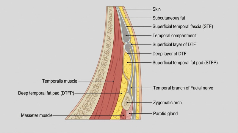
Fig. 1. Cross-sectional anatomy of the temple region depicting distinct tissue layers from superficial to deep, including skin, STF, and DTF.颞区横断面分层解剖:由浅至深为皮肤、浅表颞筋膜(STF)、疏松蜂窝层与深层颞筋膜(DTF)。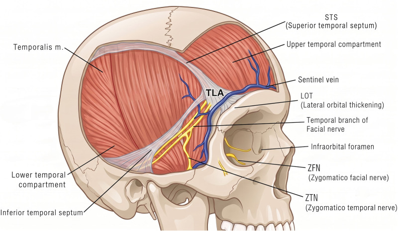
Fig. 2. Comparative illustration of upper and lower temporal compartments, emphasizing their different anatomical contents and clinical significance for thread placement.颞上/颞下间隙对比,提示不同解剖内容物与布线临床意义。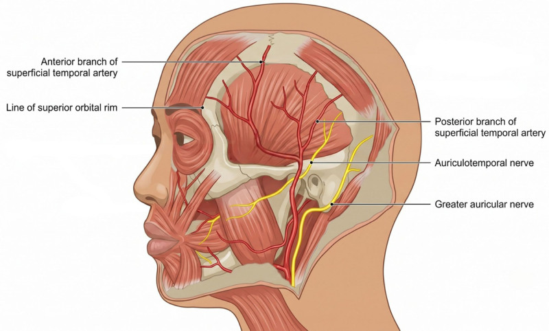
Fig. 3. Vascular anatomy mapping showing the course and relationships of the superficial temporal artery and zygomatico-orbital artery in the temporal region. The blue dotted line indicates the Pitanguy line (tragus to lateral brow), shown as a surface reference for the temporal branch danger zone in relation to the arterial pathways.颞区血管解剖(颞浅动脉与颧眶动脉走行),蓝色虚线为 Pitanguy 线(耳屏至外眉)。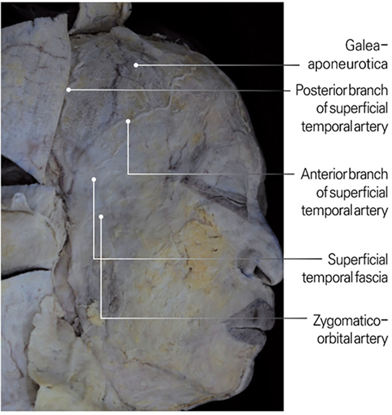
Fig. 4. Cross-sectional diagram showing the varying depths and pathways of major arteries in the temple region relative to facial planes.颞区主要动脉相对各筋膜平面的深度与路径。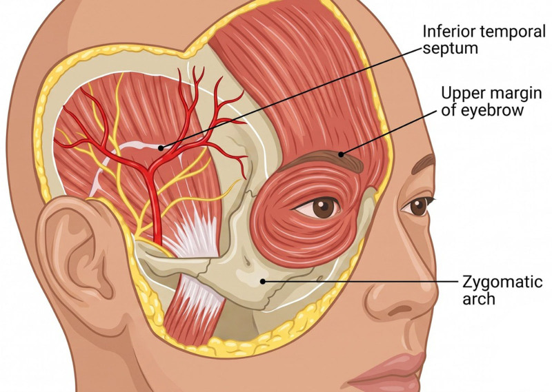
Fig. 5. Detailed illustration of the temporal branch of the facial nerve’s pathway, including key anatomical landmarks and danger zones.面神经颞支走行及危险区解剖标志。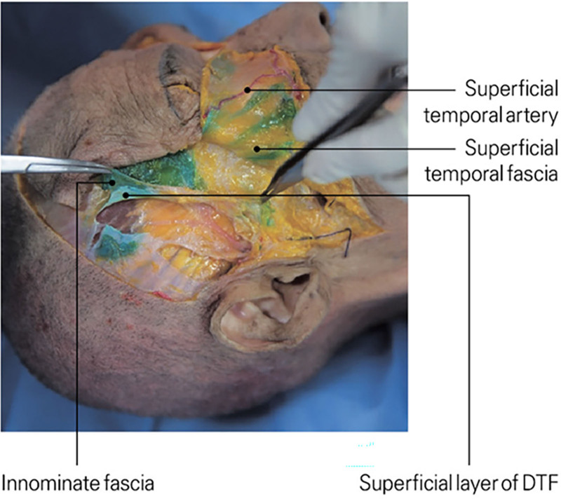
Fig. 6. Anatomical illustration showing the relationship between the innominate fascia and DTF, demonstrating their distinct planes and structural relationships.无名筋膜与 DTF 的层次关系。
With the patient seated, vascular and neural pathways are marked. The entry point—midway between the sideburn’s anterior margin and the lateral orbital rim—is placed at or above the zygomatic arch peak to avoid restrictive ligaments. Figures 7 , 8 illustrate the protected sub-STF corridor and the position of barbed threads beneath the STF and above the galeal layer. The recommended entry point and cannula orientation relative to the zygomatic arch and hairline are shown in Figure 9 .
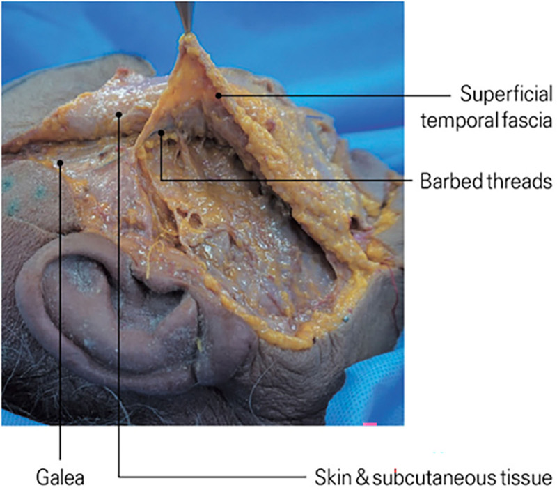
Fig. 7. Technical diagram showing optimal placement depth and directional vectors for I-type barbed threads in temple hairline lifting procedures.I 型倒刺线在颞部发际提升中的理想层次与方向向量。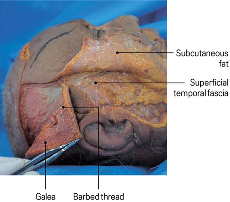
Fig. 8. Cross-sectional anatomy showing the ideal plane for barbed thread placement in temple hairline lifting, emphasizing relationship to key anatomical structures.倒刺线理想置放平面的横断面解剖(与关键结构关系)。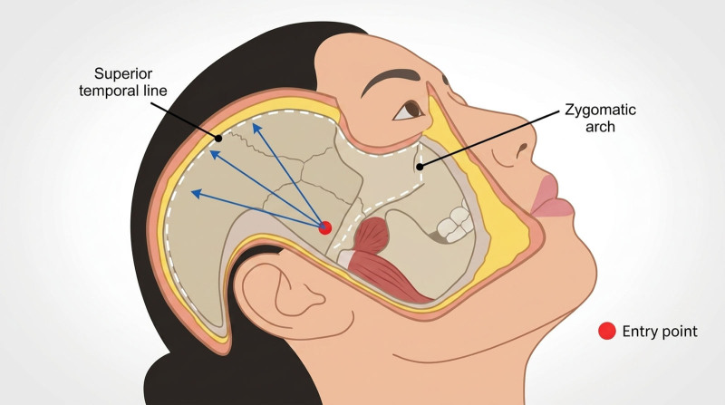
Fig. 9. Technical illustration demonstrating optimal entry points and directional vectors for I-type barbed thread insertion in temple hairline lifting.I 型倒刺线进针点与方向向量示意。A 25–27G needle creates the skin entry point, followed by a 21–22G blunt guide cannula for hydrodissection and confirmation of the target plane. Lifting threads (Sihler barbed threads, 100–160 mm) are delivered using 19–21G blunt cannulas.
The sub-STF areolar plane, located immediately deep to the STF and superficial to the DTF, serves as the ideal trajectory. Advancing the cannula in this plane minimizes resistance, avoids the temporal branch and sentinel vein, and reduces the risk of superficial thread visibility or contour irregularities. All threads are directed through this plane before transitioning into the subgaleal layer for secure fixation.
As the cannula progresses superiorly, it enters the subgaleal plane, where the STF merges with the galeal aponeurosis, providing a broad, firm surface for barb engagement and long-term vector support. Figure 10 demonstrates this transition and the anatomical relationship among the galea, STF, and upper temporal compartment.
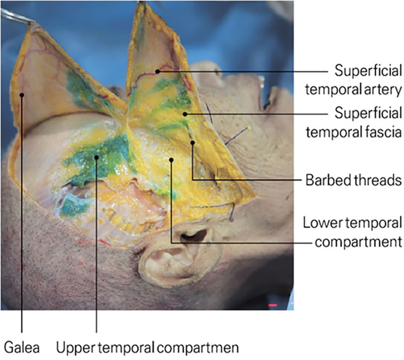
Fig. 10. Detailed representation of temporal septa and compartments, highlighting the superior and inferior temporal septum and their anatomical relationships.颞间隔与间隙(上/下颞隔)解剖关系。Threads are advanced to cross the superior temporal line (STL), where the mobile temporoparietal fascia transitions into the dense, low-mobility galeal aponeurosis. Engaging this aponeurotic layer provides a rigid anchoring substrate that resists early vector relaxation. Within the subgaleal plane, the barbs mechanically lock into this tissue, forming the primary load-bearing fixation point for sustained lift.
When correctly positioned, tissue along the arch and lateral periorbital region elevates visibly. Although the threads follow an I-type linear trajectory, the multidirectional lifting effect results from both the bidirectional orientation of the barbs—which engage tissue along anterior–posterior and superior–inferior axes—and the sequential passage through two planes (sub-STF and subgaleal), each contributing differential mechanical forces. Figure 11 illustrates the barb orientation and resulting lift vectors.
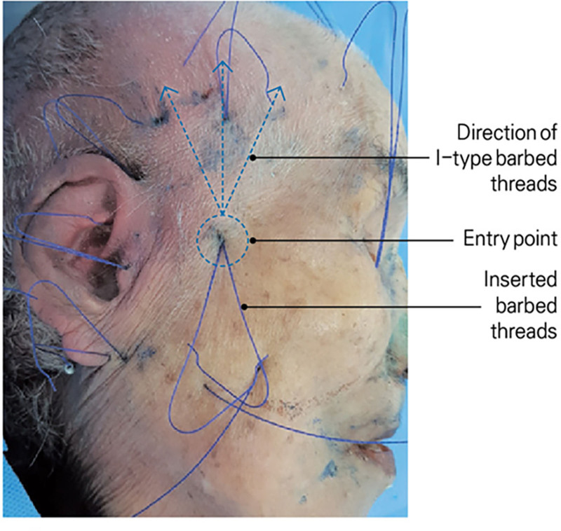
Fig. 11. Anatomical illustration demonstrating the placement technique of I-type barbed threads for temple hairline lifting, showing proper insertion angles and tissue engagement points.I 型倒刺线置放角度与组织咬合点。Typically, 1 or 2 19–21G SIHLER barbed threads (100–160 mm) are used per side, with selection based on skin thickness. Threads are oriented inward in a punching configuration for stronger grip. In this maneuver, the cannula advances slightly deeper than the intended final plane and is then withdrawn, allowing the barbs to engage the sub-STF areolar tissue from a reverse angle. This increases the number of barb–fascia contact points, enhances mechanical locking within the galeal transition zone, and minimizes early slippage, particularly in patients with thin temporal tissues. Excessively superficial placement must be avoided to prevent contour visibility.
After insertion, the patient is reseated to verify the durability and symmetry of elevation. To prevent postoperative surface irregularities or indentations, consistent maintenance of the sub-STF plane is essential, as superficial tunneling increases the risk of thread outlines or tethering in patients with thin skin. Gentle cannula advancement, avoidance of excessive barb tension, and confirmation of smooth soft-tissue gliding further reduce surface deformities. Hyperpigmentation is prevented by minimizing tissue trauma, maintaining deep-plane passage, and avoiding intradermal cannula contact. No cases of post-inflammatory hyperpigmentation or persistent contour irregularity were observed in our cohort.
Objective outcomes were assessed on standardized frontal photographs obtained at baseline, 6 months, and 12 months. Photographs were captured with fixed camera-to-subject distance, neutral facial expression, and Frankfort horizontal alignment. Images were de-identified and randomized; measurements were performed by a single assessor not involved in the procedure using ImageJ software (National Institute of Health, Bethesda, MD). 13 Pixel-to-millimeter calibration was performed using the interpupillary distance measured at each visit.
Surface landmark analysis included: (1) lateral canthal tilt (LCT), defined as the angle between the medial canthus–lateral canthus axis and the true horizontal line; (2) lateral canthus vertical displacement (LC-MPD), defined as the vertical distance change of the lateral canthus relative to a fixed midpupillary horizontal reference; and (3) brow tail height (BTH), defined as the perpendicular distance from the brow tail to a horizontal line passing through the medial canthus. Temporal upper-eyelid redundancy (“temporal hooding”) was graded on a 4-point investigator scale (0 = none, 1 = mild, 2 = moderate, 3 = severe), based on the extent of lateral upper-eyelid skin hooding over the lid margin and lateral canthal complex.
Subjective outcomes included the 5-point global aesthetic improvement scale (very much improved, much improved, improved, no change, worse) 14 and a 5-point patient satisfaction score (very satisfied to very dissatisfied). 15
Continuous outcomes were summarized as mean ± SD. Changes from baseline to 6 months and to 12 months were evaluated using paired t tests when the paired-difference SD was available; otherwise, conservative standard errors were estimated from the variance at each time point. Effect sizes (standardized mean differences) and 95% confidence intervals were reported. Statistical significance was set at P < 0.05. No a priori power analysis was performed due to the retrospective study design.
Anthropometric evaluation demonstrated measurable structural improvement after STF plane thread lifting (Table 1 ). At 6 months (n = 22), mean LCT increased from 2.1 ± 1.4 degrees to 4.3 ± 1.6 degrees (mean change +2.2 degrees, 95% CI: 1.3 to 3.1; P < 0.001) and remained improved at 12 months (n = 18), 3.6 ± 1.5 degrees (mean change +1.5 degrees, 95% CI: 0.5 to 2.5; P = 0.006).
Table 1. Surface Landmark Outcomes at Baseline, 6 Months, and 12 Months
| Outcome | Baseline | 6 Mo | 12 Mo | Change vs Baseline (95% CI), Effect Size (6/12 mo) | P (6/12 mo) |
|---|---|---|---|---|---|
| LCT (degrees) | 2.1 ± 1.4 | 4.3 ± 1.6 | 3.6 ± 1.5 | +2.2 (1.3 to 3.1), d = 1.46/+1.5 (0.5 to 2.5), d = 1.03 | <0.001/0.006 |
| LC-MPD (mm) | 0 | +1.8 ± 0.7 | +1.2 ± 0.6 | +1.8 (1.5 to 2.1), dz = 2.57/ +1.2 (0.9 to 1.5), dz=2.00 | <0.001/<0.001 |
| BTH (mm) | 0 | +2.4 ± 1.0 | +1.8 ± 0.9 | +2.4 (2.0 to 2.8), dz = 2.40/+1.8 (1.4 to 2.2), dz = 2.00 | <0.001/<0.001 |
| Temporal hooding (0–3) | 2.6 ± 0.5 | 1.4 ± 0.5 | 1.6 ± 0.6 | −1.2 (−1.5 to −0.9), d = −2.40/−1.0 (−1.4 to −0.6), d = -1.81 | <0.001/<0.001 |
LC-MPD improved by +1.8 ± 0.7 mm at 6 months (95% CI: 1.5 to 2.1; P < 0.001) and +1.2 ± 0.6 mm at 12 months (95% CI: 0.9 to 1.5; P < 0.001). BTH increased by +2.4 ± 1.0 mm at 6 months (95% CI: 2.0 to 2.8; P < 0.001) and +1.8 ± 0.9 mm at 12 months (95% CI: 1.4 to 2.2; P < 0.001).
Temporal hooding scores improved from 2.6 ± 0.5 at baseline to 1.4 ± 0.5 at 6 months (mean change −1.2, 95% CI: −1.5 to −0.9; P < 0.001) and 1.6 ± 0.6 at 12 months (mean change −1.0, 95% CI: −1.4 to −0.6; P < 0.001). Between 6 and 12 months, small regression was observed (eg, LCT −0.7 degrees; LC-MPD −0.6 mm), consistent with expected soft-tissue relaxation; these changes should be interpreted cautiously in this uncontrolled cohort.
Four patients were unavailable for the 12-month visit; available-case analysis was performed for 12-month outcomes.
These findings indicate maintenance of lateral canthal elevation, brow support, and reduction of temporal hooding through the observed follow-up period. Given the uncontrolled design and the millimetric scale of some changes, clinical significance should be interpreted alongside patient-reported outcomes.
At 6 months, 20 (90.9%) of 22 patients were rated “improved” or “much improved,” and 2 (9.1%) showed minimal change. Among 18 patients available at 12 months, 14 (77.8%) maintained their global aesthetic improvement scale rating, whereas 4 showed mild regression (Fig. 12 ).
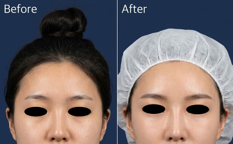
Fig. 12. Standardized pre- and 12-month postprocedure photographs demonstrating improvement in lateral canthal position and temporal hooding following I-type barbed-thread lifting in the sub-STF plane with subgaleal anchoring. A magnified inset of the lateral canthus is provided to aid visualization of the change in canthal tilt.术前与术后 12 个月标准照片,示外眦位置与颞部遮盖改善。
Patient satisfaction was high and aligned with objective findings. Mean satisfaction score was 4.5 ± 0.6 at 6 months and 4.2 ± 0.7 at 12 months on a 5-point scale, reflecting sustained perceived benefit.
Minor adverse events included transient bruising (8 of 22; 36.4%), localized edema (5 of 22; 22.7%), and transient paresthesia (3 of 22; 13.6%), all resolving within 2–3 weeks. No infections, nerve injuries, hematomas, or thread extrusions occurred. Notably, no cases of postoperative cutaneous indentation, contour deformity, or postinflammatory hyperpigmentation were observed.
Traditional thread lifting has historically emphasized midface and lower-face repositioning, with fewer detailed reports on fixation strategies specific to the temporal region. Recent reviews by Hong and colleagues describe the progression from early U-shaped vectors to more anatomically guided approaches that strategically utilize validated fascial planes such as the superficial musculoaponeurotic system–TPF continuum and the galeal expansion. 16 , 17 Consistent with these developments, our findings demonstrate that an I-type trajectory can still generate a multidirectional lifting effect through barb orientation, differential engagement across 2 anatomical planes, and the mechanical transition from the mobile temporoparietal fascia to the more rigid galeal aponeurosis.
Building on this mechanical rationale, the stability and durability of the lifting effect depend critically on the selection of the anchoring layer. Prior clinical studies have attributed variable longevity in temple thread lifts to superficial placement, weak fascial engagement, or insufficient extension beyond the STL. 4 , 18 As illustrated in Figure 2 , the compartmental transition above the STL provides a mechanically more stable region for fixation. Our technique intentionally crosses the STL into the dense galeal layer, consistent with the recommendations of Ingallina et al and Fante et al, who identified the galeal aponeurosis as a superior anchoring substrate compared with the more compliant tissues inferior to the STL. 2 , 4 The sustained elevation observed in our cohort further supports the clinical relevance of engaging this aponeurotic layer, reflecting the anchoring stability predicted by these anatomical studies.
In this context, thread lifting may provide modest structural repositioning with lower morbidity than surgery in carefully selected patients. However, our study was retrospective and uncontrolled; therefore, comparisons with surgical procedures should be interpreted as contextual discussion rather than evidence of equivalence.
The improvements in LCT, BTH, vertical canthal position, and temporal hooding reflect true vector-based fascial engagement rather than transient edema, as durable elevation was maintained at 12 months. This pattern indicates effective tension retention within low-mobility structures—particularly the galeal aponeurosis—which provides more stable support than the mobile temporoparietal fascia. 2 , 10 The reduction in temporal hooding further aligns with tightening of the periorbital retaining system and counters the inferolateral descent described in aging studies. 19
In our cohort, improvements were maintained at 12 months in most patients; longer follow-up and comparative designs are needed before making conclusions about durability beyond this time frame.
Patient selection is critical. In this series, candidates had mild-to-moderate temporal hooding and lateral canthal descent without excess skin requiring excision. Although lateral canthal ptosis is commonly age-related, younger patients may present with congenital canthal tilt, early soft-tissue descent, or asymmetry of lateral brow support; in such cases, a minimally invasive thread technique may be considered when the goal is subtle repositioning and the patient prefers to avoid surgery. Conversely, marked canthal laxity, significant dermatochalasis, or functional eyelid pathology remain better addressed with established surgical procedures (eg, canthopexy/canthoplasty and blepharoplasty).
Importantly, the technique does not advance or reposition the true temporal hairline; all visible lifting effects derive from fascial engagement within the sub-STF and subgaleal planes without altering hair-bearing scalp.
Although complication rates were low, temporal thread lifting carries predictable risks. Minor events—transient bruising, edema, superficial irregularities, or temporary paresthesia—generally result from deviation from the sub-STF areolar plane or overly superficial cannula passage. More serious complications, such as vascular or neural injury, may occur if the cannula enters deeper compartments. The favorable safety profile in this cohort likely reflects consistent adherence to the sub-STF plane, use of blunt cannulas, and avoidance of high-risk zones.
The safety outcomes also align with established anatomical principles: maintaining the sub-STF plane protects the temporal branch 7 and reduces contact with the superficial temporal artery and sentinel vein. 20 , 21 Figure 8 demonstrates clear depth separation from these structures, supporting prior recommendations that emphasize superficial fascial navigation to minimize complications. 8 , 22 The absence of persistent contour irregularities or pigmentary sequelae further supports the safety of this plane.
This study has several limitations. The retrospective design, absence of a control group, and single-operator treatment preclude causal inference and limit conclusions about durability or mechanical stability. Four patients were lost to 12-month follow-up, and no survival analysis of lift persistence was performed. All participants were Asian women with mild-to-moderate ptosis treated with a single thread type (Sihler Thread), limiting generalizability to men, non-Asian phenotypes, other thread materials, and more severe degrees of ptosis. Surface landmark outcomes were derived from standardized 2-dimensional photographs; although images were de-identified and randomized, measurements were performed by a single assessor and interobserver reliability was not evaluated. Finally, a minimal clinically important difference for small millimetric changes has not been established for these metrics; future studies should incorporate validated patient-reported outcome measures, objective 3-dimensional assessment, and comparative prospective designs.
This retrospective, uncontrolled study (Evidence Level V) suggests that engaging the sub-STF and subgaleal planes can provide an anatomically guided pathway for improving temporal hooding and supporting the lateral canthus, with measurable improvements maintained up to 12 months in this cohort. The technique’s safety was supported by use of the sub-STF protective corridor, with no serious neurovascular complications observed. Given the absence of a control group and the single-operator, all-female Asian cohort, these findings should be interpreted as preliminary. Prospective comparative studies and broader patient populations are needed before extrapolating durability, effect size, or equivalence to surgical techniques.
The authors have no financial interest to declare in relation to the content of this article.
Patients provided written consent for the use of their images.
This retrospective study was conducted in accordance with the Declaration of Helsinki and analyzed de-identified clinical data and photographs. In accordance with local regulations, the study qualified for exemption from formal institutional review board review.
声明:中文精读 · 仅供学术参考。内容来自公开文献检索,不代表本人观点,不构成诊疗建议。 医疗美容需在正规医疗机构由执业医师实施。
基于相同主题推荐 · 继续深挖
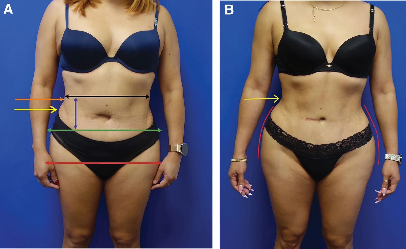
研究设计:PRS Global Open 回顾性研究,2016-11 至 2025-01 共 1999 例连续手术,旨在界定并塑形『腰腹过渡美学单元(waist transitio
背景:颞区支撑外眦与眉尾,年龄相关浅表颞筋膜(STF)与腱膜连续性减弱可致颞部遮盖与外眦下垂;双平面颞部线雕的结局数据有限。
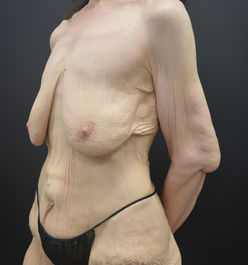
文章对躯干与四肢提出美学单元(AU)与粘连分区(ZA)的系统分类,作为身体塑形手术(BCS)切口规划的解剖基础。