重复低能量连续单极射频对面部脂肪的脂肪生成与干细胞激活作用:超声、三维影像与组织学研究
2026年9月21日26 min read
Plastic and Reconstructive Surgery Global Open
Plastic and Reconstructive Surgery Global Open
结论与实践要点
本研究为前瞻性单中心研究,纳入25名BMI<21的健康女性,接受5次115 W连续单极射频(每3周一次),于基线及18周用QuantifiCare与Morpheus三维成像及高频超声评估面部容量与软组织厚度,并取1名受试者治疗前后配对皮肤活检行CD166、CD34、CD31免疫组化,以GAIS评估临床改善。结果显示受试期间体重稳定;三维成像示软组织厚度显著增加(QuantifiCare +0.86±0.21 mm,P<0.0001;Morpheus +1.01±0.22 mm,P=0.0002);超声示总脂肪厚度由14.73±2.02 mm增至16.33±1.99 mm(P<0.00001),浅层脂肪由5.99±0.78 mm增至7.64±0.86 mm(P<0.0001)。组化见CD166与CD34表达增高(提示间充质干细胞/前体激活),CD31增高(提示血管生成)。GAIS示中面部容量、皮肤质地与弹性改善明显。作者认为脂肪层增厚更符合脂肪生成性重塑而非炎性水肿,并在治疗期后持续。
临床落地需注意:① 传统高强度脉冲射频以紧肤为主、瘦人可致脂肪萎缩,而本研究采用重复低能量连续模式旨在促脂肪增生,为“瘦脸凹陷“人群的非侵入性容量补充提供新思路;② 证据仍有限——仅1例活检、样本量小、无对照、单中心,干细胞激活为“瞬时“且长期转归不明;③ 设备参数(115 W连续单极、5次/3周)需在规范下操作,能量与间隔的安全性尚待更大样本验证。该方案适合面部偏瘦、中面部容量不足且暂不愿接受填充或手术者,作为探索性容量改善选项;临床决策应结合个体脂肪厚度基线,避免对脂肪本已丰满者过度促增生。
Background:High-intensity pulsed radiofrequency (RF) devices are effective for skin tightening but may cause facial fat atrophy in lean individuals. This study investigates whether repeated low-energy continuous monopolar RF could stimulate adipogenesis and stem cell activation, providing a noninvasive approach to facial volumization.
Methods:In this prospective, single-center study, 25 healthy women (body mass index <21 kg/m 2 ) received 5 sessions of 115-W continuous monopolar RF at 3-week intervals. Facial volume and soft tissue thickness were assessed at baseline and 18 weeks using QuantifiCare and Morpheus 3D imaging and ultrasound. One participant underwent paired skin biopsies for immunohistochemical analysis of CD166, CD34, and CD31. Clinical outcomes were evaluated using the Global Aesthetic Improvement Scale.
Results:Body weight remained stable throughout the study. Three-dimensional imaging demonstrated significant increases in tissue thickness (QuantifiCare, +0.86 ± 0.21 mm, P < 0.0001; Morpheus, +1.01 ± 0.22 mm, P = 0.0002). Ultrasound showed significant increases in total fat thickness (14.73 ± 2.02 to 16.33 ± 1.99 mm, P < 0.00001) and superficial fat thickness (5.99 ± 0.78 to 7.64 ± 0.86 mm, P < 0.0001). Histology demonstrated increased CD166 and CD34 expression, consistent with stem/progenitor cell activation, and increased CD31 expression, indicating angiogenesis. Global Aesthetic Improvement Scale scores showed high improvement in midface volume, skin texture, and elasticity.
Conclusions:This study is the first to present convergent evidence from 3D imaging, ultrasound, and histological analysis demonstrating that repeated low-energy continuous monopolar RF increased subcutaneous fat thickness and transiently activated mesenchymal stem/progenitor cells in human facial tissue. The increased fat layer was consistent with adipogenic remodeling rather than inflammatory edema and persisted beyond the treatment period.
In recent years, radiofrequency (RF)-based noninvasive treatments have been widely adopted in aesthetic medicine for skin tightening and contouring. 1 – 3 Conventional high-intensity monopolar RF devices, such as Thermage, use fractional delivery systems that deliver multiple RF pulses per second, resulting in significant energy deposition and simultaneous CO 2 gas cooling. 4 – 6 Although such systems are effective in inducing dermal collagen remodeling, they often generate considerable pain and unintended adipose tissue atrophy—resulting in sunken cheeks, especially in lean individuals with minimal subcutaneous fat. 4 , 7 Clinically, this has led to increasing concerns over facial volume loss following RF procedures, as documented by both medical reports and anecdotal patient complaints across social platforms. 4 , 8 , 9
To address these limitations, the author used a new RF delivery modality—continuous radiation monopolar RF. This next-generation device (Volnewmer) uses sustained RF output at 115 W combined with a continuous water-cooling system. Unlike traditional pulsed systems, this continuous emission allows for gradual thermal stimulation of subcutaneous tissue without inducing abrupt thermal spikes, theoretically reducing pain and adipocyte damage. 10 , 11
Emerging evidence suggests that moderate thermal stimulation may activate regenerative pathways in adipose-derived stem cells (ADSCs). Notably, studies have shown that focused ultrasound can stimulate adipogenesis by modulating primary cilia and triggering downstream signaling cascades such as Hedgehog and Wnt/β-catenin pathways. Similarly, low-intensity ultrasound has been reported to promote ADSC proliferation and adipogenic differentiation by enhancing cytoskeletal remodeling and transcriptional regulation of PPARγ, FABP4, and adiponectin genes. Given these findings, it is plausible that low-intensity thermal energy from RF may exert comparable bioeffects. 12 – 15
This clinical study aims to evaluate whether repeated exposure to 50 shots of continuous RF energy per side can induce fat tissue thickening and facial volume enhancement in lean female patients. Using high-resolution ultrasound and 3D surface scanning (QuantifiCare and Morpheus systems), the volumetric changes in facial subcutaneous tissue were investigated following a standardized treatment protocol. 16 , 17 In addition, histological evaluation of pre- and posttreatment biopsies in 1 subject will assess the expression of stem cell markers CD166, CD31, and CD34 as indicators of tissue remodeling and angiogenesis. 18 – 20
Our hypothesis is that subthermolytic, sustained RF stimulation can favorably modulate the subcutaneous microenvironment, promoting adipose regeneration rather than atrophy. This study provides a novel paradigm in aesthetic energy-based treatments, shifting from aggressive thermal injury to controlled biostimulation of adipogenic pathways, particularly in patients prone to posttreatment volume loss.
This prospective, single-center, clinical study was conducted from April 2024 to July 2025 and approved by the public institutional review board (approval no.: P01-202404-06-001). The objective was to evaluate volumetric changes and soft tissue thickening in the midface following 5 sessions of 115-W continuous monopolar RF therapy using the Volnewmer RF device (Classys, Seoul, Korea) performed at 3-week intervals. Volumetric changes were assessed with 3D imaging, and soft tissue thickness was measured via high-resolution ultrasonography.
Twenty-five healthy adult female participants (age range, 20–79 y) with a lean body habitus (body mass index [BMI] < 21 kg/m 2 ) were enrolled. All participants desired skin tightening without volume loss in the submalar area. Written informed consent, including consent for facial photography and optional biopsy, was obtained from each participant before enrollment.
Local anesthesia cream was not used. Each subject underwent 5 treatment sessions with the Volnewmer RF device at 3-week intervals. A 4 cm 2 square tip was used to deliver a total of 100 shots per session (50 shots per hemiface, midface, and lower face). The RF energy at energy level 5 was delivered using stamping mode for 2 passes, then sliding mode was performed. Postprocedural skin care included application of an ampoule (Reteenage Goldstem Ampoule, Seoul, Korea) to soothe the skin and provide hydration for 1 day after treatment.
Facial volume was quantitatively assessed at baseline and at 18 weeks using the QuantifiCare LifeViz (Quantificare SA, Sophia Antipolis, France) system and Morpheus 3D scanner (Morpheus Co., Seoul, Korea). Volumetric change was calculated in the submalar area by comparing pre- and posttreatment 3D data. ( See Video [online] , which shows 3D facial overlay images before and after repeated low-energy continuous monopolar RF treatment using Volnewmer, demonstrating volumetric improvement in the submalar region.)
补充视频 / 附件(原文未随文提供原图,可于出版方页面查看)gox-14-e7972-s001.mp4Video 1. 3D image before and after low power radiofrequency Volumer. 3D overlay image of the patients who had sunken cheek region before and after the radiofrequency, demonstrating volumetric improvement and soft tissue augmentation.
Soft tissue thickness was evaluated using high-frequency ultrasound (Samsung RS85, an 18-MHz matrix linear probe, Samsung Medison, Seoul, Korea). Measurements (0.01 mm) were taken along the midcheek vector line connecting the earlobe to the oral commissure, focusing on the submalar depression zone.
Independent evaluators assessed clinical improvements using the Global Aesthetic Improvement Scale (GAIS), focusing on improvements in midfacial volume, skin texture, and elasticity.
In a single volunteer, 2-mm punch biopsies were performed from the preauricular midcheek at 2 weeks before first treatment (control baseline, left side) and 18 weeks postfinal treatment (right side) for histological analysis. The biopsies were performed under local anesthesia, and the wounds were sutured using 5-0 Vicryl for convenience and absorption, as no long-term scarring was expected or observed in this area upon follow-up. Immunohistochemical staining was conducted to evaluate the expression of adipogenic and vascular stem cell markers, including CD166, CD31, and CD34.
All 25 enrolled participants completed the 5-session treatment protocol and posttreatment evaluations at week 18. There was no statistically significant change in body weight during the study period (pretreatment: 51.67 kg; posttreatment: 51.5 kg; no significant change), confirming that the observed volumetric changes were not attributable to systemic weight gain.
Three-dimensional imaging analysis using the QuantifiCare LifeViz system revealed a significant increase in facial soft tissue volume. The mean volumetric gain in the treated midface region was 0.78 ± 0.25 mL ( t = –0.91; P = 0.399; not statistically significant; P > 0.01). Correspondingly, 3D thickness in the same region increased to 0.86 ± 0.21 mm based on QuantifiCare measurements (1-sample t test, t = 46.56, P < 0.0001), and to 1.01 ± 0.22 mm using the Morpheus 3D system (1-sample t test, t = 10.25, P = 0.0002; P < 0.001), corroborating the volumetric data (Figs. 1 – 7 ).
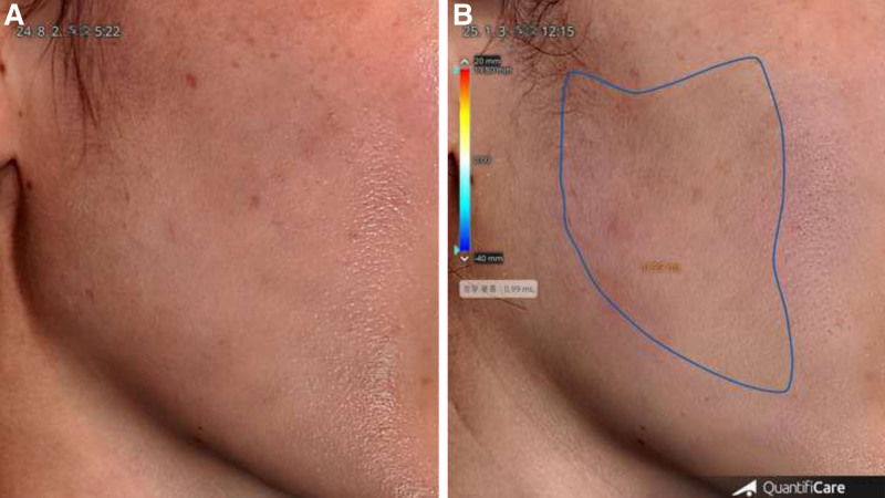
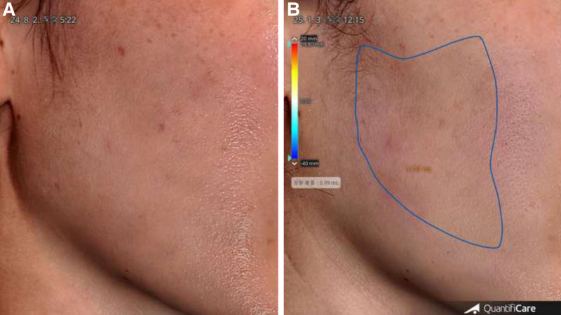
Fig. 1. QuantifiCare 3D imaging analysis of right-side midfacial volume change in a lean 45-year-old woman. A, Baseline image before treatment with monopolar RF (Volumer). B, At 18 weeks posttreatment, a volume increase of 0.99 mL is observed in the outlined region of the right midface.QuantifiCare 3D右侧中面部体积变化(45岁偏瘦女性),18周增量0.99 mL。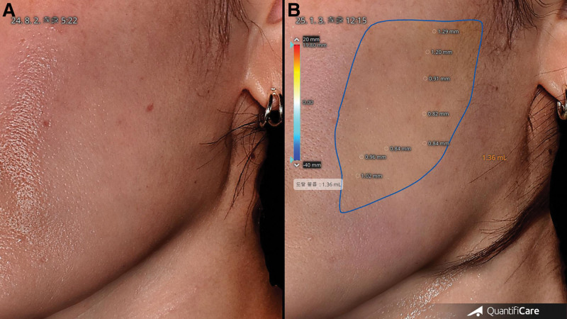
Fig. 2. QuantifiCare 3D imaging analysis of left midfacial volume enhancement in a lean 45-year-old woman. A, Baseline. B, At 18 weeks posttreatment with RF, the left midface shows a 1.36-mL volume increase, and regional thickness increases are also visualized: 1.02, 0.96, and 0.91 mm.左侧中面部3D体积+1.36 mL,区域厚度增加可视化。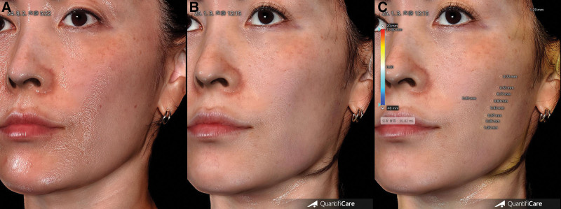
Fig. 3. QuantifiCare 3D imaging analysis of the same lean 45-year-old female patient shown in Figures 1 and 2. A, Baseline. B, At 18 weeks posttreatment, visible midfacial fullness is noted clinically. C, The 3D scan analysis demonstrates a total midfacial volume gain of 30.82 mL, with focal increases in soft tissue thickness ranging from 0.63 to 1.03 mm.同患者总中面部体积增益的3D扫描分析。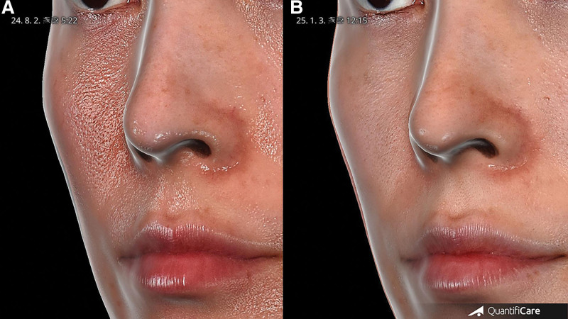
Fig. 4. Overlaid QuantifiCare 3D skin surface comparison of the same lean 45-year-old female patient shown in Figures 1–3. A, Pretreatment. B, Posttreatment surface superimposition clearly visualizes midfacial projection enhancement and volumization, and the highlighted surface bulge.同一患者3D皮肤表面叠加对比,中面部突出度增强。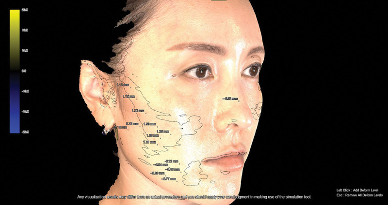
Fig. 5. Three-dimensional overlay analysis using the Morpheus imaging system of the same right cheek shown in Figures 1–4, captured from a lean 45-year-old female patient. The superimposed pre- and posttreatment images illustrate soft tissue augmentation, with a volumetric improvement of 1.32, 1.38, and up to 1.53 mm in the midcheek depression.Morpheus系统右侧颊部3D叠加,软组织增量可视化。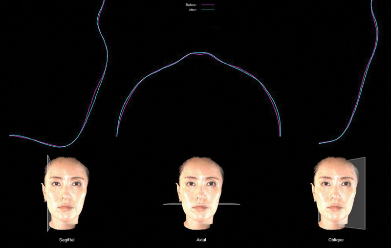
Fig. 6. Cross-sectional contour overlay analysis of the face using the Morpheus imaging system in sagittal, axial, and oblique planes. The red (magenta) lines represent the pretreatment facial contours, whereas the blue lines indicate posttreatment contours. Notable outward shift in the midcheek area reflects volumetric augmentation following treatment, particularly in regions of preexisting volume deficiency.Morpheus矢状/轴位/斜位轮廓叠加,红为治疗前、蓝为治疗后。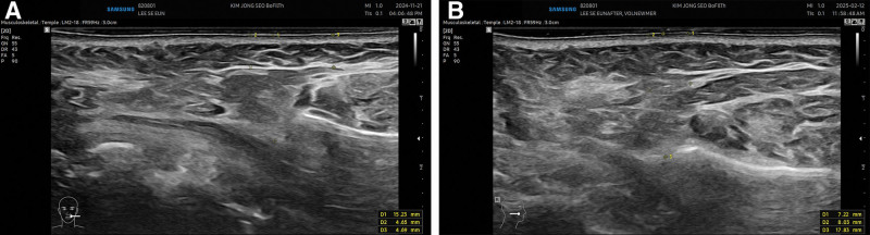
Fig. 7. Ultrasound imaging of the zygomatic hollow region from the earlobe to the oral commissure after monopolar RF treatment in a lean 45-year-old female patient. A, The pretreatment scan shows soft tissue thickness of 15.23 mm in the zygomatic depression. B, The posttreatment scan reveals a significant increase in thickness to 17.83 mm.颧凹陷区超声,治疗前15.23 mm、治疗后增厚。Ultrasonographic evaluation demonstrated significant thickening of the subcutaneous adipose tissue. The total fat thickness increased 1.60 mm, from 14.73 ± 2.02 mm at baseline to 16.33 ± 1.99 mm posttreatment ( t = 13.08, P < 0.00001). Notably, the superficial fat layer showed a marked increase of 1.65 mm, from 5.99 ± 0.78 to 7.64 ± 0.86 mm ( t = 6.67, P < 0.0001), indicating that low-intensity continuous RF therapy predominantly augmented the superficial adipose compartment (Figs 8 , 9 ).
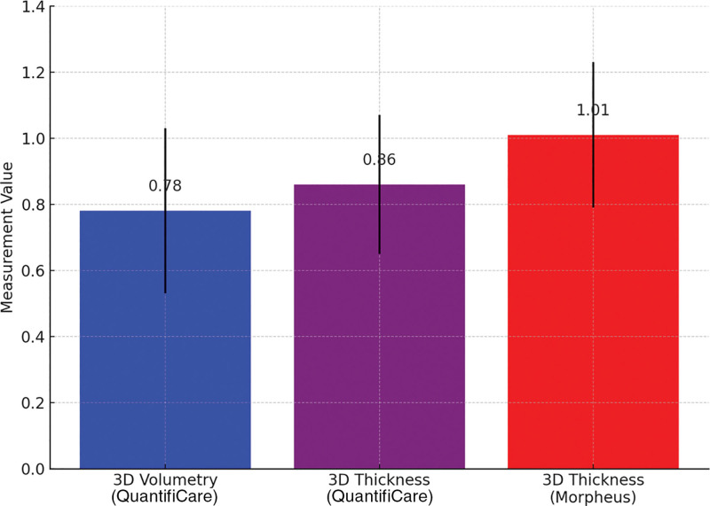
Fig. 8. Quantitative comparison of 3D analysis results measuring soft tissue augmentation after treatment: QuantifiCare and Morpheus. The blue and purple bars represent the mean volumetric increase (0.78 mm) and the mean thickness increase (0.86 mm), respectively, measured by QuantifiCare. The red bar shows the mean thickness increase (1.01 mm) measured by Morpheus, indicating a slightly higher sensitivity in detecting midcheek volumetric changes.3D分析定量对比(QuantifiCare与Morpheus)体积与厚度增量。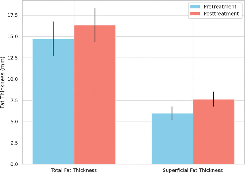
Fig. 9. Ultrasound-based evaluation of fat layer thickness before and after treatment. Total fat thickness increased from 14.7 to 16.3 mm on average, and superficial fat thickness increased from 6.0 to 7.6 mm. These findings support the volumizing effect of the treatment, particularly in the superficial fat compartment, as visualized and measured by high-resolution ultrasound imaging.超声评估脂肪层厚度,总脂肪14.7→16.3 mm、浅层6.0→7.6 mm。Subjective assessments based on the GAIS also supported these findings. The mean score for improvement in midface depression was 4.24 ± 0.54, indicating substantial volumetric restoration. GAIS scores for skin texture and elasticity were 4.48 ± 0.51 and 4.33 ± 0.48, respectively, suggesting overall enhancement in skin quality (Fig. 10 ).
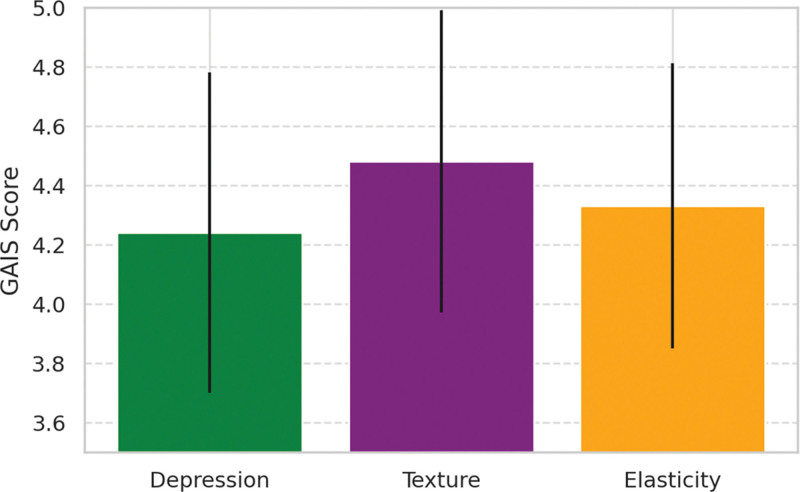
Fig. 10. GAIS scores reported by patients after treatment. The average GAIS scores were 4.23 ± 0.57 for depression, 4.48 ± 0.51 for texture, and 4.32 ± 0.50 for elasticity. These results indicate high patient satisfaction, with texture improvement rated the highest among the 3 parameters. Error bars represent the SD.患者GAIS评分(中面部容量/质地/弹性)均示较高改善。In a histological analysis of paired pre- and posttreatment biopsies from a representative subject, a significant upregulation of regenerative and vascular biomarkers was observed, providing histological evidence of subcutaneous tissue remodeling following repeated low-intensity monopolar RF stimulation. Specifically, CD166 expression, a marker of mesenchymal stemness and regenerative precursor cell populations, was markedly increased posttreatment (Fig. 11 ). CD166-positive cells were localized adjacent to mature adipocytes in the subcutaneous layer, suggesting activation or recruitment of ADSCs in response to repeated sublethal thermal stimulation. CD34, a transmembrane glycoprotein expressed in both endothelial progenitor cells and preadipocytes, also demonstrated enhanced expression in the treated specimen (Fig. 12 ). CD34-positive cells were identified both in the perivascular space and around adipocytes, supporting the hypothesis that monopolar RF stimulation not only promotes adipogenic proliferation but also augments stem cell–driven neovascular remodeling. Similarly, CD31, an endothelial marker indicative of angiogenic activity, showed increased immunoreactivity in treated tissue (Fig. 13 ). CD31-positive staining was evident around fibroblasts, adipocytes, and blood vessels, reflecting a robust vascular response to thermal stimulation and further supporting the notion of enhanced angiogenesis following RF exposure. Taken together, these histological findings demonstrate that repetitive sublethal thermal exposure delivered via continuous RF may modify the subcutaneous niche in a manner conducive to both stem cell activation and vascular remodeling, which may underlie the clinically observed improvements in volume and texture. These observations are particularly meaningful in the context of current regenerative dermatology and aesthetic medicine, as they provide direct tissue-level evidence supporting the biological plausibility of RF-induced adipogenesis. The coordinated upregulation of CD166, CD34, and CD31 represents an integrated regenerative response involving stem cell activation, preadipocyte proliferation, and neovascularization. This supports the emerging paradigm that controlled thermal stimulation can serve as a noninvasive trigger for tissue regeneration, with implications for facial volumization and dermal rejuvenation (Table 1 ). These results collectively demonstrate that low-power, continuous monopolar RF treatment induces measurable volumetric and adipose tissue augmentation in the midface, accompanied by cellular and histological signs of tissue regeneration and remodeling.
Table 1. Summary of Clinical, 3D Imaging, Ultrasound, and Histologic Outcomes Following 5 Sessions of Low-energy Continuous Monopolar Radiofrequency Treatment
| Parameter | Pretreatment | Posttreatment | Δ/Significance |
|---|---|---|---|
| Weight, kg | 51.67 | 51.5 | NS (no significant change) |
| 3D volumetry (QuantifiCare), mL | — | 0.78 ± 0.25 | P < 0.01 |
| 3D thickness (QuantifiCare), mm | 0.86 ± 0.21 | P < 0.01 | |
| 3D thickness (Morpheus), mm | — | 1.01 ± 0.22 | P < 0.01 |
| Ultrasound total fat thickness, mm | 14.73 ± 2.02 | 16.33 ± 1.99 | P < 0.01 |
| Ultrasound superficial fat thickness, mm | 5.99 ± 0.78 | 7.64 ± 0.86 | P < 0.01 |
| GAIS (depression area improvement) | 4.24 ± 0.54 | ||
| GAIS (texture improvement) | 4.48 ± 0.51 | ||
| GAIS (elasticity improvement) | 4.33 ± 0.48 | ||
| CD166 expression | ↑ stemness | ||
| CD34 expression | ↑ preadipocyte pool | ||
| CD31 expression | ↑ angiogenesis |
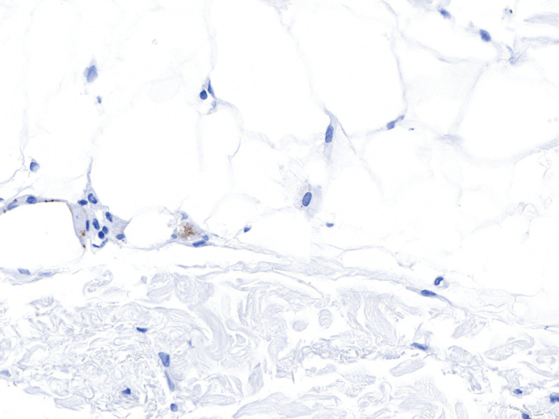
Fig. 11. Immunohistochemical staining for CD166 expression in the subcutaneous fat layer following 5 sessions of low-intensity monopolar RF treatment. CD166-positive cells (brown staining) are observed adjacent to adipocytes, suggesting activation of ADSCs in response to repeated RF-induced sublethal thermal stimulation. This histological evidence supports the hypothesis that monopolar RF may promote adipogenesis through stem cell activation in the adipose niche.CD166免疫组化,脂肪细胞旁棕染提示脂肪干细胞激活。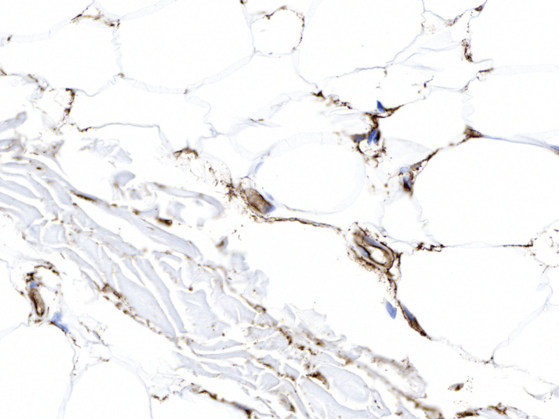
Fig. 12. Immunohistochemical staining for CD34 expression in the subcutaneous tissue following 5 sessions of low-intensity monopolar RF treatment. CD34-positive cells (brown staining) are observed both around adipocytes and adjacent to vascular structures, indicating an increased presence of endothelial progenitor and stem-like cells.CD34免疫组化,脂肪细胞旁及血管周棕染提示前体/血管激活。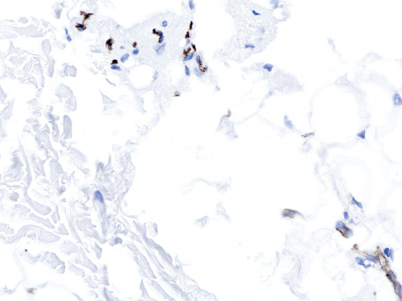
Fig. 13. Immunohistochemical staining for CD31 expression in the subcutaneous tissue following 5 sessions of low-intensity monopolar RF treatment. CD31-positive cells (brown staining) are observed surrounding fibroblasts, adipocytes, and vascular structures, indicating endothelial activation and neovascular remodeling.CD31免疫组化,血管周棕染提示血管生成。
This prospective study demonstrates that repeated application of low-power, continuous monopolar RF (Volnewmer) significantly increases subcutaneous fat thickness and midface volume in lean individuals. The findings were substantiated by multimodal evaluations, including high-frequency ultrasound, 3D volumetric imaging, and histological analyses with CD166, CD34, and CD31 immunohistochemistry. 18 – 20
The observed adipose thickening in both total and superficial layers supports the hypothesis that nonablative, sustained RF stimulation may promote adipogenesis rather than induce lipoatrophy—a common concern with traditional high-intensity pulsed RF devices. In particular, superficial fat increased by an average of 1.65 mm ( P < 0.0001), a change that is both statistically and clinically meaningful. This outcome contrasts with prior reports of volume depletion and fat atrophy following ablative or pulsed RF energy delivery, especially in patients with low baseline fat volumes. 12 – 15
At the cellular level, histological evidence from paired biopsies confirmed a posttreatment upregulation of CD166 and CD34, indicating activation of ADSCs and increased preadipocyte proliferation. CD31 expression, a vascular endothelial marker, was also elevated, suggesting concurrent neovascularization. These findings collectively imply that repetitive, sublethal thermal stress via continuous RF may modify the subcutaneous niche in favor of stem cell activation, adipogenic differentiation, and vascular remodeling. 21 – 23
This mechanistic pathway aligns with emerging literature in ultrasound-mediated tissue regeneration. Kim et al 24 demonstrated that high-intensity focused ultrasound stimulates adipogenesis in ADSCs) via primary cilia signaling, involving pathways such as Hedgehog and Wnt/β-catenin. Their study revealed that thermal and mechanical stimulation can upregulate adipogenic markers such as PPARγ, C/EBPα, and FABP4, accompanied by increased lipid droplet accumulation. 24 The authors further emphasized the role of ciliary disassembly as a necessary step in initiating adipogenic differentiation, suggesting that subcellular organelles play a key regulatory role in mechanotransduction. Clinically, this offers a paradigm shift: RF can be leveraged not only for dermal tightening but also for volumization of sunken midfacial regions—an area traditionally treated with fillers or fat grafting. Importantly, the treatment was well tolerated without anesthesia and demonstrated excellent patient satisfaction across GAIS scores, reinforcing its feasibility in everyday clinical practice.
Similarly, Hsiao et al 25 reported that low-intensity ultrasound promotes proliferation and adipogenic differentiation of ADSCs via cytoskeletal remodeling and focal adhesion signaling. They found that low-intensity ultrasound enhances the expression of early adipogenic genes such as adiponectin, FABP4, and leptin, while increasing intracellular calcium influx and extracellular signal-regulated kinase phosphorylation. This suggests a broader spectrum of bioeffects induced by mechanical-acoustic stimulation, potentially mimicked by low-power RF. The parallels between thermal and mechanical energy modalities suggest a shared biological response spectrum, involving both gene-level regulation and cytoskeletal reorganization.
Although these studies focus on ultrasound, the principle of sublethal biophysical stimulation inducing stem cell activity and adipogenesis may extend to RF. The histological findings in our study suggest that RF-mediated adipogenic stimulation is biologically plausible and aligns with this growing body of literature. Notably, CD166, an established stemness marker expressed in mesenchymal stem cells, was markedly upregulated after repeated RF treatment, further confirming activation of regenerative cell populations. 26 – 28 The mean soft tissue volume increase after 5 sessions was 1.1–1.3 mL in the zygomatic hollow area. Although individual variation existed, consistent fat proliferation was observed. The extent of fat increase seemed more closely associated with treatment frequency than with total energy dose. Excessively high RF energy may promote fat reduction rather than adipogenesis.
Clinically, this offers a paradigm shift: RF can be leveraged not only for dermal tightening but also for volumization of sunken midfacial regions—an area traditionally treated with fillers or fat grafting. Importantly, the treatment was well tolerated without anesthesia and demonstrated excellent patient satisfaction across GAIS scores, reinforcing its feasibility in everyday clinical practice.
Although the current study was limited to an 18-week observation period, the increase in subcutaneous tissue thickness and adipocyte count suggests a true adipogenic effect rather than transient edema. Adipocytes, once differentiated, are known to be long-lived, and therefore, the increase in fat volume is likely to be maintained beyond the study duration. In contrast, stem cell markers such as CD34 and CD166 peaked shortly after the final treatment session and are expected to decline thereafter, consistent with the transient activation pattern seen in tissue remodeling. In our study, older patients did show reduced responsiveness to RF stimulation. Future studies with extended follow-up and additional biopsies across age groups will help validate the durability and regenerative trajectory of RF-induced adipogenesis.
The findings of this study indicate that adipogenic differentiation occurs predominantly during the early regenerative phase, characterized by transient upregulation of CD34 and CD166 markers. The concurrent increases in adipocyte number, subcutaneous thickness, and vascular density (CD31 expression) support that low-intensity RF promotes true adipogenesis rather than transient edema. These multimodal findings provide new evidence that repeated RF stimulation may activate stem cell–driven regenerative remodeling within the subcutaneous tissue.
Several limitations merit consideration. First, the sample size for histological analysis was limited to 1 patient due to the invasive nature of biopsy. Although consistent trends were seen, broader histological validation is needed. Second, the follow-up period was limited to 18 weeks; long-term retention of volume and histological changes requires further investigation. Third, although the participants’ BMIs were stable, individual metabolic variability may still influence fat response. Finally, this study was not randomized or placebo-controlled, which may introduce bias despite objective measurements. Long-term durability of volume increase was not assessed in this study and represents a limitation. A follow-up study of 6–12 months is planned to address this.
Future research should include a randomized controlled design, long-term follow-up to assess durability, and multicenter validation. Exploration of dosing parameters (eg, energy levels, shot counts) and patient stratification by age, fat thickness, and baseline volume may further optimize clinical outcomes. Moreover, combination approaches with other noninvasive modalities or biologics (eg, exosomes, platelet-rich plasma) may synergize with RF-induced adipogenesis.
This study demonstrates that repeated low-energy monopolar RF treatment leads to adipogenesis in facial fat tissue, as supported by histological analysis, ultrasound-based fat thickness measurements, and 3D volumetric scanning. The documented increase in subcutaneous thickness reflects true adipocyte proliferation rather than transient edema or fibrosis.
Importantly, the increased fat layer persisted for several weeks posttreatment, possibly indicating a relatively stable adipogenic remodeling. Furthermore, CD34 and CD166 expression—markers for endothelial and mesenchymal progenitor cells—were significantly upregulated in the adipose tissue near fibrovascular areas, particularly after the final RF session. However, their expression is transient and typically returns to baseline after the regenerative phase concludes, consistent with endogenous stem cell activation patterns during tissue remodeling.
These findings, integrating objective and direct evidence via 3D scanning, high-resolution ultrasound, and immunohistochemistry, suggest that repeated low-energy RF stimulation may induce a regenerative cascade involving neovascularization and adipocyte progenitor activation. Further long-term studies are warranted to determine the durability and clinical utility of RF-induced adipogenesis in facial rejuvenation.
The author has no financial interest to declare in relation to the content of this article.
The author expresses sincere gratitude to Sangyup Seok for his expert insight and support in the development of this study.
声明:中文精读 · 仅供学术参考。内容来自公开文献检索,不代表本人观点,不构成诊疗建议。 医疗美容需在正规医疗机构由执业医师实施。
基于相同主题推荐 · 继续深挖
前瞻性研究:25名偏瘦女性接受5次低能量连续单极射频(每3周一次)
前瞻性单臂研究(西班牙 Asturias 伦理委员会批准),39 例健康女性志愿者(23–68 岁,平均 47 岁,Fitzpatrick II–III),腹部/腰侧 25 例、大
一项巴西前瞻性单中心研究(n=16,PASMFRS 1–4 级颔下脂肪)采用 Megaderme Duo 多层微针射频(40 针镀金微针,深度 2/4/6 mm,15–30 W,1