分段吸脂的肌群界定:一种新的解剖学入路
2026年9月23日23 min read
Plastic and Reconstructive Surgery Global Open
Background:Liposuction has shifted from empirical practice to anatomically guided surgery. Ultrasound and adipose compression enhance safety by preserving the Scarpa fascia and tailoring techniques to each region.
Methods:A retrospective cohort study included 615 patients (461 women, 154 men; aged 18–60 y) treated in Bogotá (2021–2024). Exclusion criteria included smoking, body mass index greater than 30 kg/m 2 , and major comorbidities. Variables included demographics, surgical details, complications, and satisfaction (analog visual scale, 0–10). Statistical analysis included measures of central tendency, SD, and the Shapiro–Wilk test.
Results:Mean age was 35.4 years, body mass index was 24.6 kg/m 2 , and mean volume aspirated was 3.4 L. Complications included 3 seromas (0.49%), 2 hematomas (0.32%), and 1 infection (0.16%).
Conclusions:Segmental liposuction with ultrasound and Scarpa fascia preservation provides safe contouring, low morbidity, and high satisfaction. Mastery of 3-dimensional adipose anatomy is essential for reliable, natural outcomes.
Liposuction is the most frequently performed aesthetic procedure worldwide. 1 According to the International Society of Aesthetic Plastic Surgery and the American Society for Aesthetic Plastic Surgery, more than 15.8 million aesthetic procedures were reported in 2023, with liposuction leading and demonstrating a 40% increase during a 4-year period and a 3.4% rise compared with 2022. In 2023, liposuction surpassed 5.1 million cases, nearly doubling the 2.6 million reported in 2009. 2 Since its initial description in 1921 using a uterine curette, liposuction has evolved through advancements in instrumentation, minimally invasive techniques, and technologies such as ultrasound, laser, and radiofrequency, enhancing safety, reducing complications, and improving outcomes. 3 , 4
Several factors influence liposuction results, with the operative plane remaining debated. 5 , 6 Superficial-plane liposuction has gained popularity for reported benefits, including skin contraction, contour improvement, and enhanced muscular definition. 5 – 7 However, it also carries risks such as fibrosis, adhesions, contour irregularities, and visible cannula track markings. 8 , 9
The challenge in superficial liposuction stems not from the technique itself but from inadequate execution, insufficient anatomical understanding, and failure to preserve the superficial fascia (Scarpa fascia). When performed without energy-assisted devices, liposuction may injure the Scarpa fascia, compromising uniform aspiration and producing contour irregularities. Repeated transfixion can lead to adhesions, intercompartmental communication, and mixing of adipocyte populations, resulting in nodules and fibrosis, as described by Mera. 8 In this context, the present study aimed to describe the clinical and morphological outcomes of superficial- and deep-plane liposuction with systematic preservation of the Scarpa fascia, using the previously reported technique 8 performed by the principal author.
Liposuction involves 2 primary fat compartments: superficial and deep. In the abdomen, the Scarpa fascia separates these layers, continuing as Colles fascia in the pelvis and the fascia lata in the lower limbs. 9 , 10 This superficial fascial system also extends to the upper extremities, breasts, and back, with regional variations.
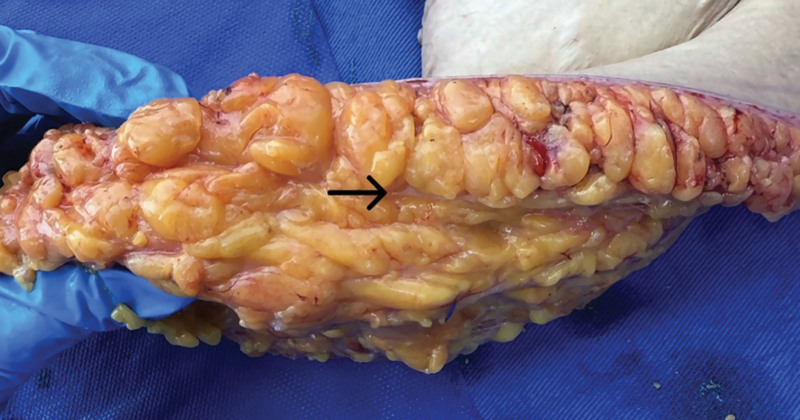
For clarity, subcutaneous tissue can be divided into 3 planes (Fig. 1 ): superficial, intermediate, and deep. The superficial layer lies between the dermis and Camper fascia, and the intermediate layer, between the Camper fascia and Scarpa fascia; both contain densely packed fat with strong fibroseptal attachments. 11 , 12 The superficial layer is particularly important, as its rich vascularity increases the risk of contour irregularities if not properly managed.
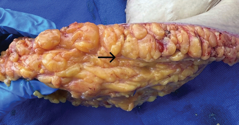
Fig. 1. Subcutaneous tissue (panniculus adiposus): superficial, intermediate, and deep planes. The black arrow indicates the Scarpa fascia.皮下脂肪三层(浅、中、深)示意,黑箭头指示 Scarpa 筋膜所在。The deep adipose layer, located between the Scarpa fascia and the premuscular (Gallaudet/innominate) fascia, 13 contains larger, loosely organized lobules, making aspiration easier and less prone to contour irregularities. 14 This layer is prioritized for emulsification and suction because of its greater regenerative capacity, 15 which reduces postoperative nodules. 8 For this article, the intermediate layer is considered part of the superficial plane.
Adipose tissue varies by anatomical region. In the supraumbilical area, the superficial layer is thicker in more than 70% of patients, whereas in the infraumbilical region it is approximately 55%–60% thicker than the deep layer. 16 Accordingly, the principal author routinely uses transcutaneous ultrasound to determine which plane requires greater emulsification or liposuction. In the back, deep fat is nearly 3 times thicker and more compact and fibrous than the superficial layer. In contrast, buttock and hip fat show little variation in thickness and feature small lobules with dense fibrous septa, 13 whereas medial-thigh fat is looser and less fibrous, increasing the risk of contour irregularities. 17
A retrospective cohort of 615 liposuction patients (aged 18–60 y, body mass index [BMI] < 30 kg/m 2 , nonsmokers) treated between January 2021 and December 2024 at the practice of the principal author in Bogotá, Colombia, was analyzed. Patients with incomplete records or comorbidities, including diabetes, coagulopathies, connective tissue or autoimmune disorders, respiratory disease, or biopolymer-related allogenosis, were excluded (n = 102).
Postoperative follow-up consisted of daily evaluations during the first 10 days, then monthly for 6 months. At the 6-month visit, patient satisfaction with the aesthetic result was assessed using the ANA scale (0–10) developed by Funk et al 18 (Table 1 ).
Table 1. Surgical and Postoperative Satisfaction Characteristics of the Studied Population (N = 615)
| Satisfaction Scale: ANA | n (%) |
|---|---|
| 0: Insufficient | 0 (0) |
| 1: Poor | 0 (0) |
| 2: Unsatisfied | 0 (0) |
| 3: Enough | 0 (0) |
| 4: Neutral | 0 (0) |
| 5: Acceptable | 0 (0) |
| 6: Satisfied | 6 (0.9) |
| 7: As agreed | 30 (4.9) |
| 8: Harmonic | 144 (23.5) |
| 9: Highly satisfied | 397 (64.5) |
| 10: Perfect | 38 (6.2) |
Data collected included demographics (sex, age), medical history, prior surgery, BMI, infiltration volume, technology used, and aspirated fat volume. Postoperative follow-up assessed complications and patient satisfaction.
For qualitative variables, absolute and relative frequencies (percentages) were calculated. Quantitative variables were summarized using the mean and SD, as they exhibited a normal distribution, which was assessed with the Shapiro–Wilk test. A P value less than 0.05 was considered statistically significant.
In accordance with the characteristics of the study and the guidelines established by the Colombian Ministry of Health Resolution 8430 of 1993, 19 as well as the ethical principles of the Declaration of Helsinki, 20 this research was classified as minimal risk. Respect for participants’ privacy, confidentiality of data, and personal integrity was ensured at all times.
Preoperative markings were performed with the patient in the standing position. ( See Video [online] , which demonstrates preoperative surgical marking, where the reader can see the anatomical areas to be delimited [in straight black lines] and the areas with excess fat deposits to be removed [black circles].) Subsequently, ultrasound was used to assess the thickness of the adipose tissue in the abdomen (both supra- and infraumbilical regions), waist, and back. This imaging evaluation provided precise and objective information regarding the depth and distribution of adipose tissue across the different anatomical planes, facilitating surgical planning for segmental liposuction and enabling tissue homogenization with ultrasound according to the specific characteristics of each area.
The procedure begins with the patient in the prone position. Access ports are made at the posterior axillary fold, over the sacrum, and along the upper two-thirds of each gluteal border. An additional port may occasionally be placed along the midline of the upper back, just below the bra-strap clasp.
With the patient in the supine position, 3 lower access ports are used; the central port is located on the midline, 6 cm from the vaginal introitus, allowing full access to the abdomen. At the lateral insertion of the rectus abdominis fascia and its transition toward the oblique muscles, 5 mm above the inguinal fold, a second and third port are created, positioned 1 cm lateral to the semilunar lines. Finally, 2 additional ports are placed in the inframammary fold, aligned longitudinally with the semilunar lines. All access ports are positioned within concealed anatomical areas (Figs. 2 , 3 ).
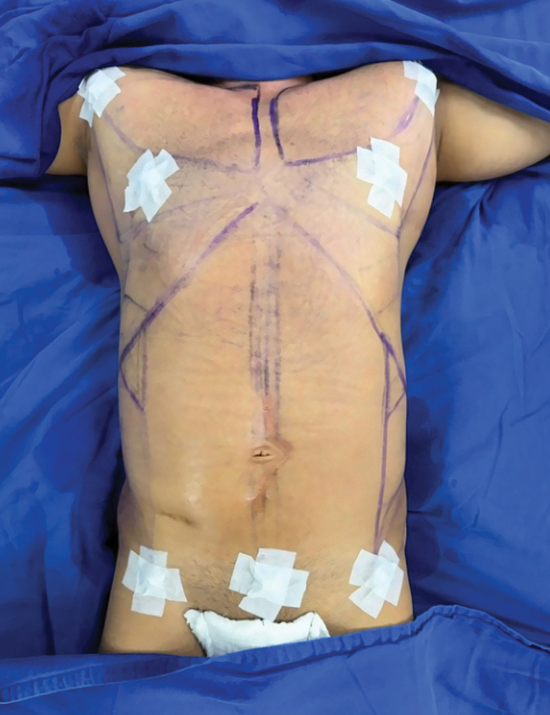
Fig. 2. Surgical ports on the anterior aspect of the patient.患者前面观的手术入路(穿刺)位点分布。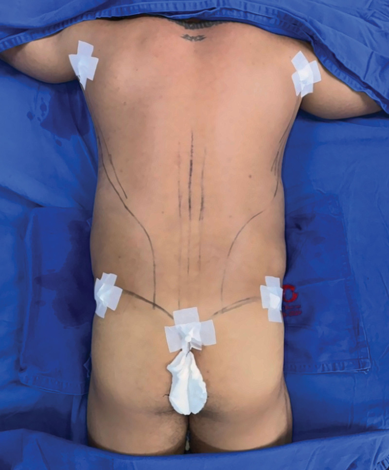
Fig. 3. Surgical ports on the posterior aspect of the patient.患者后面观的手术入路(穿刺)位点分布。A super-wet tumescent solution (per 1000-mL saline: 100-mg tranexamic acid, 1-mg epinephrine, 3 mL of 1% lidocaine without epinephrine [30 mg], and 1 mL [1 mEq] of sodium bicarbonate) was used. With a 3.5-mm multiport cannula and maintaining an angulation not exceeding 35 degrees, infiltration of the superficial plane is performed. For infiltration of the deep plane, the angulation is adjusted to 45 degrees, making a single perforation through the Scarpa fascia and maintaining this angle throughout the maneuver to avoid injury to the Scarpa fascia and the innominate fascia.
A minimum wait time of 10 minutes after infiltration is required to achieve an adequate vasoconstrictive effect. Emulsification of the adipose tissue is then performed using ultrasound-assisted technology (HEUS) with the FOCUS cannula, set at a power range between 40% and 80%, beginning in the superficial plane and subsequently treating the deep plane. Longitudinal strokes no longer than 20 cm are made from proximal to distal, maintaining the previously described angulations for each plane, with gentle movements and spacing between tunnels not exceeding 5 mm.
If the objective is adipose emulsification, ultrasound power should exceed 60%. In contrast, if the goal is fat dispersion and tissue homogenization—such as in areas with lower density, including the hips or abdomen with minimal adipose tissue and low fat percentage—power should remain less than 60%. 21 It is essential to emphasize that a 5-mm safety margin of subdermal tissue must always be preserved from the skin surface when working in the superficial plane.
Aspiration of the emulsified fat is then performed to reduce volumes and enhance body contour according to the initial surgical plan, using the MicroAire device (Charlottesville, VA) with a 4-mm cannula featuring 3 anterior Mercedes-type ports. Cannula trajectories are executed with suction applied from distal to proximal, beginning in the superficial plane and maintaining no more than 5 mm of spacing between each tunnel (Fig. 4 ).
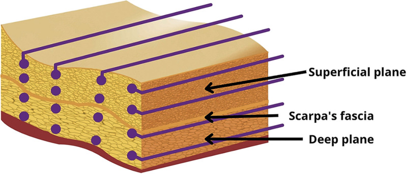
Fig. 4. Three-dimensional graphic representation of the management of superficial and deep planes during emulsification and liposuction. Courtesy of HEUS.浅层与深层在乳化及抽吸时的三维操作示意(HEUS 提供)。After aspiration, symmetry is verified. A sacral Penrose drain is placed for postoperative drainage, secured appropriately with 2-0 silk sutures, and is typically removed around postoperative day 5.
补充视频 / 附件(原文未随文提供原图,可于出版方页面查看)gox-14-e7790-s001.mp4Video 1 shows preoperative surgical marking, where the reader can see the anatomical areas to be delimited (in straight black lines) and those areas with excess fat deposits that are to be removed (black circles).术前标记视频:黑色直线标示待界定的解剖区,黑圈标示需去除的脂肪堆积区。
In the supraumbilical region, the superficial adipose layer is thicker; therefore, both emulsification and suction are performed in similar proportions. In contrast, in the deep plane, ultrasound emulsification is carried out predominantly, with minimal liposuction. These decisions are based on the morphological characteristics and cellular compartment distribution of the adipose tissue in this area. 22 , 23
In the infraumbilical region, the thickness of adipose tissue is, in most cases, similar in both the superficial and deep planes; however, in some patients, the deep plane may predominate. It is essential to perform emulsification and liposuction in equal proportions while preserving a dermoadipose flap less than 1.5 cm between the dermis and the muscular fascia.
In the waist and flank regions, the proportion of adipose tissue between the superficial and deep planes is similar; however, in these areas, the primary goal is to achieve greater skin contraction, which requires performing a higher proportion of superficial-plane liposuction. Symmetrical emulsification is carried out in both the superficial and deep planes.
In the dorsal region, specifically in the upper back, approximately 80% of the deep-plane thickness should be treated with ultrasound emulsification, with the remaining portion managed through liposuction. In the superficial plane, ultrasound and liposuction should be combined in equal proportions, aiming to preserve roughly 30% of the adipose tissue in the deep plane and 20% in the superficial plane. This approach reduces volume while creating a visual effect of increased trunk length and a slimmer contour.
In the lower back, at the lumbar segment, both emulsification and liposuction should be performed in the superficial and deep planes to achieve greater definition. Homogenization must always be performed using ultrasound.
For the anatomical marking of the linea alba and semilunar lines, emulsification is performed in the superficial plane to a greater extent than in the deep plane, accompanied by superficial-plane liposuction. Subdermal lacerations and perforations of the Scarpa fascia must be avoided, as any disruption of this fascia may lead to adherence zones and the formation of fibrotic cords, which are difficult to manage in both the early and late postoperative periods. 8
To conclude, the thickness of the superficial and deep subcutaneous planes is assessed using intraoperative ultrasound in both the supraumbilical and infraumbilical regions. This evaluation determines where additional suction or emulsification is required to achieve symmetry and homogenization.
A total of 615 patients were included: 461 women and 154 men; all of them underwent liposuction at the senior author’s private practice between January 2021 and December 2024 (Table 2 ). The mean age was 35.4 (±9.09) years, and the mean BMI was 24.6 (±3.23) kg/m 2 , both within the normal range. Overall, 65.8% of patients had a prior body-contouring procedure within 10 years.
Table 2. Demographic Characteristics of the Studied Population (N = 615)
| Variable | N = 615 |
|---|---|
| Sociodemographics | |
| Sex, n (%) | Female: 461 (75); male: 154 (25) |
| Age, * y | 35.4 (9.09) |
| BMI, kg/m 2 | 24.6 (3.23) |
| No. previous surgeries, n (%) | 405 (65.8) |
| Surgical features | |
| Tumescent solution infiltration volume, * mL | 6627 (1454) |
| Liposuction volume, * mL | 3426.3 (1224.2) |
| Use of Vaser (Vaser y HEUS), n (%) | 615 (100) |
| Complications, n (%) | |
| Seroma | 3 (0.49) |
| Hematoma | 2 (0.32) |
| Infection | 1 (0.16) |
| Necrosis | 0 (0) |
The mean tumescent volume infiltrated was 6627 (±1454) mL. All cases used ultrasound-assisted liposuction (VASER and HEUS), with power-assisted aspiration (MicroAire). Mean aspirated fat volume was 3426.3 (±1224.2) mL.
Postoperative follow-up included daily visits for the first 10 days and monthly thereafter (Figs. 5 A– 6 B). A low complication rate was observed, including 3 cases of seroma, 2 abdominal wall hematomas, and 1 mild superficial infection. All cases were managed conservatively with favorable outcomes. The hematomas, measuring approximately 4 × 7 cm, were diagnosed in the immediate postoperative period and treated successfully with drainage and tranexamic acid irrigation, without sequelae. No transfusions were required.
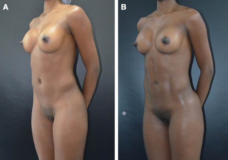
Fig. 5. Pre- and postoperative photographs of a female patient who underwent muscle definition by segmental liposuction. A, Presurgical photograph of a 30-year-old woman with a BMI of 19.6 kg/m2. B, Photograph taken 1 month postoperatively after segmental liposuction in the same patient.女性患者分段吸脂肌肉塑形术前与术后 1 个月对比。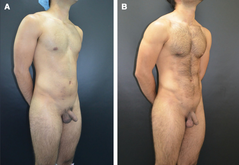
Fig. 6. Pre- and postoperative photographs of a male patient who underwent muscle definition by segmental liposuction. A, Presurgical photograph of a 26-year-old man with a BMI of 21.5 kg/m2. B, Photograph taken 1 month postoperatively after segmental liposuction in the same patient.男性患者分段吸脂肌肉塑形术前与术后对比。Regarding satisfaction with the surgical outcomes, assessed using the ANA scale, 100% of patients assigned scores greater than 6 out of 10; a rating of 9 out of 10 (high satisfaction) was the most frequently reported (Fig. 7 ). It should be noted that the extent of muscular etching was individualized for each patient, according to the preferences expressed during the preoperative evaluation.
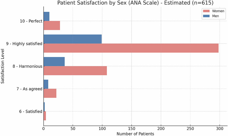
Fig. 7. Distribution of patient satisfaction (ANA scale).患者满意度(ANA 量表,0–10)分布,多数评分达 9 分。In the practice of the principal author, 103 lipectomy flaps from patients treated exclusively with this technique were examined histopathologically. Specimens consistently showed minimal fibrosis at the Camper fascia and Scarpa fascia (Figs. 8 A–D), reflecting normal repair. Fibrosis in this context should be understood as part of normal tissue repair. Although this is an isolated (single-center) analysis, the finding suggests that the technique preserves the integrity of the subcutaneous anatomical planes (superficial and deep), indicates favorable wound-healing dynamics, and aligns with the low complication rate observed in this cohort.
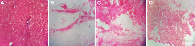
Fig. 8. A–D, Histological findings from an abdominal flap after segmental liposuction using the described technique: skin and Camper fascia (1); fibrous septa within adipose tissue (2); deep fibrosis of the Scarpa fascia (3); superficial fibrosis of the Camper fascia (4).术后腹部皮瓣组织学:Camper 与 Scarpa 筋膜仅见轻度纤维化。The pathology report is appended for the reader’s reference. ( See appendix, Supplemental Digital Content 1 , which shows the pathology report, https://links.lww.com/PRSGO/F91.)
Liposuction has progressed alongside improved understanding of adipose-tissue anatomy and regional variation, shifting from a subjective technique to a procedure grounded in anatomical and scientific principles. Systematic assessment of outcomes and complications has refined the technique, enabling safer, more natural, and longer lasting results.
Both superficial and deep-plane liposuction play essential roles in body contouring; when properly performed, each reduces volume while maintaining aesthetic harmony. 3 Neither plane should be considered inferior, as the optimal approach depends on regional anatomy.
Adipose tissue is organized into superficial and deep layers, each with distinct regional characteristics that vary by anatomical region. 13 Understanding these differences enables plane-specific strategies for safe and effective contouring. In the deep plane, liposuction or ultrasound-assisted emulsification should reduce volume while preserving a residual adipose thickness of approximately 5–7 mm (Figs. 9 A–D). The superficial plane, being more variable, demands individualized management: some areas may benefit from more aggressive liposuction, whereas others require only ultrasound, leaving residual subcutaneous fat less than 5 mm.
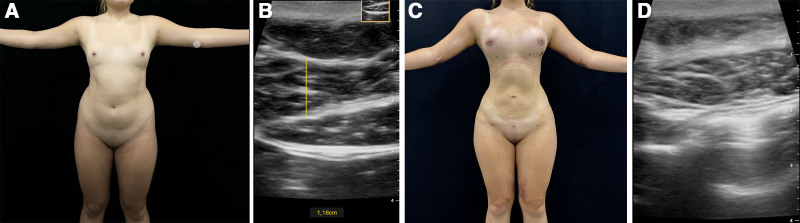
Fig. 9. Pre- and postoperative ultrasound images of a patient who underwent muscle delimitation by segmental liposuction. A and B, Ultrasound images demonstrating the superficial and deep planes of subcutaneous tissue in the supraumbilical abdominal region, obtained from a 21-year-old woman before surgery. C and D, Ultrasound images demonstrating the superficial and deep planes of subcutaneous tissue in the postsurgical supraumbilical abdominal region.术前术后超声图像,展示上腹浅层与深层脂肪厚度的变化。Structural differences between superficial and deep adipose layers require region-specific management, known as segmental liposuction. This approach applies tailored protocols to prevent lipodystrophic zones and avoid dermoadipose flaps greater than 1.5 cm. In patients with higher BMIs, residual thickness should be adjusted to baseline fat, skin laxity, and perfusion while preserving Scarpa fascia and ensuring uniform suction. This technique reduces irregularities and enables clear schematization and standardization, making it reproducible for plastic surgeons.
A key element is preserving the superficial (Scarpa) fascia, a dense collagenous layer interlaced with areolar tissue and elastic fibers. It allows the Camper fascia to glide over abdominal muscles, supports wall movement, 24 resists tensile forces, and stabilizes vessels and nerves.
Scarpa fascia is a key factor in reducing seroma formation after abdominoplasty. A systematic review by Van der Sluis et al 25 reported that its preservation lowers seroma incidence and allows earlier drain removal. Along with its functional properties, this underscores the importance of maintaining the Scarpa fascia during liposuction to decrease adhesions, seromas, and fibrosis, while ensuring homogeneous results and structural support. 8
In the supraumbilical abdomen, the superficial fat layer usually exceeds the deep layer. Thus, greater ultrasound emulsification is recommended in the superficial plane, combined with liposuction in both planes for adequate debulking. In the infraumbilical region, anatomical variability requires balancing ultrasound and liposuction across both planes to achieve definition, maintain residual fat less than 1 cm, and preserve skin perfusion.
In the flanks and waist, fat is evenly distributed between planes. Therefore, ultrasound should address both, whereas liposuction is confined to the superficial plane to preserve support and enhance skin retraction.
In the back, treatment varies by region. In the upper back, where thicker fat deposits create folds and broaden the trunk, ultrasound emulsification should reduce both adipose layers by more than 50%, followed by superficial-plane liposuction to refine contour. 22 This decreases fat volume and weight of fat on the skin, eliminating folds and producing a slimmer, elongated, more youthful appearance, as illustrated in cases. These results were consistently achieved with preoperative external ultrasound.
In the lumbar region, the surgical approach differs because the objective is greater definition with thinner dermoadipose flaps. Accordingly, ultrasound emulsification and liposuction are performed in both fat planes, bearing in mind that the deep plane is thicker. 14 Importantly, in body segments where the deep panniculus exceeds 1.5 cm and the goal is an etched, anatomically defined contour, deep-plane liposuction should be carried out but confined to its most superficial stratum immediately deep to the Scarpa fascia.
In liposuction, especially in high-definition liposuction, the objective is to enhance an athletic appearance by highlighting specific muscle groups. This is achieved through superficial-plane etching and suction with cannulas. 26 , 27 However, despite its popularity, techniques seeking maximal definition have been linked to complications, such as skin dyschromia, nodules, contour irregularities, seromas, hematomas, and even burns. 27 , 28
Complications in high-definition procedures highlight the need to reassess techniques and redefine paradigms to optimize outcomes and minimize adverse events. Thus, liposuction should be limited to the superficial plane, avoiding the subdermal plane.
Superficial-plane liposuction must preserve a subdermal dermoadipose flap thicker than 5 mm to prevent complications described in the literature. 8 Deep-plane liposuction remains essential given regional thickness variations and its role in muscular definition, with fewer fibrosis-related issues. Techniques should therefore be adapted to anatomical differences rather than applied uniformly, ensuring natural and anatomically accurate results.
Ultrasound is crucial in liposuction, enabling greater than 50% adipose reduction through emulsification while minimizing vascular injury and contour irregularities. Cannula-based liposuction complements this by removing residual fat and refining contours, provided the correct suction plane and fascial integrity are maintained.
Liposuction requires a substantial learning curve, as misidentifying adipose planes increases complication risk. This study presents our experience and a technique yielding low complication rates and high satisfaction for both patients and surgeons.
Segmental liposuction allows precise muscular contouring with fewer complications and improved aesthetic outcomes. Its effectiveness depends on an anatomically tailored approach, as adipose distribution varies by region. Preserving the Scarpa fascia is essential because its disruption increases the risk of adhesions, contour irregularities, and other complications.
For optimal skin contraction, liposuction should remain superficial to the Scarpa fascia; perforation, particularly in areas lacking muscular support (linea alba, supraumbilical, mid-abdomen), predisposes to thickening and “guitar-string” deformities. 8 In deeper regions (supraumbilical and infraumbilical regions, waist), ultrasound emulsification with controlled suction less than or equal to 60% is recommended for safety and consistency. In superficial liposuction, maintaining an approximately 5-mm subdermal dermoadipose flap is critical to protect the Camper fascia and prevent complications such as telangiectasias or livedo reticularis.
The authors have no financial interest to declare in relation to the content of this article.
本文报道一项 615 例回顾性队列,系统描述了「分段吸脂」技术:依据浅层(Camper/Scarpa 筋膜浅面)与深层脂肪的不同解剖与再生特性,分区域定制超声乳化与机械抽吸的比例,并全程借助术中超声评估层次厚度。其核心安全要点是保护 Scarpa 筋膜——该层损伤会增加粘连、血清肿与轮廓不规则的风险;浅层操作建议保留约 5 mm 的皮下皮脂瓣。
研究中总体并发症率较低(血清肿 0.49%、血肿 0.32%、感染 0.16%),且 103 例组织学标本仅见轻度纤维化,提示该技术可较好地维持皮下层次完整性;患者满意度(ANA 量表)普遍较高。需注意本研究为单中心回顾性、缺乏对照组,结论更宜视为术者经验总结。临床落地时应重视学习曲线与层次辨识,避免过度追求极致雕刻而增加风险。
声明:中文精读 · 仅供学术参考。内容来自公开文献检索,不代表本人观点,不构成诊疗建议。 医疗美容需在正规医疗机构由执业医师实施。