脂肪的机械处理:从抽吸物到基质血管成分浓缩物
2026年9月25日26 min read
Plastic and Reconstructive Surgery
结论与实践要点
本文介绍了一套围手术期即可完成的机械性脂肪处理流程,将传统需要酶消化、耗时且引入外源物质的SVF获取方式,简化为纯机械分馏与离心。流程分三步:抽取的脂肪经洗涤离心得到Coleman脂肪;再通过鲁尔锁接头反复推挤(增加剪切力)制成nanofat;最后对nanofat行短时高速离心沉降出SVF浓缩物。
流式结果显示,SVF浓缩物每毫升活ASC数量约为轻柔处理Coleman脂肪的2倍,同时保留血管内皮细胞与巨噬细胞;增殖实验中nanofat来源细胞汇合时间(19±5天)略长于Coleman脂肪(16±4天,P<0.001),但差异较小、仍具增殖能力。组织学与细胞学证实SVF浓缩物富含细胞、核密度高,并存在毛细血管断片附着的“微移植物”。
临床提示:该浓缩物既可单独注射用于再生,也可回混入未处理脂肪制成“干细胞富集脂肪”移植,且无需特殊设备、易于开展。需注意“蛋黄酱问题”——含水较多的抽吸物易乳化而难以离心,作者给出补救操作;本研究为离体实验,活细胞功能与体内转归仍需进一步验证。
Background:The adipose stromal vascular fraction (SVF) is a mesh of connective tissue, capillaries, stem cells, and supportive cells that may have regenerative properties. Attention is shifting from enzymatic isolation and expansion of the stem cell population alone to the benefit of preserving the SVF as a functional unit. Enzymatic breakdown of lipoaspirate is time-consuming and involves use of foreign substances. The authors have established a simple procedure for perioperative mechanical isolation of SVF. This study investigates how mechanical fractionation and centrifugation changes the qualities of lipoaspirate.
Methods:Three derivatives of manipulated fat were defined: washed and centrifuged lipoaspirate (Coleman fat), mechanically fractionated Coleman fat (nanofat), and SVF concentrate obtained by hard centrifugation of nanofat. The end products were characterized by flow cytometry, proliferation capacity measured as time to confluence, and histologic and cytologic characteristics.
Results:Viable stem cells are preserved during vigorous mechanical fractionation of dry lipoaspirate to nanofat. A sedimented SVF concentrate from centrifugated nanofat has a high stem cell concentration. A nanofat “mayonnaise problem” may require a salvage procedure to enable centrifugal separation. Proliferation is significantly but marginally delayed in SVF cells from fractionated fat versus nonmanipulated lipoaspirate. Histology and cytology demonstrate the presence of micrografts in SVF concentrate (ie, capillary fragments with SVF cells attached).
Conclusions:SVF concentrate manufactured by mechanical fractionation and centrifugation contains viable cells capable of proliferation. SVF concentrate contains vascular micrografts that may be injected alone for regenerative purposes or added to nonmanipulated lipoaspirate for stem cell–enriched fat transfer.
Clinical Relevance Statement:The procedures described in this article, and the illustrative videos, are relevant to all plastic surgeons involved in regenerative fat grafting. The methods require no special equipment and can be instantly adopted.
When Sidney Coleman introduced the concept of structural fat grafting in 1995, 1 he stressed the importance of an atraumatic technique to preserve the vulnerable adipocytes. Early users of the Coleman technique noted that in addition to correcting volume defects, fat grafts also exerted a regenerative effect on damaged tissues. The therapeutic softening and increased mobility of fibrosis in irradiated tissue was particularly noteworthy. 2 , 3 When Zuk et al. 4 demonstrated the abundance of mesenchymal stem cells in fat in 2001, the regenerative properties of fat grafts were attributed to the adipose-derived stem cells (ASCs). Devices for enzymatic breakdown of fatty tissue to isolate the ASC population soon ensued, along with laboratory procedures for ASC expansion. However, the addition of allogeneic products such as collagenase to fat grafts was questioned by regulatory authorities, and ASC isolation requiring advanced devices or transfer to laboratories proved expensive and time consuming, and had a risk of contamination and loss of cell “stemness.” 5 , 6
Rigotti et al. 7 postulated that rather than isolating single ASCs, an intact natural scaffold of the regenerative precursor and supportive cells of the stroma—the stromal vascular fraction (SVF)—might provide synergistic regenerative cell units. To isolate the SVF, Rigotti et al. 7 disrupted the adipocytes by means of hard centrifugation of the fat. After discarding fluids and the surplus oil, the resulting SVF was named “purified lipoaspirate,” although very few adipocytes remained intact. Injections into radiation ulcers in patients with breast cancer demonstrated neovascularization and a regenerative effect of the connective tissue, with significant clinical improvement of these otherwise untreatable ulcers. 7
In 2013, Tonnard et al. 8 suggested a nomenclature for mechanically manipulated fat grafts. Macrofat refers to regular lipoaspirate from regular harvesting cannulas; microfat is lipoaspirate obtained with smaller-diameter harvesting cannulas, providing small enough grafts to pass through narrow inserting needles or cannulas. Tonnard et al. 8 described the use of a 3-mm multiport harvesting cannula with sharp 1-mm openings, which has since been referred to as the Tonnard cannula. Tonnard et al. 8 further fractionated the microfat by vigorous mechanical shifting between syringes, using small-diameter Luer lock connectors until emulsification occurred. After filtration of the emulsified microfat, the resulting liquid was named nanofat and could be injected through even very small needles. The nanofat had few surviving adipocytes but would contain fragments of the SVF. These fragments have been termed micrografts, as they may consist of regenerative SVF cells adhering to fragments of extracellular matrix scaffolding and capillaries capable of microintegration and neovascularization.
Condensed lipoaspirate can be fractionated, followed by centrifugation to further concentrate the SVF, described in the literature under the terms fractionation of adipose tissue (FAT)–SVF , 9 SVF gel , 10 lipoconcentrate , 11 and tissue SVF . 12 We suggest the phrase SVF concentrate as a precise description of this product.
The purpose of this study is to analyze how different degrees of mechanical manipulation affects both adipocytes and the SVF in lipoaspirate, thereby providing a guide on how to mechanically process fat grafts in various clinical settings.
The study received funding from the Aleris Research Fund and St. Olav’s Research fund and was approved by the Regional Committee for Medical and Health Research Ethics in Mid Norway. Lipoaspirate was obtained from 20 consenting patients undergoing abdominoplasties at St. Olav’s University Hospital and Aleris Medical Center, in Trondheim, Norway. Age, sex, and body mass index (BMI) were registered for each patient.
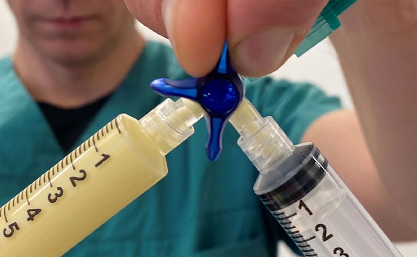
When liposuction was part of the abdominoplasty, the lipoaspirate was collected. When liposuction was not part of the procedure, lipoaspirate was obtained by dry suction of the resected abdominal tissue. Coleman fat was obtained by twice rinsing lipoaspirate with equal volumes of saline followed by centrifugation for 5 minutes at 850 × g . The superior oily fraction from centrifuged Coleman fat was stored for salvage of the potential “mayonnaise problem,” described in the following. Nanofat was obtained by subjecting Coleman fat to 20 vigorous single passages through an open Luer lock stopcock (10 times back and forth), followed by another 20 single passages through a partially closed Luer lock stopcock (Fig. 1 ). Note that Coleman fat in our setting is condensed fat with the aqueous fraction removed, whereas for the original nanofat, there was no centrifugation of the lipoaspirate before emulsification. SVF concentrate was obtained through centrifugation of the nanofat for 5 minutes at 1200 × g . Various combinations of force and duration 10 , 11 were used to achieve the desired outcome—effective separation and avoidance of the mayonnaise problem. ( See Video 1 [online] , which demonstrates the process of obtaining SVF concentrate from lipoaspirate [https://youtu.be/Zgsz3zvvu4g]. Copyright © Kjersti Ausen.)
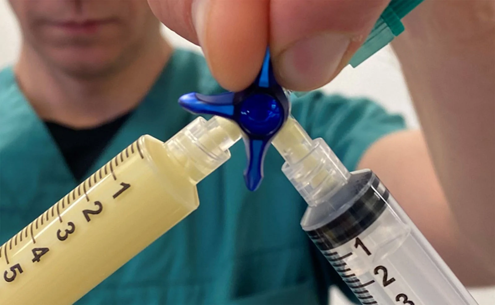
Fig. 1. A Luer lock stopcock used for fat fragmentation. Fat is transferred through a partially closed Luer lock stopcock, which reduces the lumen diameter and produces finer fragmentation of the adipose tissue.用于脂肪分馏的鲁尔锁接头:部分关闭以缩小管腔,使脂肪被剪切成更细颗粒。
Processing lipoaspirate into nanofat as described by Tonnard et al. 8 involves vigorous shifting of fat through a narrow lumen. This fractionation of the fat may lead to flocculation or emulsification (Fig. 2 ). A flocculated fluid will separate into oil, SVF concentrate, and aqueous portions upon centrifugation, whereas an emulsified substance remains as a homogenous, mayonnaise-like creamy layer above the aqueous fraction (Fig. 3 ). The mayonnaise problem occurs very frequently when using lipoaspirate with a significant water content, but only rarely occurs when using condensed (dry) lipoaspirate that has been sufficiently centrifuged before fractionation. When this phenomenon occurred despite having condensed Coleman fat by centrifugation for 5 minutes at 850 × g , we used a salvage procedure as published by Yao et al. 10 by adding 0.5 mL of oil obtained from the previous Coleman fat centrifugation. By gently shifting the added oil and the emulsion 6 to 8 times through a stopcock, flocculation occurred and centrifugation was repeated. If nanofat seemed to have turned to mayonnaise before centrifugation, leaving the sample standing for a short while could clarify whether the emulsion would spontaneously separate or whether a salvage procedure was needed. If the fat showed signs of spontaneous flocculation and moved like fluid, then we would attempt centrifugation. If the fat remained homogenous and creamy, and did not move like fluid, then the salvage procedure was performed immediately before the first centrifugation (Fig. 2 ). ( See Video 2 [online] , which demonstrates the mechanical manipulation of lipoaspirate to SVF concentrate and the salvage procedure for the mayonnaise problem; https://youtu.be/fupn36oL0K8. Copyright © Kjersti Ausen.)
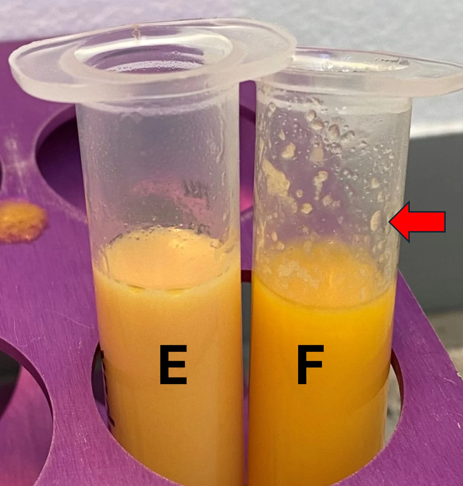
Fig. 2. A photograph of 2 fat samples illustrating outcomes after shifting Coleman fat through a stopcock. On the left, E illustrates emulsified lipoaspirate, which is whitish and creamy and appears stiff when the syringe is tilted. On the right, F represents flocculated lipoaspirate, which moves more like fluid when tilted, and the fluid will leave behind flocks adhering to the sides of the syringe (red arrow).经接头推挤后两种脂肪外观:左为乳化状(奶油样、倾斜发硬),右为絮凝状(流动、挂壁)。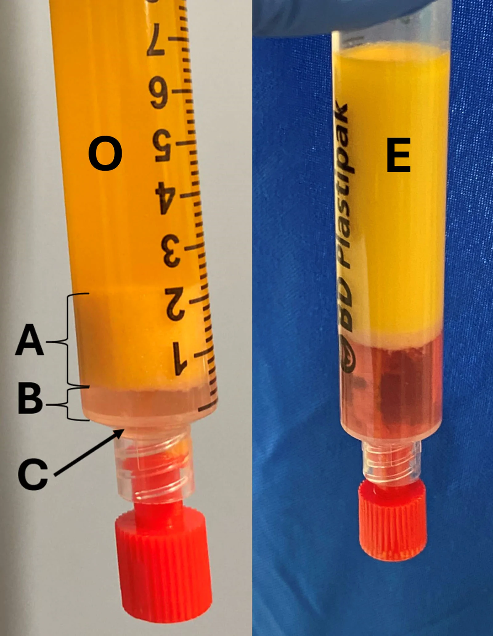
Fig. 3. A photograph of 2 separate syringes showing flocculated fat (left) and emulsified fat (right) after centrifugation for 5 minutes at 1200 ×g. The flocculated fat (left) has separated into an upper oily fraction (O), a fraction of microfragmented SVF and cellular debris (A), an aqueous fraction (B), and a small pellet of single cells from the SVF (C). The emulsified fat (right) has separated into an aqueous fraction; oil, cells, and debris remain a stable emulsification (E).离心后絮凝脂肪分层为油相/SVF碎屑/水相/细胞沉淀;乳化脂肪维持稳定乳状。The samples were submitted to several investigations, described in the following.
The release of oil from ruptured adipocytes was used as a surrogate measure of adipocyte resilience to mechanical manipulation. Samples of Coleman fat from the same patient were subjected to increasing degrees of mechanical manipulation. The supernatant oil fraction obtained after centrifugation of the samples for 5 minutes at 1200 × g was assumed to be proportional to adipocyte damage.
Counting and characterization of the SVF cell population from Coleman fat, nanofat, and SVF concentrate from 13 different patients was performed through enzymatic isolation, immunophenotypic staining, and flow cytometry analyses of SVF cells according to a protocol based on Zuk et al. 4 and published by Alstrup et al. 13 SVF cells were stained with 7-aminoactinomycin D for viability and with antibodies for cell surface markers CD31, CD34, CD45, CD73, CD90, and CD105, following the manufacturer’s recommendations. In the live cell population, ASCs, endothelial cells, and macrophages were chosen for final presentations. ASCs were defined as cells with CD90+/34+/73+/105+/45−/31− surface staining, endothelial cells were defined as CD31+CD45− cells, and macrophages were defined as cells with CD45+/side scatter–high. Samples were run on a FACSCanto 2 flow cytometer and analyzed with Infinicyt 2.0 software (Cytognos).
Enzymatically isolated cells from Coleman fat and corresponding nanofat from 6 different donors were filtered through a 100-µm cell strainer. Without any further immunophenotypic identification or live/dead analyses, 75,000 cells in mesenchymal stem cell culture medium (15% fetal bovine serum in MEM-α + 1% penicillin/streptomycin solution) were seeded per well in 6-well plates, with a total of 33 parallel samples, to measure time to confluence, defined as the time needed for the cells to cover 80% of the well surface assessed by visual microscopy. The protocol was run separately for the parallel samples obtained from each donor.
Histologic slides of Coleman fat and SVF concentrate were made from formaldehyde-fixed samples embedded in paraffin blocks to create hematoxylin & eosin (H&E)–stained slides. CD31 staining was performed for better illustration of vascular structures. Smears of SVF concentrate were subjected to H&E staining for visualization of micrografts.
补充视频 / 附件(原文未随文提供原图,可于出版方页面查看)prs-158-435e-s002.mp4Video 2. This video demonstrates the mechanical manipulation of lipoaspirate to SVF concentrate and the salvage procedure for the mayonnaise problem; https://youtu.be/fupn36oL0K8. Copyright © Kjersti Ausen.
Hanks balanced salt solution, collagenase type IV, fetal bovine serum, phosphate-buffered saline, minimum essential medium (MEM alpha), and penicillin–streptomycin (10,000 U/mL penicillin/10,000 µg/mL streptomycin [15140-122]) were obtained from Thermo Fisher Scientific. Bovine serum albumin, RPMI-1640 growth medium, and EDTA were obtained from Sigma. Viability dye (7-aminoactinomycin D) and the following monoclonal, fluorescence-conjugated antibodies were obtained from BD Biosciences: CD31-FITC (555445), CD73-PE (550257), CD90-PE-Cy7 (561558), CD105-APC (562408), CD34-BV421 (562577), and CD45-HRZ V500-C (655873).
Statistical analyses were performed using IBM SPSS Statistics version 26. Descriptive continuous variables are reported as median (range) or mean ± SD, as appropriate depending on distribution. Comparison of continuous variables from paired samples of fat was done using the Wilcoxon signed rank test. Correlation between continuous variables was assessed using Spearman correlation. Two-tailed P values less than 0.05 were considered significant.
补充视频 / 附件(原文未随文提供原图,可于出版方页面查看)prs-158-435e-s001.mp4Video 1. This video demonstrates the process of obtaining SVF concentrate from lipoaspirate (https://youtu.be/Zgsz3zvvu4g). Copyright © Kjersti Ausen.
Sex, age, and BMI of participating patients, as well as total cell count, ASC cell count, ASC fraction of total cell count, and SVF concentrate volume from Coleman fat from participating patients, is presented in Table 1 .
Table 1. Patient and Coleman Fat Characteristicsa
| Patient Characteristics | Values |
|---|---|
| Female sex | 18 (90) |
| Age, yrs | 45 (20–67) |
| BMI, kg/m 2 | 25.8 (21.0–31.1) |
| Coleman fat characteristics ( n = 13) a | |
| Live total cell count, cells ×10 6 /mL | 1.32 ± 0.24 |
| ASCs (CD90+/34+/73+/105+/45−/31−), cells ×10 6 /mL | 0.18 ± 0.03 |
| ASCs part of total cell count, % | 48.9 (23.3–73.1) |
| SVF concentrate obtained from 10 mL of Coleman fat, mL | 2.0 (1.4–3.1) |
Slow shifting of 10 mL of Coleman fat 6 times 1 way through an open Luer lock stopcock, as one could do to homogenize or reduce the size of fat particles before grafting of the lipoaspirate, did not significantly increase the oily fraction after centrifugation compared with unmanipulated Coleman fat (representative samples illustrated in Fig. 4 , samples A and B). Quick shifting of Coleman fat 20 times 1 way through an open Luer lock stopcock significantly increased the oily fraction after centrifugation, but approximately half of the fatty tissue still had intact adipocytes (Fig. 4 , sample C). Nanofat was produced by shifting the fat first 20 times 1 way through an open Luer lock stopcock and then an additional 20 times through a partially closed stopcock. Centrifugation of nanofat resulted in a large oily fraction with a bottom sediment, the SVF concentrate, pellet, and fluid containing water-soluble cytokines and growth factors from ruptured macrophages (Fig. 4 , sample D). When discarding the oil only and keeping SVF concentrate, pellet, and remaining fluid, the mean volume obtained from 10 mL of Coleman fat was 2.1 mL (1.5 to 3.3 mL). The volume of this SVF concentrate was negatively correlated with increasing age (Spearman correlation rs = −0.72, P = 0.005). Increasing BMI also showed a trend toward being correlated with less SVF concentrate, but this was not statistically significant (rs = −0.50; P = 0.08). Age and BMI were not correlated (rs = 0.12, P = 0.62).
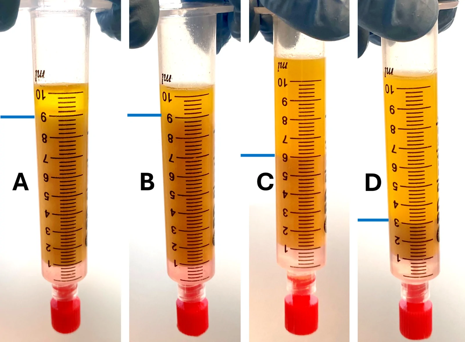
Fig. 4. Four samples of fat from the same patient subjected to increasing shear forces. Oil fractions (oil above the blue line) represent ruptured adipocytes after 5 minutes of centrifugation at 1200 ×g. Sample A, Coleman fat; sample B, Coleman fat after an additional 6 slow one-way open passes through an open Luer lock stopcock; sample C, Coleman fat after adding 20 swift one-way open passes; sample D, nanofat, having added another 20 one-way passes through a partially closed stopcock.同一患者脂肪接受递增剪切力,油相(破裂脂肪细胞)随处理增强而增多。
The mechanical fragmentation to turn Coleman fat into nanofat resulted in a lower number per milliliter of live ASCs and macrophages identified by flow cytometry, whereas the number of endothelial cells increased (Fig. 5 ). After centrifugation of nanofat for 5 minutes at 1200 × g , the resulting SVF concentrate contained approximately twice as many live ASCs per milliliter as gently manipulated Coleman fat. The highest number of macrophages per milliliter was found in Coleman fat. Macrophage concentration per milliliter was larger in SVF concentrate than in nanofat. The number of ASCs per milliliter in Coleman fat did not significantly decrease with increasing age (Spearman correlation rs = −0.19, P = 0.54). There was a not significant trend toward lower ASC count per milliliter with increasing BMI (rs = −0.45, P = 0.13).
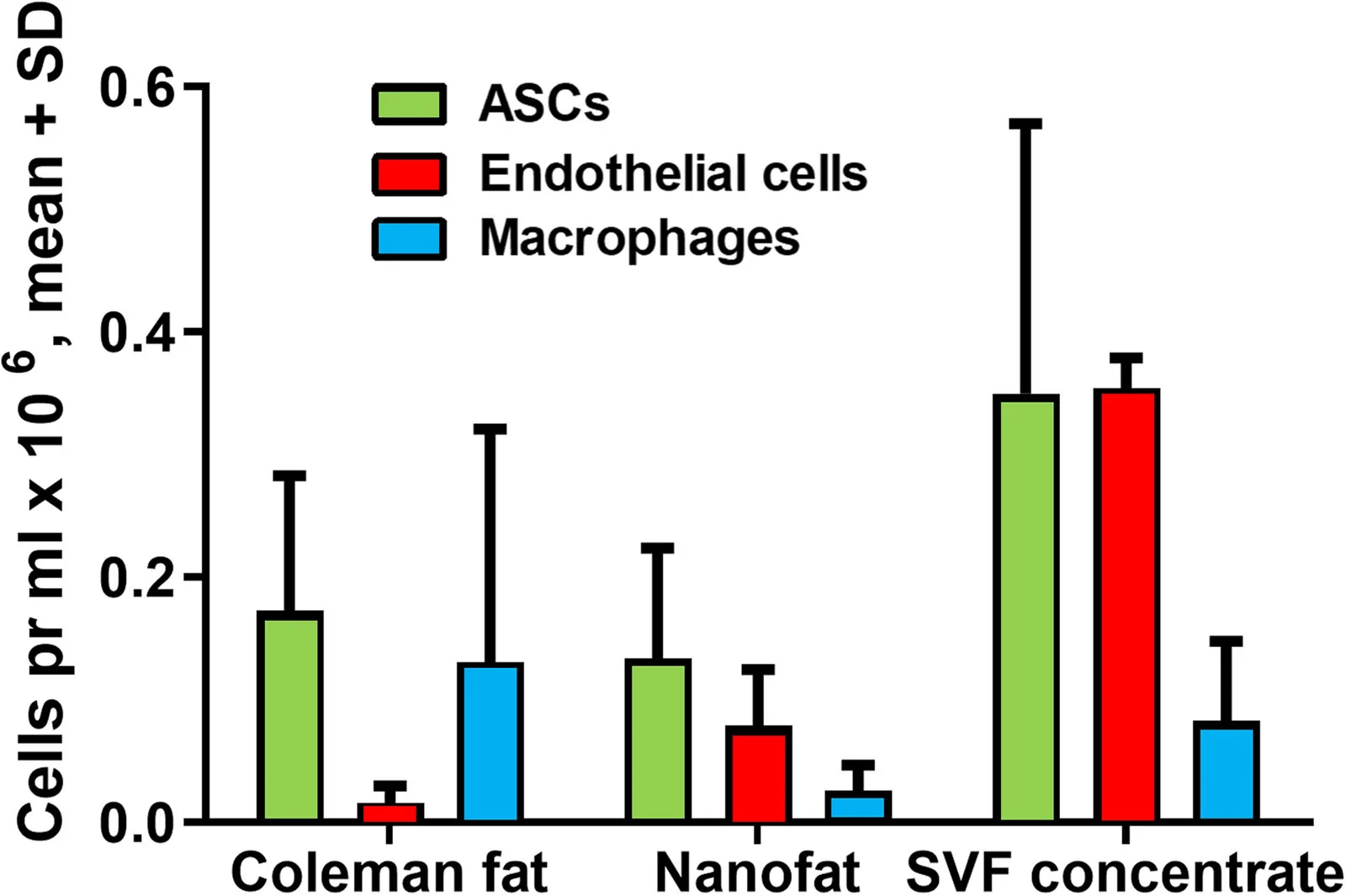
Fig. 5. The number of ASCs, endothelial cells, and macrophages per milliliter of Coleman fat, nanofat, and SVF concentrate as counted by flow cytometry. Parallel samples are from 13 different donors.流式细胞术计数Coleman脂肪、nanofat与SVF浓缩物中ASC、内皮细胞与巨噬细胞密度。
Cells derived from Coleman fat reached confluence in 16 ± 4 days on average, whereas cells derived from nanofat required 19 ± 5 days, a significantly longer period ( P < 0.001), although substantial interindividual differences were observed (Fig. 6 ).
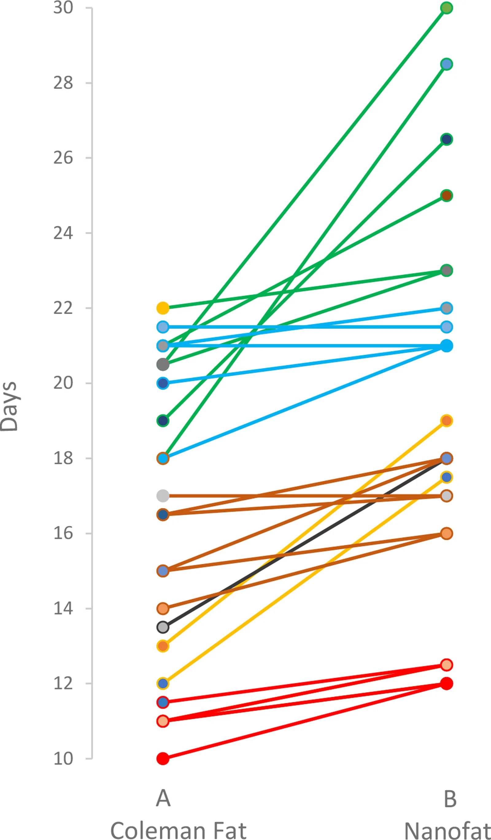
Fig. 6. Days needed for seeded single cells from Coleman fat and nanofat to reach confluence, defined as coverage of 80% of well surface as determined by microscope inspection. A total of 33 parallel samples are from 6 different patients. Samples from the same patient have the same color.种植细胞达汇合所需天数:Coleman脂肪平均16±4天,nanofat 19±5天。
Histological slides were obtained from formaldehyde-fixed Coleman fat and SVF concentrate (Fig. 7 ). Coleman fat displayed an intact structure of the adipose tissue, whereas SVF concentrate consisted of fragmented tissue with ruptured adipocytes but a much higher density of cell nuclei and blood vessels. Cytological smears of SVF concentrate revealed a high concentration of cells and the presence of micrografts (ie, capillary fragments with both larger preadipocytes and smaller SVF cells attached) (Fig. 8 ).
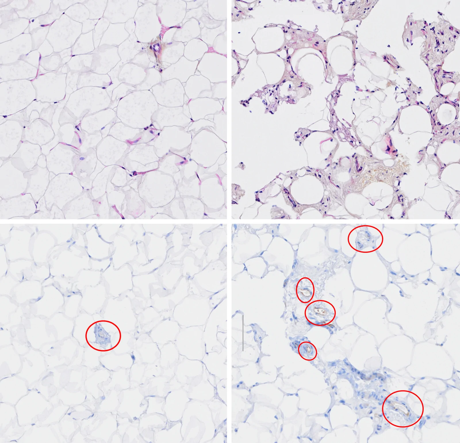
Fig. 7. Histological slides. (Above) Slides on the left show Coleman fat; slides on the right show SVF concentrate (H&E staining; magnification ×20). (Below) CD31 staining has been added to the slides to mark blood vessels (red circles).组织学切片:SVF浓缩物结构破碎、核与血管密度更高,CD31标记血管(红圈)。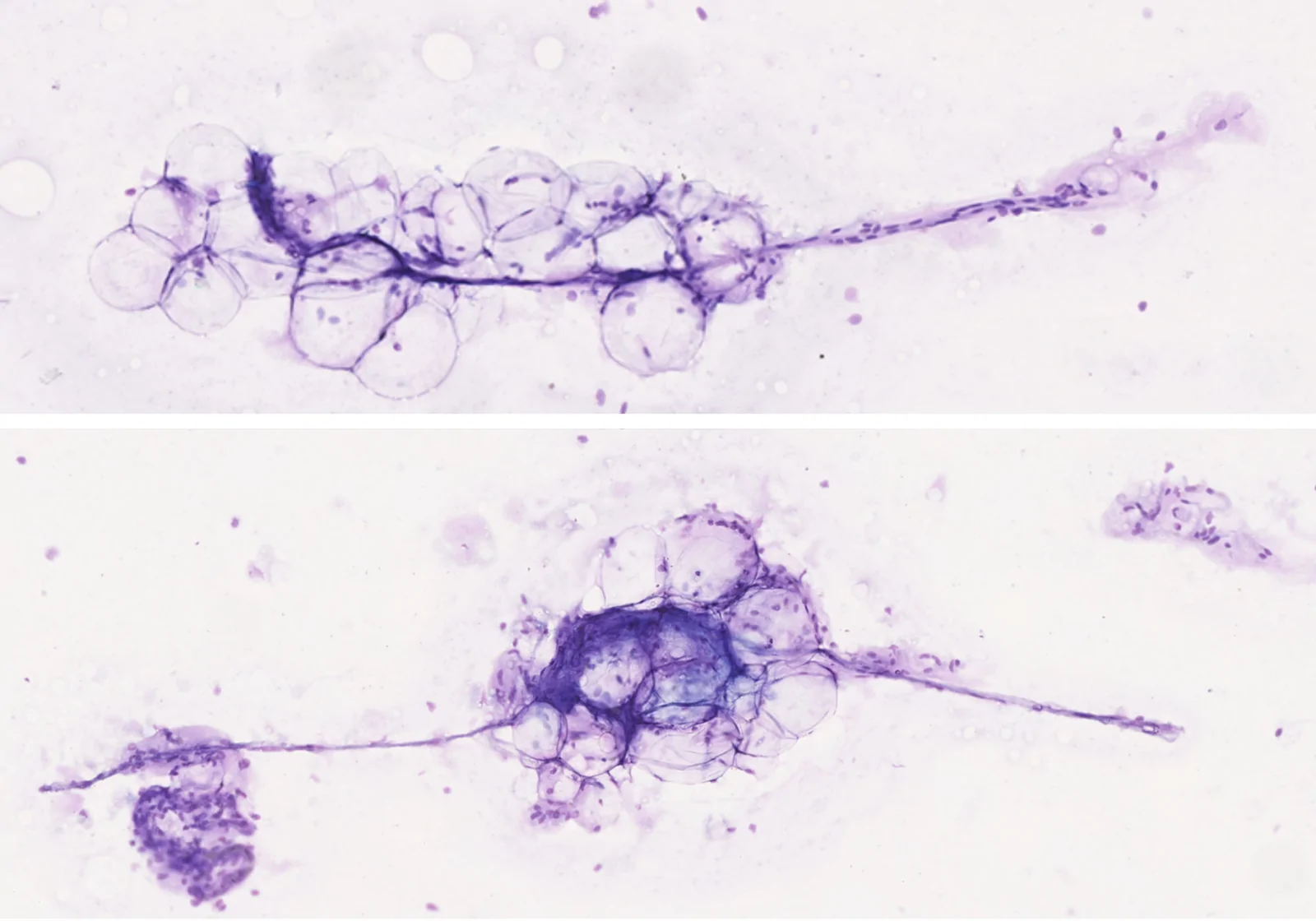
Fig. 8. Cytological smears of SVF concentrate (H&E staining; magnification ×10). Note the density of cell nuclei and the presence of micrografts (ie, capillary fragments with attached adipocytes and SVF cells).SVF浓缩物细胞学涂片,可见高细胞密度与附壁毛细血管断片(微移植物)。
The purpose of this study was to characterize and compare the different versions of fat grafts plastic surgeons can obtain during a surgical procedure by mechanical manipulation of the lipoaspirate, without time-consuming advanced manipulation requiring laboratory facilities, expensive kits, or additives, such as collagenase. Except for flow cytometry for cell characterization, the analyses performed in this study are fairly crude and basic. This study did not aim to compete with the plethora of high-quality laboratory studies investigating the survival and proliferative qualities of isolated ASCs and SVF. 12 , 14 The study was limited by the lack of clinical outcome measures, which should be investigated in a clinical trial. Because SVF has a resilient structure and no particular means of preparation seems superior to others, the therapeutic potential of our SVF concentrate is likely similar to that reported in other clinical trials. 14 , 15 The findings of this study can serve as a practical guide for everyday fat grafting practice.
Mechanical fragmentation by passing lipoaspirate through a narrow lumen, thereby tearing the cellular structures apart, is undoubtedly traumatic to the tissue. The number of macrophages, which are large and fragile cells, was particularly reduced. Endothelial cell count was increased in nanofat, likely reflecting how mechanical disruption of capillaries results in small vessel fragments that are more accessible to collagenase digestion, yielding a higher number of single cells. 13
Similar to the original publication by Tonnard et al., 8 we found that this vigorous processing destroyed most adipocytes and significantly reduced the number of ASCs. However, although the cells of nanofat may be traumatized, obtaining SVF concentrate from centrifugation of nanofat still resulted in a product with twice the ASC count per milliliter compared with Coleman fat.
Gentler mechanical distortion of lipoaspirate—for example, using a laboratory tissue dissociator, as demonstrated by Alstrup et al. 16 —can increase ASC yield, as smaller fat fragments are more completely dissolved by collagenase, resulting in greater release of single cells. The potential to increase the single cell yield of ASCs by gentle mechanical dissociation before enzymatic digestion is of interest, primarily in laboratory stem cell research, but is beyond the scope of this article.
The mayonnaise problem may present a challenge. In the original publication by Tonnard et al., 8 an illustrative photograph of nanofat demonstrates a mayonnaise-like emulsion, and there is no mention of further centrifugation of the nanofat. Some authors have pointed out the lack of top oil after centrifugation of emulsified fat (Fig. 3 ), interpreting this as indicating that the adipocytes are intact 17 and that water content in the lipoaspirate may protect adipocytes during fractionation. However, the “mayonnaise” is not composed of intact adipocytes but is rather a stable emulsion of particulate matter, oil, and water droplets. Shifting the balance of contents (eg, by removing water or adding oil) can alter the balance of an emulsion, and emulsification occurs less frequently if the lipoaspirate has been sufficiently condensed (eg, centrifuged for at least 3 to 5 minutes at 850 × g to 1200 × g to remove the water content). Because the mayonnaise problem can still occur despite such centrifugation, “salvage oil” can be prepared at the beginning of the procedure by hard centrifugation of unmanipulated lipoaspirate. Adding this oil can convert the emulsion into a centrifugable flocculated mixture. 10
This study demonstrates that vigorous mechanical fractionation of fat results in a concentration of micrografts containing viable cells capable of proliferation. We found that cell suspensions subjected to trauma needed significantly more time to proliferate to confluence. However, in this experiment, we merely counted and seeded an equal number of the cells achieved after collagenase digestion. Furthermore, for the proliferation studies, we neither differentiated cell types nor ran a live/dead assay; hence, among the 75,000 cells seeded from crushed nanofat, some “cells” may have been nonviable, contributing to a slower time to confluence. Nanofat cells from the first 3 patients needed 4 to 7 days more than Coleman fat cells to reach confluence; nanofat cells from the last 3 included patients only needed 1 to 3 extra days.
We demonstrated that adipocyte rupture is proportional to the degree of mechanical fragmentation (Fig 4 ). Furthermore, with vigorous fractionation combined with hard centrifugation, the functional units of SVF micrografts are ripped apart. Yao et al. 10 demonstrated that the scaffolding (extracellular matrix) of SVF micrografts will deteriorate with increasing centrifugation. In this study, we adhered to multiple passages of the original nanofat protocol. 8 Using a gentler fragmentation technique may yield an SVF concentrate with better-preserved scaffolding or more intact micrograft units. Sample C in Figure 4 illustrates an intermediate mixture containing both SVF and viable adipocytes. By removing the oil fraction from sample C, the resulting product contains more concentrated SVF and a higher cell count per milliliter compared with Coleman fat.
Interindividual differences with regard to total cell count, ASC fraction, and cell proliferation ability are significant. SVF concentrate volume obtained from a set volume of aspirated fat can be expected to be inversely correlated with increasing BMI, as more of the fat volume will consist of enlarged adipocytes. The SVF may also degenerate, and the numbers of ASCs may decrease with increasing age, although this is debated. 18 – 20 There was a trend in our study toward these findings, but larger populations are needed to determine statistical significance.
We found variable cell counts and variable volumes of SVF fraction in parallel samples from the same patient. Short centrifugation performed to remove fluids in the production of Coleman fat will also produce a gravitational cell gradient in the remaining fat, with the heavier SVF accumulating toward the bottom of the fat pillar. When adding fat from different syringes to make study samples of 10 mL of “pure” Coleman fat, one sample may contain a majority of “top level,” more oily fat, whereas another sample may contain the more cell-rich bottom fat from 2 centrifuged syringes and, hence, contain more cells. Therefore, 2 samples of fat from the same patient may not be quite comparable. In this study, we did not attempt to homogenize the Coleman fat, as this would have required further and potentially traumatic manipulation. When working with this type of biologic material, strict scientific measurement of characteristics and clinical effect is therefore difficult, both intraindividually and interindividually.
Fat grafting is performed both to increase volume and to achieve regenerative effects. Immunolabelling of the transplanted cells has shown that both the transplanted adipocytes and ASCs proliferate and contribute to the increased volume seen in fat grafting. 21 Therefore, when the primary aim is to increase volume, rough manipulation that traumatizes the adipocytes should be avoided. Our study suggests that gentle shifting of fat through a stopcock for careful homogenization does not significantly harm the adipocytes. However, when the primary aim is to achieve regenerative neovascularization in fibrotic tissue, only small volumes can be injected, and the removal of adipocyte volume in favor of micrografts/SVF concentrate may be beneficial.
Our study demonstrates that vigorous mechanical shifting of fat produces micrografts, in which capillary fragments and regenerative SVF cells capable of proliferation are maintained in their natural 3-dimensional scaffold. Centrifugation of fragmented fat to produce SVF concentrate provides an SVF-enriched product that may promote neovascularization and contribute to tissue regeneration. The possibility to intraoperatively adjust the components of fat grafting to clinical needs may be useful for plastic surgeons.
The authors have no financial interest in any of the products, devices, or drugs mentioned in this article.
The study was funded by research grants from The St. Olav’s University Hospital Research Fund and Aleris Research Fund. The authors thank Kathrin J. G. Torseth, Ingunn Nervik, and Unn Sophie Granli at the Cellular and Molecular Imaging Core Facility, Norwegian University of Science and Technology; and Patricia G. Mjønes, Alexandra B. Kepka, and Pavla Sustova at the Department of Pathology and Kirsti Sørås and Gøril Bakken at the Clinical Research Facilities of St. Olav’s University Hospital, in Trondheim, Norway.
声明:中文精读 · 仅供学术参考。内容来自公开文献检索,不代表本人观点,不构成诊疗建议。 医疗美容需在正规医疗机构由执业医师实施。
基于相同主题推荐 · 继续深挖
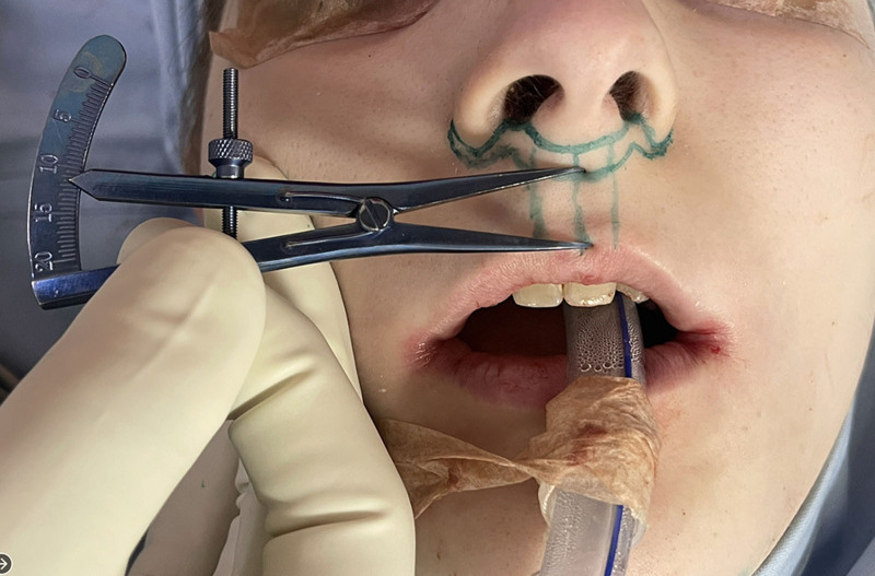
1997-2023年共988例出生分配为男性的性别焦虑者接受唇女性化与年轻化,累计1789例次手术,年龄22-75岁(平均39.5岁),79.8%小于50岁。
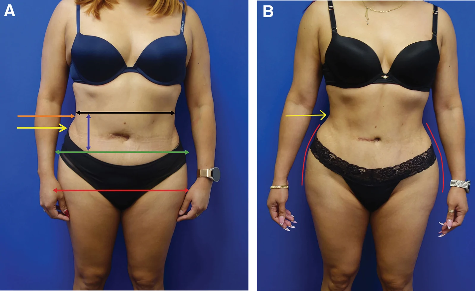
回顾性大样本病例系列(2016年11月至2025年1月),共1999例连续手术,中位年龄36岁(IQR 31至42),中位BMI 29(IQR 26至32),最短随访6个月。
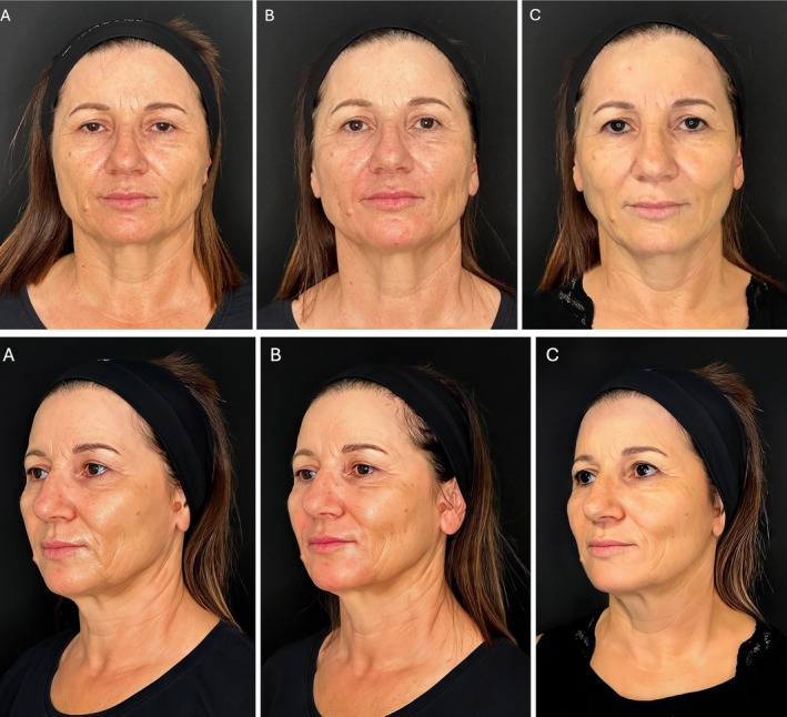
研究设计:意大利团队提出的三层(深层脂肪、浅层脂肪、真皮皮下层)超声引导中面部年轻化技术,属观察性/概念验证研究;15 例患者(13 女 2 男,50±8 岁,范围 42-65 岁