微创机器人内镜吸脂腹壁成形术(MIRELA)治疗腹直肌分离:临床与人口学病例系列
2026年9月28日28 min read
Plastic and Reconstructive Surgery Global Open
Plastic and Reconstructive Surgery Global Open
结论与实践要点
腹直肌分离常伴随腹壁轮廓改变与功能困扰。传统腹壁成形术创伤较大,而内镜与机器人平台的引入,使「在保留现代吸脂腹壁成形原则的同时完成中线重建」成为可能。本研究报告了作者前 50 例 MIRELA 的初步经验。
技术要点值得关注:经 3 处既有的 8 mm 耻骨上切口建立皮下操作空间并置入 3 个机器人套管,在 da Vinci Xi 系统下完成皮下隧道分离至剑突区,并采用所谓的 Sanfurgo 手法——以单极钩状电刀进行靶向肌肉刺激,准确定位腹直肌内侧缘后再行连续缝合折叠。该设计的核心价值在于改善视野与缝合精度,同时缩小切口创伤。
结果层面,全部手术按计划完成、无中转开放,短期安全性得到支持,合并脐疝者可同期修复。但研究性质为描述性病例系列,缺少对照与长期随访,作者亦明确指出需前瞻性大样本研究来验证长期结局与比较效果。
临床启示:该术式对术者机器人操作经验、设备可及性与患者筛选(BMI、分离宽度、皮肤松弛度、是否合并疝)均有要求,学习曲线与费用是现实考量;对以轻中度分离、皮肤弹性尚可、希望避免长切口瘢痕的患者,可作为讨论选项之一,但不应被理解为可替代经典腹壁成形术的普适方案。
Background:Rectus diastasis is frequently associated with abdominal contour deformity and functional impairment of the abdominal wall. Although lipoabdominoplasty has progressively evolved toward less invasive approaches, the incorporation of robotic assistance may further improve visualization, precision, and surgical control. This study describes the demographic and clinical characteristics of patients undergoing minimally invasive robotic endoscopic lipoabdominoplasty (MIRELA) for rectus diastasis and reports the author’s initial clinical experience with this robotic-assisted approach.
Methods:A descriptive case series was conducted involving 50 patients who underwent MIRELA with robotic correction of rectus diastasis. Demographic data, clinical characteristics, diastasis severity, associated umbilical hernia, and patient-reported preoperative complaints were analyzed. Rectus diastasis was classified as mild (<3 cm), moderate (3–5 cm), or severe (>5 cm). Concomitant umbilical hernia repair was performed when indicated.
Results:The mean age was 39.2 ± 4.9 years, and the mean body mass index was 21.8 ± 3.4 kg/m 2 . The mean rectus diastasis width was 3.27 ± 1.0 cm. Diastasis severity was mild in 32.0%, moderate in 48.0%, and severe in 20.0% of patients. Umbilical hernia was present in 20.0%. The most frequent preoperative complaint was abdominal bulging or contour deformity (62.0%), followed by other aesthetic and functional concerns related to the abdominal wall.
Conclusions:MIRELA appears to be a feasible robotic-assisted minimally invasive approach for rectus diastasis repair in selected patients, allowing correction of abdominal wall defects while preserving the principles of modern lipoabdominoplasty.
Robotic surgery has evolved significantly since its introduction in the late 1990s, enhancing conventional laparoscopic approaches through improved visualization, precision, and surgeon ergonomics. Robotic platforms provide immersive 3D imaging with independent control of the camera and instruments, combined with robotic tremor filtration, enabling superior depth perception and operative accuracy. 1 – 4 In addition, articulated instruments allow a range of motion that surpasses human wrist capabilities, facilitating precise and stable movements during complex and delicate procedures. 5 – 7
Although robotic technology has been widely adopted across multiple surgical disciplines, its integration into plastic and reconstructive surgery has progressed more slowly, despite its clear advantages for minimally invasive and high-precision procedures. Although robotic surgery has gained widespread acceptance in several surgical disciplines, its incorporation into plastic and reconstructive surgery remains in its early stages, particularly in aesthetic surgery, where its clinical application is still limited. 8 – 12 Given the global demand for body contouring procedures, the application of robotic platforms to abdominal wall reconstruction represents an important area for further development.
Rectus diastasis, frequently associated with multiparity and obesity, is characterized by pathological widening of the linea alba with separation of the rectus abdominis muscles, resulting in functional impairment and aesthetic deformity. This condition weakens the anterior abdominal wall and has been associated with altered visceral mechanics, lumbar pain, and reduced quality of life. 13 , 14 From an aesthetic perspective, rectus diastasis contributes to abdominal wall bulging, loss of waist definition, and contour irregularities, significantly impacting patient satisfaction and body image in aesthetic plastic surgery. Conventional treatment options include open abdominoplasty, mini-abdominoplasty, laparoscopic repair, and robotic-assisted techniques; however, the morbidity and scarring associated with open approaches often limit patient acceptance. 15
In response, minimally invasive strategies have evolved, transitioning from minimally invasive lipoabdominoplasty (MILA) to minimally invasive robotic endoscopic lipoabdominoplasty (MIRELA). The MIRELA technique integrates robotic precision with endoscopic principles to achieve effective diastasis repair while minimizing surgical trauma, reducing visible scarring, accelerating recovery, and enhancing aesthetic outcomes. 16 The objective of this study was to characterize patients undergoing MIRELA-assisted rectus diastasis repair through analysis of chief complaints, obstetric history, and relevant physical and anthropometric characteristics.
This retrospective descriptive study analyzed the principal clinical and demographic characteristics of 50 female patients who underwent MIRELA for rectus diastasis repair at 2 surgical centers specialized in robotic surgery in São Paulo, Brazil, between September 2023 and June 2025, representing the first 50 consecutive MIRELA cases performed by the author. All patients underwent this procedure and met the following inclusion criteria: age 18–50 years; aesthetic dissatisfaction with abdominal contour; and consent for use of audiovisual information and images for the study. Exclusion criteria included diastasis exceeding 6 cm; visceral fat exceeding 12% as determined by InBody 120 analysis using InBodyR20 software; and moderate-to-severe abdominal flaccidity (Matarasso scale II or greater). Each patient presented with dissatisfaction concerning abdominal contour and a desire for body contouring improvement. Following a comprehensive assessment, MIRELA was selected as the treatment for rectus diastasis repair and body contouring. Initial consultations recorded parity, age, weight, height, body mass index, and visceral fat quantification.
The study was conducted at 2 specialized centers in robotic surgery, one of which was Hospital e Maternidade de São Luiz. Ethical approval for the study was obtained from the institutional ethics committee of this institution. The study consisted of a retrospective review of medical records, surgical video archives, and postoperative follow-up data obtained from outpatient clinic visits. As this was a retrospective chart review, formal authorization was obtained from the participating institutions for access and review of the digital medical records and archives. Additionally, the institutional surgical consent forms signed by all patients included authorization for the use of clinical information and images for academic and research purposes related to studies such as this article. Furthermore, this study was conducted in accordance with the ethical principles outlined in the Declaration of Helsinki. Preoperative consultations included ultrasonographic measurements of diastasis size and assessment of the presence and size of the umbilical hernia.
Before the intervention, and following preoperative marking by the plastic surgeon, patients underwent general anesthesia. As part of a standardized perioperative medication protocol, all patients received 2 g of intravenous tranexamic acid (TXA) administered at anesthetic induction. In addition, TXA was incorporated into the infiltration solution at a concentration of 1 g/L, according to our institutional protocol aimed at reducing intraoperative blood loss.
Small incisions were then made, consisting of three 8-mm incisions in the pubic region (robotic trocar ports) and 6 additional 4-mm incisions strategically placed in the inframammary sulcus, umbilicus, posterior axillary line, and superior intergluteal sulcus (liposuction access sites). This meticulous incision placement minimized surgical invasiveness.
Subsequently, infiltration was performed using a solution of normal saline prepared according to a standardized protocol. Each liter of solution contained 1 ampule of 2% lidocaine (20 mL; 400 mg), corresponding to a final concentration of 400 mg/L; 1 ampule of epinephrine adjusted to a final dilution of 1:1,500,000; and 2 ampules of TXA (500 mg), resulting in a final concentration of 1 g/L. Vaser technology was applied for liposculpture, delivering 10 minutes of energy to the posterior trunk and 10 minutes to the anterior abdomen, followed by deep liposuction. The procedure was initiated in the posterior trunk, addressing the lower lumbar region and posterior flanks to reduce the posterior diameter and establish the structural “frame” of the waist, creating a defined transition toward the upper gluteal region while preserving support zones. The lateral flanks were then treated using a progressive deep-to-superficial technique to narrow the transverse diameter and refine the thorax-to-pelvis transition. Finally, the anterior abdomen was approached conservatively, respecting the midline and cutaneous vascularity, with controlled liposuction to enhance definition without compromising skin retraction. Dynamic intraoperative assessment was performed throughout to ensure symmetry and allow fine adjustments, prioritizing 3D trunk harmonization while avoiding over-resection and preserving the natural anatomical contour.
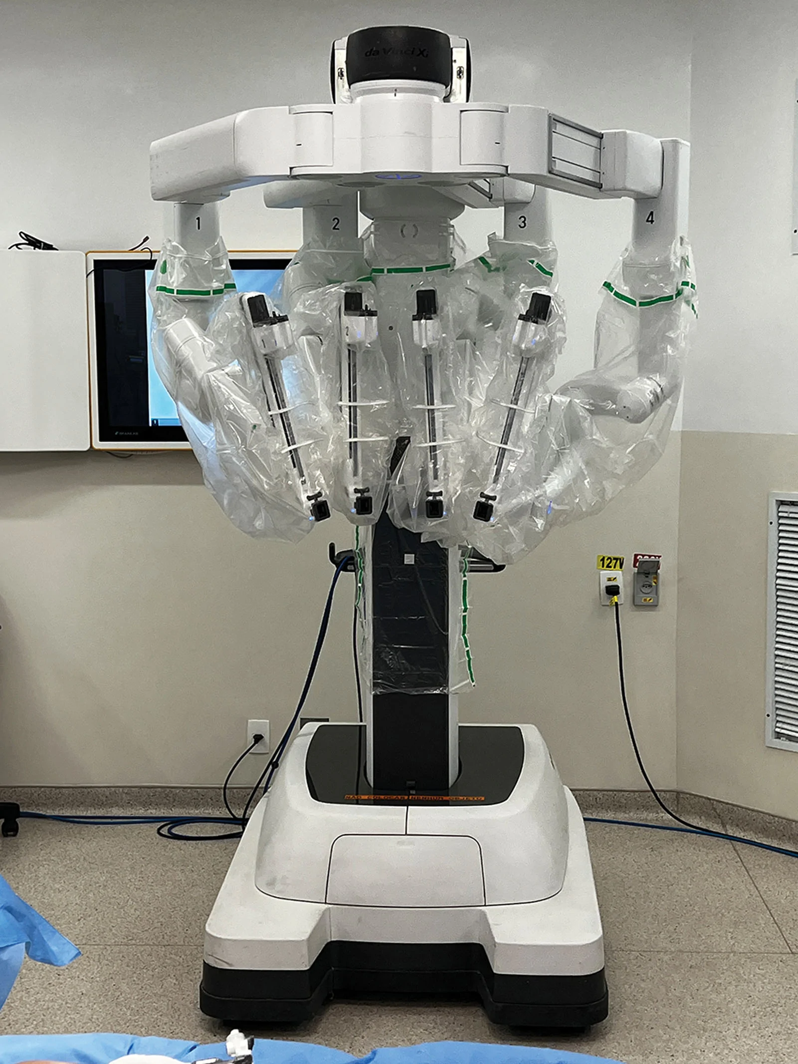
Once this process was completed, we continued with the use of the da Vinci XI robotic platform. The robotic arms connected to the 3 trocars were positioned in the pubic incisions. Through these trocars, robotic instruments were introduced: a high-definition camera for precise visualization; a grasper for tissue manipulation, electrocautery, and dissection; and a Maryland forceps for coagulation and delicate suture assistance.
All procedures were performed at 2 specialized surgical centers in operating rooms equipped with the da Vinci Xi robotic surgical system, which is essential for the precise execution of the MIRELA technique. Surgical access was established through the creation of a subcutaneous working space for 3 robotic trocars using 3 preexisting 8-mm pubic incisions: 1 midline and 2 lateral, spaced approximately 6–8 cm apart (Figs. 1 – 3 ).
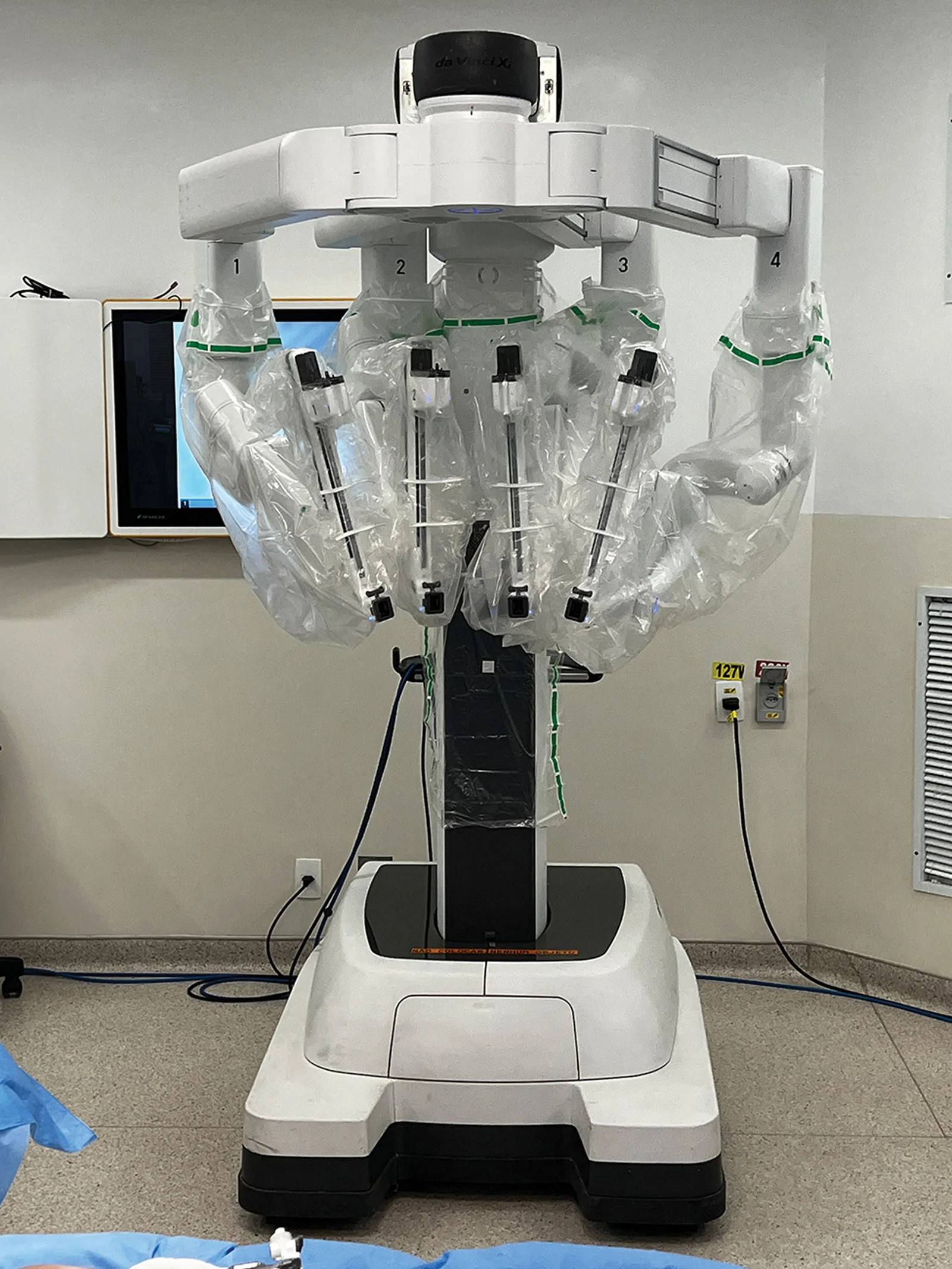
Fig. 1. Robotic-assisted surgical platform with multiple articulated arms, enabling precise instrument control, stable positioning, and optimized ergonomics in the operating room.机器人手术平台与多关节机械臂,提供稳定定位、精细器械控制与良好的人机工效。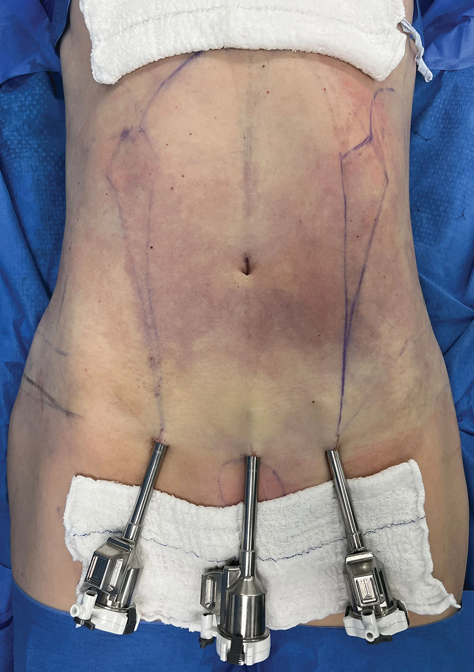
Fig. 2. Intraoperative view of the abdomen showing preoperative markings and percutaneous placement of trocars for a minimally invasive approach.腹部术野:可见术前标记与经皮置入的套管位置,体现微创入路。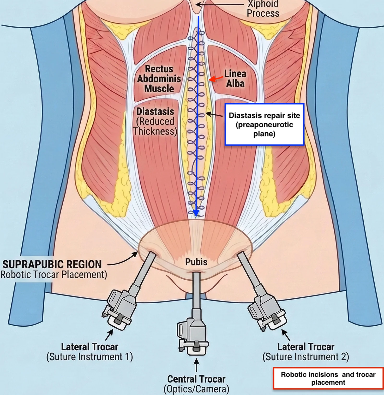
Fig. 3. Schematic representation of robotic trocar placement for the camera and suturing instruments during rectus diastasis repair with MIRELA. The blue arrow indicates the direction of continuous plication along the linea alba from the xiphoid process to the suprapubic region, whereas the red arrow identifies the rectus diastasis defect before repair.机器人套管(镜头与缝合器械)布置示意图:蓝箭头示自剑突至耻骨上区沿腹白线的连续折叠缝合方向,红箭头示修复前的腹直肌分离缺损。After trocar insertion, robotic docking and arm positioning were completed. A 0-degree endoscope was placed through the central port, with a monopolar hook introduced on the right and a Maryland forceps on the left; the hook was subsequently exchanged for a needle holder during the suturing phase. Subcutaneous dissection was then carried out down to the aponeurosis, following a mini-abdominoplasty–like plane extending from the pubic region to the xiphoid process. The working space for plication was developed in the preaponeurotic plane, between the posterior aspect of the subcutaneous tissue and the anterior rectus sheath, extending from the subxiphoid area to the suprapubic region along the midline.
Midline closure was performed using a continuous barbed suture (Stratafix 0) in a single-layer running fashion along the entire xiphopubic extent of the diastasis. After completion of the plication, the umbilical stalk was reattached to the aponeurosis using an externalized 3-0 nylon suture. The needle was passed from the skin surface into the subcutaneous plane, through the aponeurosis, and then exteriorized again through the umbilical scar, allowing external knot tying. The external suture was removed approximately 15 days postoperatively after adequate local wound care.
A 10-Fr Blake drain was placed at the conclusion of the procedure. All incisions were then closed in a standard fashion, the drain and umbilical knot were secured, and appropriate dressings and postoperative wound care were applied. ( See Video [online] , which is a step-by-step demonstration of the MIRELA technique, illustrating patient positioning, port placement, endoscopic dissection, robotic-assisted abdominal wall management, contour refinement, and execution of the Sanfurgo maneuver.)
补充视频 / 附件(原文未随文提供原图,可于出版方页面查看)gox-14-e8020-s001.mp4Video 1. This video is a step-by-step demonstration of the Minimally Invasive Robotic Endoscopic Lipoabdominoplasty (MIRELA) technique, illustrating patient positioning, port placement, endoscopic dissection, robotic-assisted abdominal wall management, contour refinement, and the execution of the Sanfurgo maneuver.
Following complete subcutaneous tunnel dissection extending to the xiphoid region, monopolar hook electrocautery was used to perform targeted muscle stimulation, allowing accurate identification of the medial borders of the rectus abdominis muscles. Once identified, these anatomical landmarks were marked using the tip of a Codman surgical marker to ensure precise intraoperative orientation.
Marking was systematically performed at predefined anatomical reference levels to guide subsequent suturing. Initial reference points were placed at the superior medial borders of the right and left rectus muscles, establishing the starting point for the plication. Additional paired markings were then created between the xiphoid process and the umbilical scar, at the level of the umbilicus, and between the umbilicus and the trochanteric line. These reference points were subsequently connected to delineate the true extent and configuration of the muscular separation. The planned suture line followed this predefined marking, ensuring anatomically accurate realignment of the rectus muscles and standardized restoration of the linea alba ( see Video [online] ).
A total of 50 patients underwent MIRELA with robotic correction of rectus diastasis (Table 1 ). All procedures were completed as planned, with no need for conversion to open surgery.
Table 1. Monthly Enrollment and Follow-up Summary
| Month | Cases Enrolled | Cumulative | Follow-up Range, mo | Median Follow-up, mo (IQR) |
|---|---|---|---|---|
| September 2023 | 2 | 2 | 26–27 | 27 (26–27) |
| October 2023 | 1 | 3 | 25–26 | 26 (25–26) |
| November 2023 | 3 | 6 | 24–25 | 25 (24–25) |
| December 2023 | 2 | 8 | 23–24 | 24 (23–24) |
| January 2024 | 2 | 10 | 22–23 | 23 (22–23) |
| February 2024 | 3 | 13 | 21–22 | 22 (21–22) |
| March 2024 | 2 | 15 | 20–21 | 21 (20–21) |
| April 2024 | 3 | 18 | 19–20 | 20 (19–20) |
| May 2024 | 2 | 20 | 18–19 | 19 (18–19) |
| June 2024 | 3 | 23 | 17–18 | 18 (17–18) |
| July 2024 | 2 | 25 | 16–17 | 17 (16–17) |
| August 2024 | 3 | 28 | 15–16 | 16 (15–16) |
| September 2024 | 2 | 30 | 14–15 | 15 (14–15) |
| October 2024 | 3 | 33 | 13–14 | 14 (13–14) |
| November 2024 | 2 | 35 | 12–13 | 13 (12–13) |
| December 2024 | 3 | 38 | 11–12 | 12 (11–12) |
| January 2025 | 2 | 40 | 10–11 | 11 (10–11) |
| February 2025 | 2 | 42 | 9–10 | 10 (9–10) |
| March 2025 | 2 | 44 | 8–9 | 9 (8–9) |
| April 2025 | 2 | 46 | 7–8 | 8 (7–8) |
| May 2025 | 2 | 48 | 6–7 | 7 (6–7) |
| June 2025 | 2 | 50 | 6 | 6 (6–6) |
| Total | 50 | — | 6–27 | 17 (12–22) |
The mean age was 39.2 ± 4.9 years (range, 29–50 y), with a mean body mass index of 21.8 ± 3.4 kg/m 2 . The median number of pregnancies was 2 (interquartile range [IQR], 1–2). The median body fat percentage was 23.9% (IQR, 20.5%–29.7%), and the median visceral fat percentage was 6.0% (IQR, 4.0%–7.0%). No patient presented with preoperative comorbidities (Table 2 ). The mean width of rectus diastasis was 3.27 ± 1.0 cm (range, 1.2–6.0 cm). Based on severity, 16 (32.0%) patients presented with mild diastasis, 24 (48.0%) patients presented with moderate diastasis, and 10 (20.0%) patients presented with severe diastasis. Umbilical hernia was identified in 10 (20.0%) patients, with defect sizes up to 1.6 cm, all of which were repaired during the same robotic procedure (Table 3 ). All procedures were successfully completed (100%). No major intraoperative or postoperative complications were recorded in the medical records during the follow-up period of this study. There were no reinterventions, no hospital readmissions, and no postoperative complaints related to surgical morbidity (Table 3 ).
Table 2. Demographic, Clinical, and Perioperative Characteristics of the Study Population (n = 50)
| Variable | Value |
|---|---|
| Demographic characteristics | |
| Age, y | 39.2 ± 4.9 (range 29–50) |
| Body mass index, kg/m 2 | 21.8 ± 3.4 (range 19.1–26.1) |
| No. pregnancies | 2 (IQR, 1–2) |
| Body fat percentage, % | 23.9 (IQR, 20.5–29.7) |
| Visceral fat percentage, % | 6.0 (IQR, 4.0–7.0) |
| Preoperative comorbidities | None |
| Previous abdominal surgery | None |
| Intraoperative data | |
| Total operative time, min | 260 ± 22 (range 220–310) |
| Robotic component, min | 80 ± 12 (range 55–105) |
| Liposuction component, min | 180 ± 18 (range 140–220) |
| Estimated blood loss, mL | 185 ± 48 (range 80–310) |
| Intraoperative transfusion | 0/50 (0%) |
| Conversion to nonrobotic approach | 0/50 (0%)—defined as any unplanned deviation from the intended robotic technique requiring change of operative approach or instrumentation |
| Device failure or aborted steps | 0/50 (0%) |
| Perioperative protocols | |
| Antibiotic prophylaxis | Cefazolin 2 g IV at anesthetic induction; second dose at 8 h postoperatively (2-dose protocol) |
| Penicillin-allergic patients: clindamycin 600 mg IV (per ASPS guidelines) | |
| VTE prophylaxis | Intermittent pneumatic compression devices applied before induction and maintained throughout surgery; enoxaparin 40 mg SC initiated 12 h postoperatively and continued until full ambulation (per Caprini risk-stratified protocol) |
| Postoperative garment/binder | Abdominal compression binder applied in the operating room; maintained continuously for 4 wk, then daytime use for an additional 2 wk |
| Early mobilization protocol | Assisted ambulation initiated within 24 h of surgery; progressive ambulation thereafter |
| Return-to-activity protocol | Sedentary activity and light daily tasks: 2 wk; light aerobic activity (walking): 4 wk; core exercise and full physical activity: 6–8 wk |
| Drain management | |
| Drain placement | 50/50 (100%) closed-suction Jackson-Pratt drain placed in all cases |
| Drain removal criteria | Output < 30 mL/24 h on 2 consecutive days, absence of signs of seroma or infection, and surgeon clinical assessment |
| Mean drain duration, d | 7.0 ± 2.0 (range 5–9) |
| Postoperative outcomes | |
| Length of hospital stay, d | 1.4 ± 0.6 (range 1–3) |
| Discharge destination | Home in 100% of cases |
Table 3. Abdominal Wall Findings and Surgical Outcomes and Safety
| Variable | Value |
|---|---|
| Abdominal wall findings | |
| Rectus diastasis width, cm, mean ± SD (range) | 3.27 ± 1.0 (1.2–6.0) |
| Mild diastasis (<3 cm), n (%) | 16 (32.0) |
| Moderate diastasis (3–5 cm), n (%) | 24 (48.0) |
| Severe diastasis (>5 cm), n (%) | 10 (20.0) |
| Umbilical hernia (present), n (%) | 10 (20.0) |
| Umbilical hernia size, cm | ≤1.6 |
| Hernia repair | Robotic (all cases) |
| Intraoperative and postoperative adverse events | |
| Procedure completion | 100% |
| Conversion to open surgery | 0 (none) |
| Intraoperative complications | 0 (none) |
| Reintervention | 0 (none) |
| Hospital readmission | 0 (none) |
| Patient complaints related to morbidity | 0 (none) |
When stratified by diastasis severity, the mean age was 37.8 ± 4.3 years in the mild group, 39.5 ± 4.6 years in the moderate group, and 41.1 ± 5.2 years in the severe group. Mean body mass index was 21.1 ± 3.1, 22.0 ± 3.3, and 22.6 ± 3.8 kg/m 2 , respectively. The median number of pregnancies was 2 (IQR, 1–2) in the mild group, 2 (IQR, 1–2) in the moderate group, and 2 (IQR, 2–3) in the severe group. Median body fat percentage was 26.0% (IQR, 20.7%–31.4%), 23.0% (IQR, 20.1%–27.9%), and 23.0% (IQR, 20.2%–23.6%), whereas the median visceral fat percentage was 6.0% (IQR, 4.0%–7.2%), 6.0% (IQR, 4.0%–7.0%), and 4.0% (IQR, 3.5%–5.5%), respectively. The mean diastasis width was 2.1 ± 0.4, 3.9 ± 0.6, and 5.6 ± 0.5 cm, respectively. Umbilical hernia was present in 12.5%, 20.8%, and 30.0% of mild, moderate, and severe cases, respectively. No complications were observed in any subgroup (Table 4 ).
Table 4. Demographic and Clinical Characteristics Stratified by Diastasis Severity
| Variable | Mild (<3 cm, n = 16) | Moderate (3–5 cm, n = 24) | Severe (>5 cm, n = 10) |
|---|---|---|---|
| Demographics | |||
| Age, y, mean ± SD | 37.8 ± 4.3 | 39.5 ± 4.6 | 41.1 ± 5.2 |
| BMI, kg/m 2 , mean ± SD | 21.1 ± 3.1 | 22.0 ± 3.3 | 22.6 ± 3.8 |
| Pregnancies, n, mean ± IQR | 2 (1–2) | 2 (1–2) | 2 (2–3) |
| Body composition, %, mean ± IQR | |||
| Body fat | 26.0 (20.7–31.4) | 23.0 (20.1–27.9) | 23.0 (20.2–23.6) |
| Visceral fat | 6.0 (4.0–7.2) | 6.0 (4.0–7.0) | 4.0 (3.5–5.5) |
| Abdominal wall | |||
| Diastasis width, cm, mean ± SD | 2.1 ± 0.4 | 3.9 ± 0.6 | 5.6 ± 0.5 |
| Umbilical hernia, % | 12.5 | 20.8 | 30.0 |
| Safety | |||
| Complications, n | 0 | 0 | 0 |
The most frequently reported preoperative complaint was abdominal bulging or contour deformity, reported by 31 (62.0%) patients. Other complaints included diastasis-related concerns (8.0%), persistent pregnancy-like abdominal appearance (6.0%), desire for a flatter abdomen (6.0%), umbilical deformity (6.0%), and loss of waist definition (4.0%). Additional complaints included clothing-related dissatisfaction, functional abdominal bulging during effort, and abdominal flaccidity. Some patients reported more than 1 complaint (Table 5 ).
Table 5. Patient-reported Complaints (Preoperative)
| Complaint | n (%) |
|---|---|
| Abdominal bulging/contour deformity | 31 (62.0) |
| Diastasis-related complaint | 4 (8.0) |
| Pregnancy-like abdominal appearance | 3 (6.0) |
| Desire for a flatter abdomen | 3 (6.0) |
| Umbilical deformity | 3 (6.0) |
| Loss of waist definition | 2 (4.0) |
| Clothing-related dissatisfaction | 1 (2.0) |
| Functional abdominal bulging on effort | 1 (2.0) |
| Abdominal flaccidity | 1 (2.0) |
Robotic surgery has emerged as a transformative modality across multiple surgical disciplines, including plastic and reconstructive surgery. Although laparoscopy represented a major milestone in the evolution of minimally invasive techniques, robotic platforms provide additional advantages that are particularly relevant in procedures requiring high levels of precision, controlled tissue handling, and 3D visualization. 1 – 6 In the setting of rectus diastasis repair, robotic-assisted surgery represents a significant advancement that addresses several limitations inherent to conventional laparoscopic approaches, including MILA. 17 , 18
In the present series, the mean rectus diastasis width was 3.27 ± 1.0 cm, encompassing mild, moderate, and severe presentations, with 20% of patients exhibiting separations greater than 5 cm. Importantly, the robotic-assisted approach allowed consistent completion of the procedure across this entire spectrum of severity without an increase in morbidity. When stratified by severity, patients with more advanced diastasis demonstrated a progressive increase in age, body mass index, number of pregnancies, and prevalence of umbilical hernia, findings that are consistent with the multifactorial etiology of abdominal wall dysfunction described in prior literature. The absence of complications across all subgroups suggests that robotic-assisted plication may provide sufficient control and precision even in anatomically challenging cases.
One of the principal advantages of robotic-assisted techniques, such as MIRELA, is the enhanced precision and stability afforded by articulated instruments and tremor filtration. These characteristics are particularly relevant given the anatomical delicacy of the abdominal wall and the need for accurate midline reconstruction. In this cohort, 48% of patients presented with moderate diastasis and 20% presented with severe diastasis, yet no intraoperative or postoperative complications were observed. This finding supports the concept that robotic technology may facilitate controlled tissue approximation and reliable suture placement in cases where conventional laparoscopy may be technically limited. 19 – 21
Umbilical hernia was identified in 20% of patients, with defect sizes up to 1.6 cm, and was repaired concomitantly during the same robotic procedure. Notably, the prevalence of umbilical hernia increased with diastasis severity, reaching 30% in the severe subgroup. Despite this association, simultaneous repair did not result in additional morbidity, reintervention, or hospital readmission. This observation highlights the potential advantage of robotic surgery in enabling precise correction of both rectus diastasis and associated midline hernias through a single minimally invasive approach, optimizing anatomical restoration while avoiding staged procedures. 21 , 22
A critical technical challenge in robotic-assisted rectus diastasis repair is the accurate identification of the true borders of the rectus muscles, especially in abdomens previously subjected to surgical intervention. This challenge is accentuated by the camera’s parallel orientation to the muscular plane and by prior liposuction, which may obscure anatomical landmarks. To mitigate this limitation, the Sanfurgo maneuver was systematically incorporated into the surgical workflow. Selective electrostimulation of the rectus muscle borders enabled reliable identification of the true muscular margins, even in cases of asymmetrical dissection or altered anatomy. By providing a stable visual reference for suture placement, this maneuver may contribute to improved alignment of muscle fibers and consistent midline reconstruction.
Electrostimulation was applied with caution, as inappropriate use may compromise aponeurotic integrity and limit the feasibility of secure suture anchoring, particularly with barbed sutures such as Stratafix. Strategic marking at selected points preserved tissue quality and may have contributed to the absence of suture-related complications in this series. Although comparative conclusions cannot be drawn, the uniform absence of adverse events supports the potential role of this maneuver as an adjunct to procedural safety and reproducibility.
Patient-reported complaints further contextualize the clinical relevance of these findings. Abdominal bulging or contour deformity was reported by 62% of patients, making it the predominant indication for surgical intervention. Additional complaints included diastasis-related concerns (8%), a pregnancy-like abdominal appearance (6%), the desire for a flatter abdomen (6%), umbilical deformity (6%), and loss of waist definition (4%). Functional manifestations, such as bulging during exertion, were also reported. These data underscore that rectus diastasis is associated with both aesthetic dissatisfaction and functional impairment, reinforcing the importance of precise anatomical correction.
From a safety perspective, the absence of intraoperative complications, postoperative complications, reinterventions, and hospital readmissions is noteworthy. However, these outcomes must be interpreted in the context of careful patient selection. The study population was characterized by a mean body mass index of 21.8 ± 3.4 kg/m 2 , a low median visceral fat percentage (6.0%; IQR, 4.0%–7.0%), and the absence of significant comorbidities, factors known to influence surgical risk and recovery. These characteristics likely contributed to the favorable short-term outcomes and should be considered when extrapolating results to broader populations (Figs. 4 – 6 ).
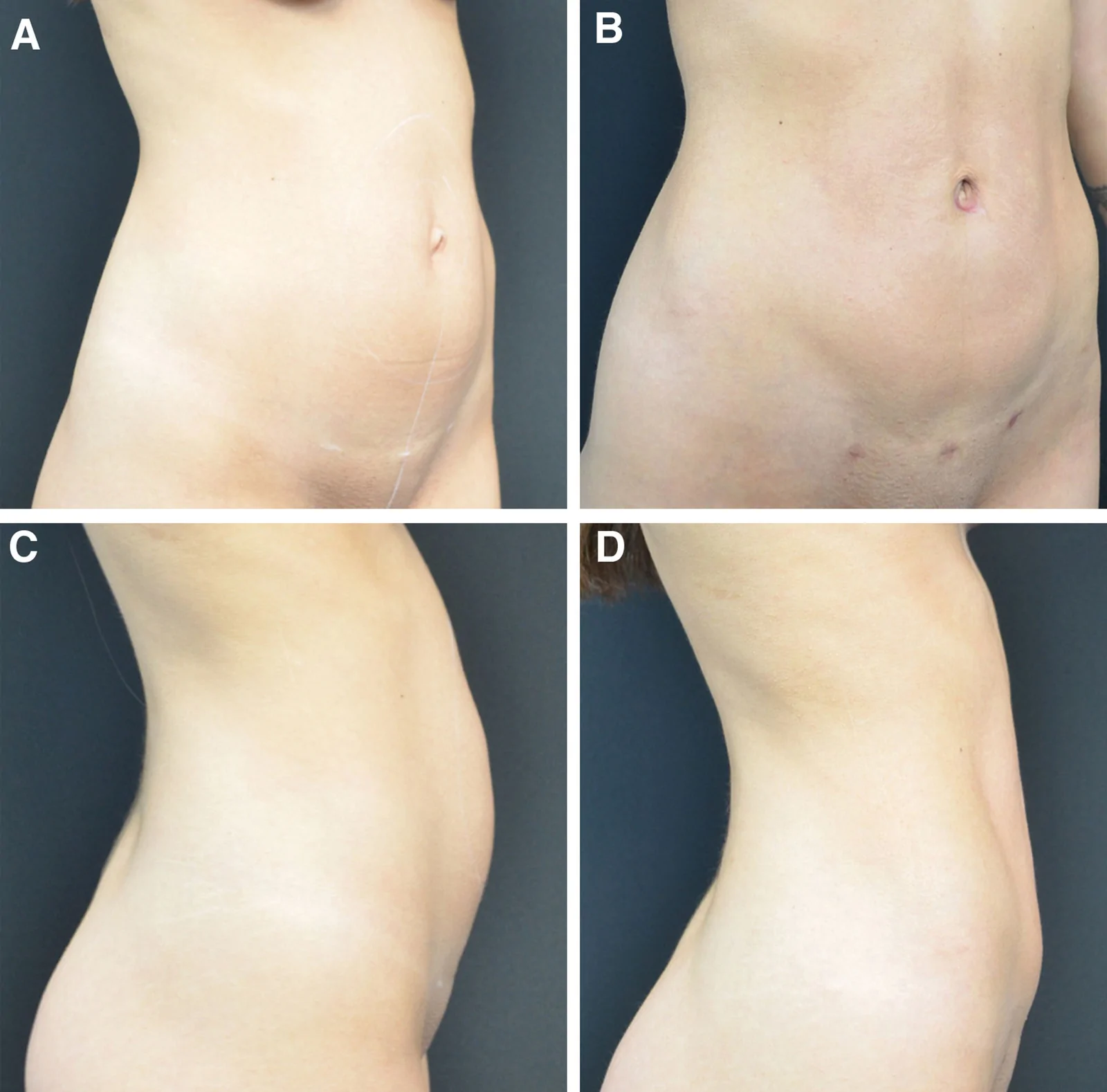
Fig. 4. A 29-year-old woman with a history of twin gestation and abdominal protrusion underwent MIRELA for a 3.4-cm rectus abdominis diastasis and 3% visceral fat. Preoperative semifrontal (A) and lateral (C) views show increased anterior abdominal contour; 3-month postoperative semifrontal (B) and lateral (D) views demonstrate reduced abdominal projection and improved body contour.29 岁女性(双胎妊娠史、腹部膨隆),腹直肌分离 3.4 cm、内脏脂肪 3%。A/C 为术前半正面与侧面,B/D 为术后 3 个月,腹部前突减小、轮廓改善。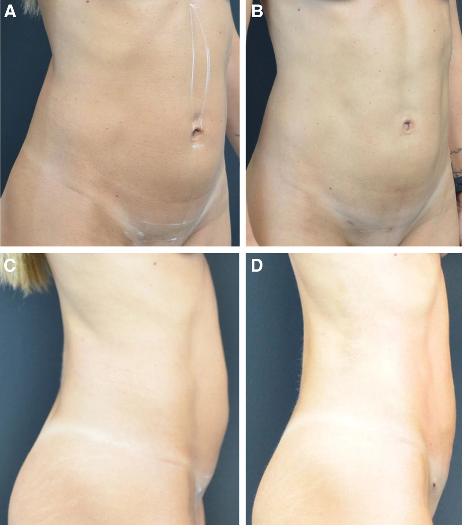
Fig. 5. A 43-year-old woman with 2 previous pregnancies and exercise-related abdominal protrusion underwent MIRELA for a 3.3-cm rectus abdominis diastasis and 5% visceral fat. Preoperative semifrontal (A) and lateral (C) views show increased anterior abdominal contour; 6-month postoperative semifrontal (B) and lateral (D) views demonstrate reduced abdominal projection and improved body contour.43 岁女性(2 次妊娠史、运动相关腹部膨隆),分离 3.3 cm、内脏脂肪 5%。A/C 术前,B/D 术后 6 个月。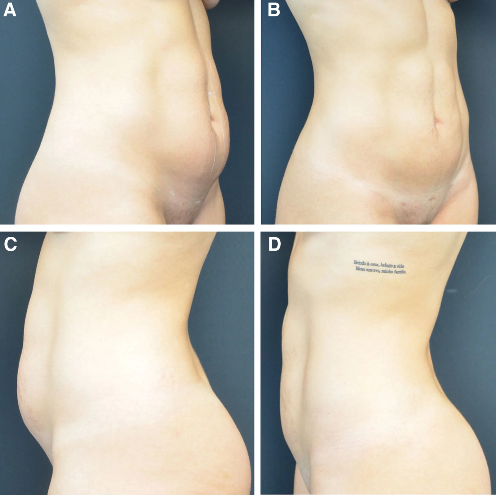
Fig. 6. A 31-year-old patient with 2 children presented with abdominal protrusion. Preoperative evaluation showed a 4.1-cm rectus abdominis diastasis and 5% visceral fat. The patient underwent MIRELA. Preoperative semifrontal (A) and lateral (C) views demonstrate increased anterior abdominal projection. One-year postoperative semifrontal (B) and lateral (D) views show sustained reduction in abdominal projection and improved body contour.31 岁患者(2 孩),分离 4.1 cm、内脏脂肪 5%。A/C 术前,B/D 术后 1 年,腹部前突的改善得到维持。The selection between MILA and MIRELA should therefore be individualized, considering surgeon experience, case complexity, patient anatomy, and institutional resources. 23 Although robotic surgery involves greater logistical complexity and higher initial costs, its potential advantages in precision, reproducibility, and safety may justify its use in selected cases. Further prospective comparative studies with adequate follow-up are required to clarify the relative benefits of each technique across different clinical scenarios and to evaluate long-term functional and aesthetic outcomes. 24 , 25
Surgical costs vary between institutions and across countries due to differences in infrastructure, reimbursement systems, technology investment, and operating room resource use. In our institutional setting, the MIRELA approach was approximately 1.5–2 times more costly than MILA and conventional abdominoplasty, primarily due to robotic platform use and disposable instrumentation; however, dedicated cost-effectiveness analyses are required to more accurately define these economic variables.
The primary limitations of this study are inherent to its descriptive design and the absence of a control group, which precludes causal inference and direct comparison with alternative techniques. Additionally, long-term outcomes and objective functional assessments were not evaluated. Nevertheless, this series provides preliminary quantitative evidence supporting the technical feasibility, reproducibility, and short-term safety of robotic-assisted correction of rectus diastasis and associated umbilical hernia when combined with lipoabdominoplasty.
This case series demonstrates the technical feasibility and short-term safety of MIRELA for rectus diastasis repair in selected patients. Robotic assistance allowed precise midline reconstruction and concomitant umbilical hernia repair. Further prospective studies with larger cohorts are needed to evaluate long-term outcomes and comparative effectiveness.
The author has no financial interest to declare in relation to the content of this article.
Patients provided written consent for the use of their images.
声明:中文精读 · 仅供学术参考。内容来自公开文献检索,不代表本人观点,不构成诊疗建议。 医疗美容需在正规医疗机构由执业医师实施。