耳部微血管皮瓣用于鼻重建的美学与患者报告结局:28 例回顾性队列
2026年9月29日26 min read
Plastic and Reconstructive Surgery Global Open
Plastic and Reconstructive Surgery Global Open
结论与实践要点
鼻位于面中部且结构精细(皮肤、软骨、黏膜衬里与骨性支架多层复合),重建难度高。额部皮瓣等局部/区域皮瓣色泽质地匹配良好,但供区瘢痕明显且常需多次手术与二期软骨移植;远位游离皮瓣则在质地与厚度上往往难以匹配鼻部亚单位。耳部微血管皮瓣提供了另一种思路。
耳廓作为供区的优势在于「以类补类」:皮肤薄、色泽接近,且自带软骨可用于支撑,血管蒂(颞浅血管)解剖相对恒定。本研究中,术者依缺损范围选择供耳——鼻翼缺损取对侧耳并包含耳轮根部与耳前皮肤,累及双侧鼻翼与鼻小柱时则沿耳轮缘向尾侧扩展,以上脚重建鼻小柱;亦可与 TAPAS 皮瓣等组合成嵌合皮瓣处理复杂缺损。
结果层面,医师评估的鼻形态、颜色与耳廓外形多位于「良好」区间,而面部毁容程度为「中度」,这提示:在需要显微外科的复杂鼻缺损中,该法可获得可接受的外观,但仍难完全恢复正常面容——术前沟通时应如实说明这一落差。患者报告的 FACE-Q 满意度较高,供区耳廓对称性被评为可接受。
风险提示:本组有 2 例需二次耳部皮瓣(一例前次皮瓣三分之二坏死、一例完全失败),提示显微血管吻合相关的部分坏死与皮瓣丢失风险客观存在,应在术式选择时充分告知。该术式尤其适合不愿接受额部瘢痕、对外观期望较高且缺损较复杂的筛选后患者。
Background:This study evaluates the aesthetic results and patient-reported outcomes following the use of microvascular flaps from the ear in nasal reconstruction.
Methods:A retrospective cohort study was conducted on patients who underwent nasal reconstruction with a microvascular ear flap between 2011 and 2021. Eleven plastic surgeons evaluated postoperative photographs using the validated observer-rated disfigurement scale and 5-point scales assessing the shape and color of the nose and donor ear appearance. Patient satisfaction with the reconstructed nose was assessed with the FACE-Q rhinoplasty module and satisfaction with the donor ear with 6-point scales.
Results:Twenty-eight patients with microsurgical ear flaps were identified. One nasal subunit was reconstructed in 16 patients and the total nose in 3. Photographs were available for 25 patients. The shape and color of the reconstructed nose, and the shape of the ear, were assessed as good by the surgeons (median scores 2 IQR 2–3]; 2 [IQR 1–2], and 2 [IQR 1–3], respectively), whereas facial disfigurement was assessed as moderate (median score 3 [IQR 3–4]). Fifteen patients completed the questionnaire, reporting high levels of satisfaction with the appearance of the nose (median score 60; IQR 54–65) and nostrils (median score 64; IQR 42–81), and acceptable symmetry of the ears (median score 2; IQR 1–3).
Conclusions:Microvascular flaps from the ear are a good alternative for complex nasal reconstruction, often yielding aesthetically favorable results.
Question: Do noses reconstructed with microvascular ear flaps achieve good aesthetic outcomes, as judged by surgeons and patients?
Findings: Microvascular ear flaps provide favorable aesthetic results for both the nose and the donor ear, as well as good patient-reported outcomes.
Meaning: Microvascular ear flaps present a good option for nasal reconstruction in selected patients, particularly for defects involving the ala or columella.
The nose is the central and one of the most noticeable features of the face. The intricate shape and the layers of skin, cartilage, and mucosal lining, as well as the bony framework, make the nose a challenging structure to reconstruct. 1 – 8 Local and regional flaps, such as the forehead flap, offer a good color and texture match but are disadvantaged by visible donor-site scarring and multiple operations, including the potential need for cartilage grafts at later stages. 1 – 4 , 7 , 9 – 12 Free flaps from distant sites, such as the radial forearm flap, provide larger coverage but, if used for purposes other than internal lining, can yield suboptimal aesthetics due to color and texture mismatches and may be of limited use by their inherent bulkiness. 3 – 8 , 13 Composite grafts from the ear can achieve good aesthetic results but are restricted to about 1.5 cm in diameter due to the lack of blood supply and are disadvantaged by their uncertain graft take. 14 , 15 Microvascular flaps from the ear present a solution to many of the limitations of the other flaps. 3 – 5 , 7 , 11 , 12 , 16 – 18
The ear, with its supportive cartilage covered by thin, hairless skin, provides an excellent color and texture match for the nose, and the helical rim closely resembles the aesthetic contour of the alar rim. 3 , 5 , 6 , 8 , 11 , 16 The ear is well vascularized, making it an optimal site for harvesting microvascular flaps capable of covering small to medium defects on the nose. 4 Moreover, the reconstruction can often be completed in 1 stage. 3 The donor-site morbidity is minimal when the donor site is primarily closed, and even with larger flaps, the resulting aesthetic outcome is generally acceptable. 6 , 19 The donor-site scar can often be hidden in the natural preauricular skin lines and the postauricular sulcus, or concealed with hair. 3 – 5 , 7 , 16 However, the aesthetic outcomes of nasal reconstructions using microvascular flaps from the ear have not been formally assessed.
The aim of this study is to critically evaluate the use of microvascular flaps from the ear in nasal reconstruction. The aesthetic outcomes of the reconstructions were assessed by both surgeons and patients. We hypothesized that microvascular ear flaps would yield aesthetically favorable results.
The ear was selected based on the extent of the defect. For nasal alar defects, the contralateral ear was used, and the flap was designed to include the root of the helix and the preauricular skin. For defects involving both alae and the columella, either ear could be selected, and the flap was extended caudally along the helical rim, with the superior crus used for reconstruction of the columella. The superficial temporal vessels were approached through a preauricular incision and dissected proximally to the maxillary branches. Progressing distally, small branches to the tragus were ligated. From the level of the root of the helix, all subcutaneous tissue was preserved between the vessels and the flap to include the small branches supplying the flap. Cranial to the helix, the superficial temporal vessels were ligated or used for a longer retrograde pedicle. The pedicle and the subcutaneous tissue were dissected along the avascular plane superficial to the deep temporal fascia. A branch of the auriculotemporal nerve adjacent to the pedicle was included when a sensate flap was desired. The flap was then raised off the helix, and the flap inset was performed with minimal reshaping. The donor site was closed directly or reconstructed with an Antia-Buch chondrocutaneous advancement flap.
A retrospective cohort study was conducted among all adult patients who underwent nasal reconstruction with a microvascular ear flap at the Helsinki University Hospital Department of Plastic Surgery between 2011 and 2021. These patients were previously included in published case series on the use of microvascular flaps from the ear for various facial defects. 3 , 4 , 19 No exclusion criteria were applied. The study was approved by the Helsinki University research ethics committees and the Helsinki University Hospital institutional review board. Written informed consent was obtained for the publication of patient photographs.
The collected preoperative data consisted of patient sex and age, and the etiology of the defect. The perioperative data included flap type and size, recipient vessels, potential vein grafts, potential neurorrhaphy, operation time, and donor-site management. The postoperative data comprised complications and the number and types of touch-ups. Patients lost to follow-up were included in the surgical outcome analysis but excluded from the patient-reported outcomes assessment.
The aesthetic results of the reconstructions were evaluated by surgeons of the plastic surgery department on anonymized photographs of patients who had undergone nasal reconstruction. None of the assessing plastic surgeons were involved in the surgery or patient care. The shape and color of the reconstructed area, as well as the appearance of the donor ear, were rated using a purpose-built, nonvalidated, 5-point Likert scale, in which a lower score indicated a better outcome. ( See appendix, Supplemental Digital Content 1 , which displays the purpose-built, nonvalidated, 5-point Likert scale and the validated 9-point observer-rated disfigurement scale used to evaluate reconstruction outcomes by 11 surgeons, https://links.lww.com/PRSGO/F250.) The overall aesthetic outcome was evaluated using a validated 9-point observer-rated disfigurement scale, with 1 representing minimal disfigurement and 9 representing severe disfigurement. 20
Patients fluent in Finnish were invited to participate in a postal survey study to assess their satisfaction with the reconstruction, using the validated Finnish version of the FACE-Q questionnaire. 21 – 24 The included FACE-Q questionnaire modules were Satisfaction With Nose, Satisfaction With Nostrils, Adverse Effects of the Nose, Satisfaction With Facial Appearance Overall, Satisfaction With the Outcome, and Appearance-related Psychosocial Distress. Answers were converted into a 0 to 100 scale using the Rasch conversion, as per the questionnaire instructions, except for Adverse Effects of the Nose, which is scored from 4 to 16. 21 , 24 Missing responses were imputed using the average score of the answered questions within that module, if less than 50% of the answers were missing. The appearance of the donor ear was assessed using a purpose-built, nonvalidated, 6-point Likert questionnaire with 4 questions, in which a lower score indicated a better outcome. ( See appendix, Supplemental Digital Content 2 , which displays the purpose-built, nonvalidated, 6-point Likert questionnaire used by 15 patients who had undergone nasal reconstruction with a microvascular ear flap to assess the donor ear site. The questionnaire consists of 4 questions, with a lower score indicating greater satisfaction, https://links.lww.com/PRSGO/F251.)
The questions addressed the appearance of the ear from the front and profile views, symmetry with the contralateral ear, and the ability to wear glasses. Information on the study, a consent form, and a prepaid return envelope were provided along with the questionnaire. Nonresponders were excluded from the patient-reported outcome analysis but were included in the rest of the study.
Descriptive analysis was performed for all quantitative variables, and median values with interquartile ranges (IQR) were reported. The complications were assessed using the Clavien-Dindo classification. 25 The data were analyzed using Rstudio version 2024.04.1 + 748 with the ggplot2 package, and the Wilcoxon rank-sum test was used to compare outcomes between groups. 26 , 27 The STROBE guidelines were applied in this study. 28
A total of 28 patients who had undergone nasal reconstruction with a microvascular ear flap were identified (Table 1 ). Two patients received ear flaps twice: 1 due to partial necrosis affecting two-thirds of the previous flap, and another due to complete flap loss. Thus, a total of 30 ear flaps used for nasal reconstruction were identified. ( See appendix, Supplemental Digital Content 3 , which displays the reconstruction of nasal defects, including subunits, flap types, complications, and revisions. POD, postoperative day; TAPAS, temporal artery posterior auricular skin flap, https://links.lww.com/PRSGO/F252.)
Table 1. Patient Demographics and Clinical Characteristics (n = 28), and Survey Respondent Data (n = 15), for Patients Undergoing Nasal Reconstruction With a Microvascular Flap From the Ear
| Variable | Summary |
|---|---|
| Sex | Male: 16 patients Female: 12 patients |
| Age during primary reconstruction | Median 59 y (range 21–79 y) |
| Primary or secondary operation | Primary: 16 patientsSecondary: 12 patients |
| Indication for reconstruction | Posttumor resection: 19 patientsTrauma: 5 patientsBurn: 4 patients |
| Follow-up time | Median 2 y and 3 mo (range 34 d to 7 y, IQR 11 mo to 4 y) * |
| Time from primary surgery to postoperative photographs | Median 2 y and 3 mo (range 5 mo to 7 y, IQR 1 y and 1 mo to 4 y and 1 mo) |
| Survey respondents | Male: 4 patientsFemale: 11 patients |
| Age at completing the survey | Median 68 y (range 57–80 y) |
| Time from primary surgery to completing the survey | Median 7 y (range 20 mo to 12 y and 3 mo, IQR 5 y and 4 mo to 10 y and 4 mo) |
In 23 patients, the reconstruction was limited to the nose, whereas both nasal and adjacent areas were involved in 5 patients. The median number of nasal subunits reconstructed was 1 (range 1–7, representing the whole nose), with 3 patients undergoing complete nasal reconstruction. The most commonly reconstructed region was the nasal alar region, involved in 22 patients, whereas the area least often affected was the lateral wall of the nose, involved in 5 patients (Table 2 ).
Table 2. Flaps From the Ear Used to Reconstruct Nasal Defects in 28 Patients
| Reconstructed Nasal Subunit(s) (n) | Flap type (n) |
|---|---|
| Ala (13) | Helix (11) Helix with a skin graft (1)Helix flap extended with TAPAS * flap (1) |
| Columella (3) | Helix (3) |
| Ala and columella (2) | Helix flap combined with hair-bearing scalp flap used for eyebrow reconstruction (1)Helix flap combined with forehead flap (1) |
| Ala and dorsum (1) | Helix with a skin graft (1) |
| Columella and tip (1) | Helix (1) |
| Dorsum and tip (1) | Extended helix (1) |
| Dorsum and lateral wall (1) | Concha flap combined with TAPAS * flap (1) |
| Ala bilaterally, columella and tip (1) | Extended helix (1) |
| Ala, columella, dorsum, and tip (1) | Helix flap combined with radial forearm flap used for cheek reconstruction (1) |
| Ala and lateral wall bilaterally, dorsum and tip (1) | Extended helix (1) |
| Total nose: ala and lateral wall bilaterally, dorsum, tip, and columella (3) | Extended helix (1) † Concha flap combined with TAPAS * flap (1) ‡ Helix flap extended with TAPAS * flap (1) § |
The helix flap was the most frequently used flap (n = 24), either on its own (n = 19) or in combination with other flaps (n = 5). The second most frequently used flap was the extended helix flap (n = 4). Flap combinations were used in 7 reconstructions (Table 2 ).
Flap dimensions were available for 19 flaps. The median length of the ear flaps was 35 mm (range 20–75 mm), and the median width was 30 mm (range 15–100 mm). The largest flap used was an extended helix flap, with dimensions of 40 × 100 mm. The smallest flap used was a helix flap, with dimensions of 30 × 25 mm.
The nasolabial artery and vein were used as the recipient vessels for 25 flaps, and the facial vessels in the neck were used for 3 flaps. Intraoral anastomosis was performed for 1 flap. Additionally, an arteriovenous loop from the neck was used for 1 flap when no suitable vessels were present in the face due to extensive defects, and a flow-through anastomosis was used with the radial forearm vessels. An infraorbital nerve neurorrhaphy was performed in 2 flaps.
The donor site was closed directly in 17 flaps, whereas an advancement and rotation of the ear was performed in 11 flaps. An advancement and rotation flap of the scalp was used for 1 donor site.
The operation time was available for 25 reconstructions, with a median of 505 minutes (range 335–730 min, IQR 402–588 min). Unilateral alar reconstructions were among the shortest, with a median operation time of 479 minutes, whereas total nose reconstructions were among the longest, with a median of 641 minutes. Operation time included additional facial or intraoral reconstructive surgery.
The median number of touch-ups was 1 (range 0–4). Twenty-one flaps had minor corrections, typically done under local anesthesia. Three flaps required an additional cartilage graft. One flap was treated with fat grafting and scar revision.
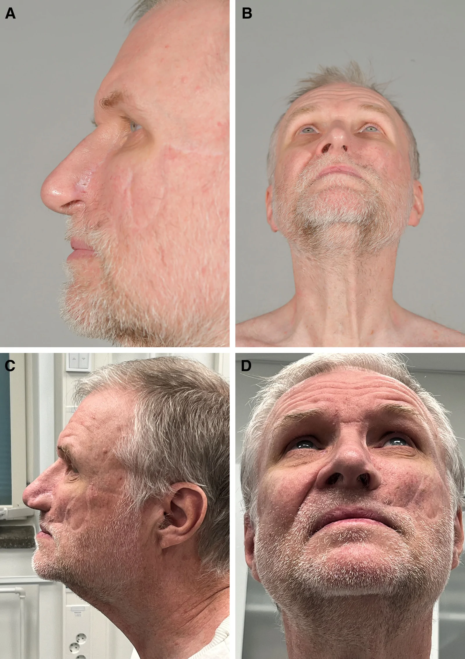
Postoperative photographs were available for 25 patients, taken at their last appointment (Table 1 , Figs. 1 – 3 ). The median rating for the shape of the nose was 2 (IQR 2–3), indicating a good outcome. The color of the nose was assessed to be a close match to the surrounding skin, with a median rating of 2 (IQR 1–2). The shape of the donor ear was deemed good, with a median rating of 2 (IQR 1–3). However, the disfigurement of the face was moderately visible, with a median rating of 3 (IQR 3–4) (Fig. 4 ).
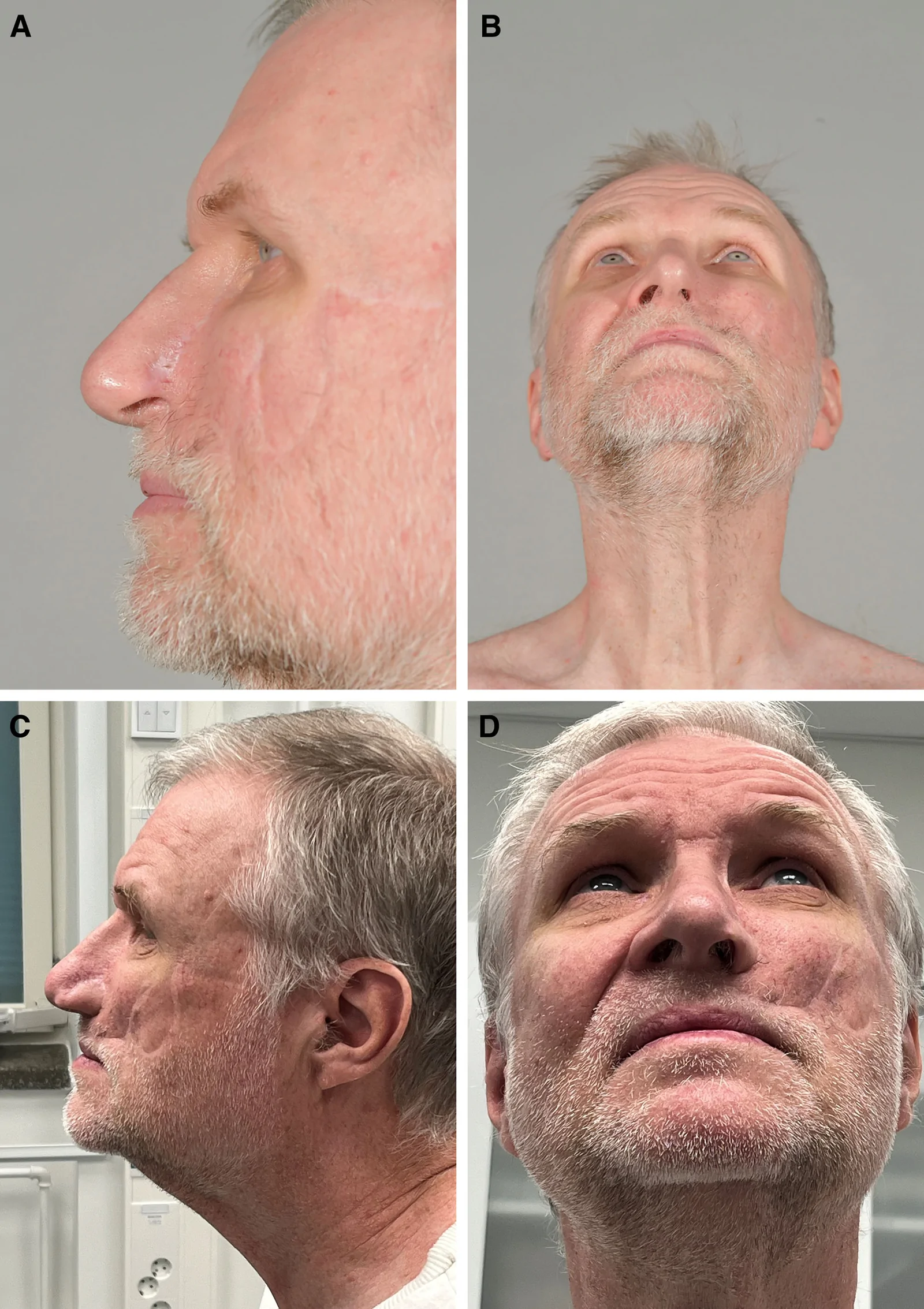
Fig. 1. Left alar reconstruction with a microvascular helix flap. A and B, Prereconstruction photographs after basal cell carcinoma resection. C and D, Postoperative photographs after 2 touch-up surgeries.左侧鼻翼重建(微血管耳轮皮瓣):A、B 为基底细胞癌切除术后重建前照片;C、D 为两次修整手术后的术后照片。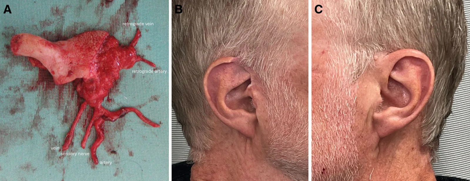
Fig. 2. Donor site. A, The flap, which includes antegrade and retrograde vessels and a sensory nerve. B, Donor site. The defect was closed by advancement of the remaining upper earlobe. C, Contralateral ear.供区情况:A 为包含顺行与逆行血管及感觉神经的皮瓣;B 为供区缺损,以剩余上耳郭推进闭合;C 为对侧耳。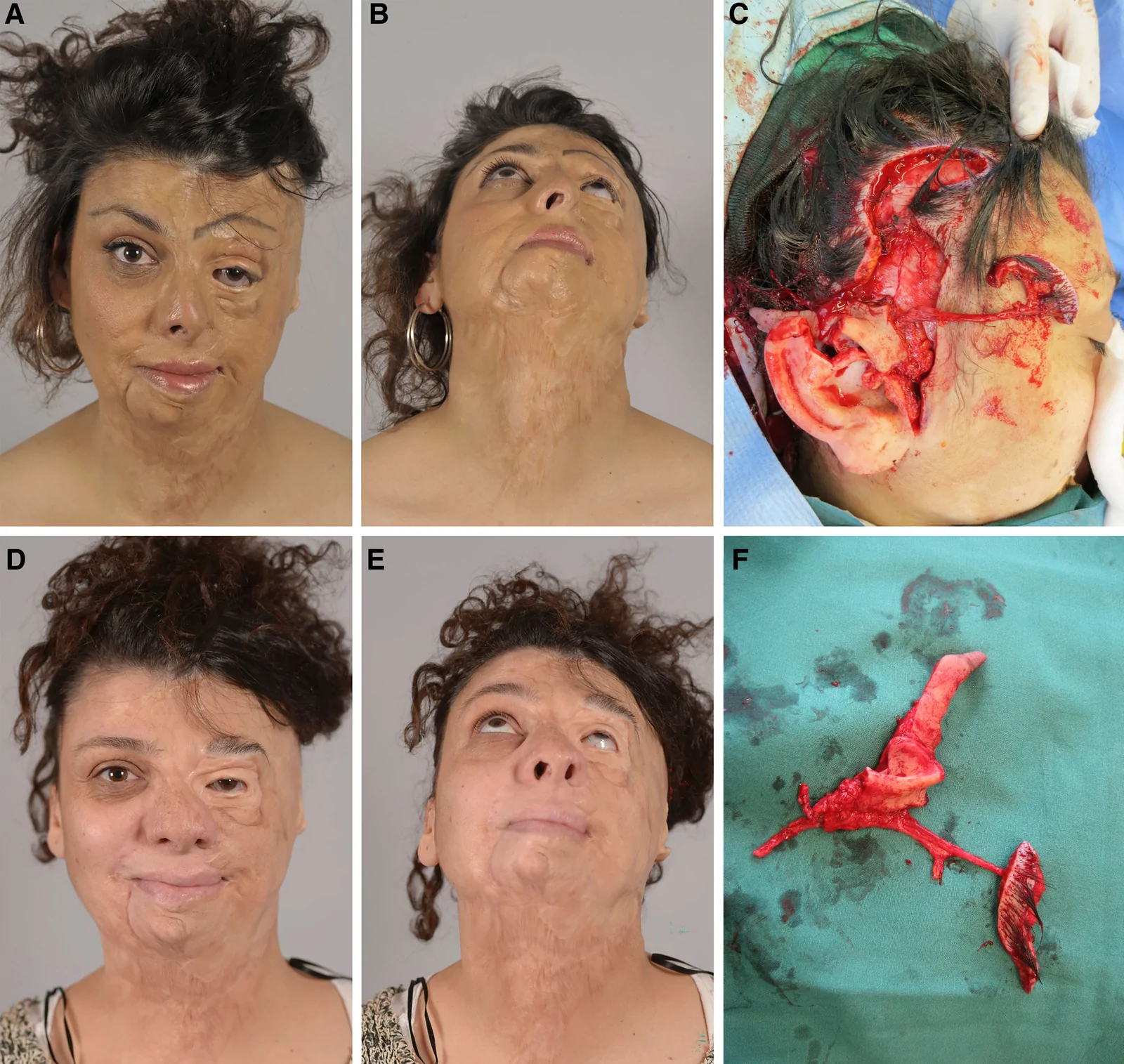
Fig. 3. Left alar, nasal dorsum, and brow reconstruction with a microvascular helix and TAPAS flap after burn injury. A and B, Prereconstruction photographs after burn injury. C and F, Chimeric helix flap with a scalp island based on the posterior temporal artery. D and E, Postoperative photographs after 2 touch-up surgeries. TAPAS, temporal artery posterior auricular skin flap.烧伤后左侧鼻翼、鼻背与眉部重建(微血管耳轮皮瓣联合 TAPAS 皮瓣):A、B 为重建前;C、F 为以颞后动脉为蒂的嵌合耳轮皮瓣带头皮岛;D、E 为两次修整术后。TAPAS 指颞动脉耳后皮瓣。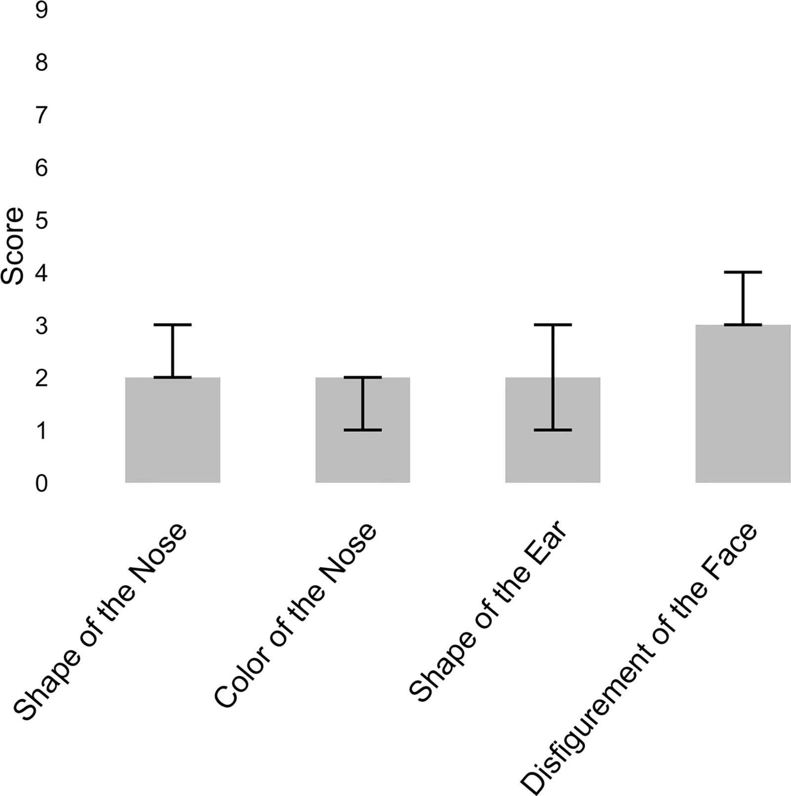
Fig. 4. The aesthetic results of the reconstructions. Median values and interquartile ranges of the aesthetic results for the reconstructions, as evaluated by the 11 plastic surgeons. The shape of the nose, color of the nose, and shape of the ear are scored on a scale from 1 to 5, whereas the disfigurement scale is from 1 to 9,20 all with a lower score indicating a better aesthetic outcome of the reconstruction.重建的美学结果:11 位整形外科医师评估的中位数与四分位距。鼻形态、鼻颜色、耳廓外形为 1–5 分,毁容量表为 1–9 分,分值越低代表美学结果越好。
Twenty-four patients were Finnish speakers and alive at the time of the study, making them eligible for the postal survey study. Fifteen patients responded to the questionnaire containing the FACE-Q scales and the purpose-built 6-point Likert questionnaire addressing the donor ear (Table 1 ).
The patients reported being satisfied with the nose (median score 60, IQR 54–65) and the nostrils (median score 64, IQR 42–81). The adverse effects on the nose were reported as minimal (median score 5, IQR 5–6). The patients reported being satisfied with their overall facial appearance (median score 61, IQR 56–74) and the outcome (median score 63, IQR 47–74). The appearance-related psychosocial distress was reported as low (median score 20, IQR 3–45) (Fig. 5 ).
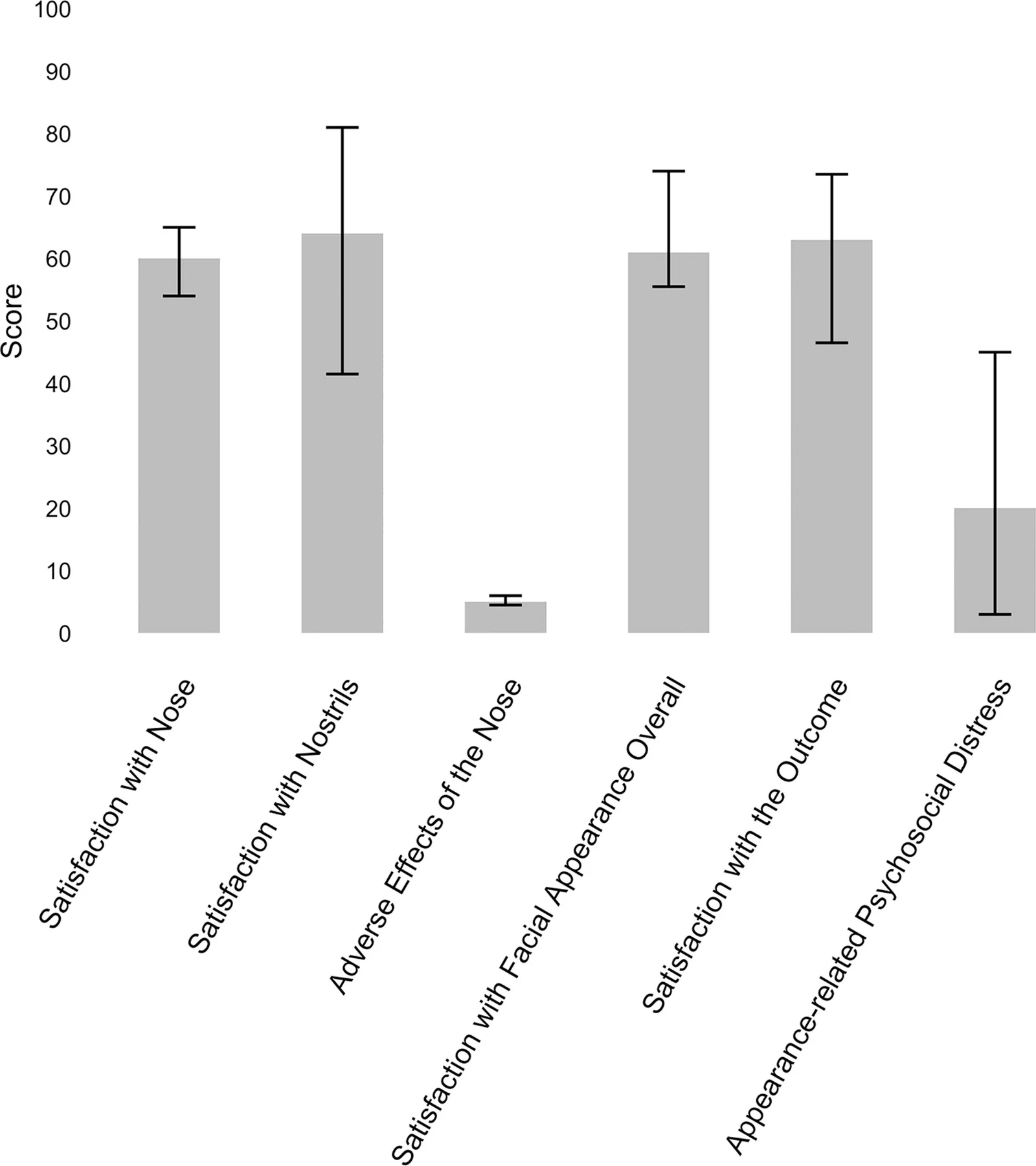
Fig. 5. FACE-Q results. Median values and interquartile ranges of the FACE-Q scores, as evaluated by the 15 patients.FACE-Q 结果:15 例患者自评的中位数与四分位距,反映对鼻外观与鼻孔的满意度。Patients were satisfied with the appearance of the donor ear when viewed from both the front and side (median score 1, IQR 1–2 for both). The symmetry of the donor ear with the contralateral ear was deemed satisfactory (median score 2, IQR 1–3). Most patients did not report difficulties wearing glasses (median score 1, IQR 1–2).
Twenty-two patients had no complications within 30 days of the operation (Clavien-Dindo grade 0). One patient had transient frontal nerve paresis (Clavien-Dindo grade I). One flap experienced partial dehiscence that was repaired with a glabellar flap under local anesthesia, and 1 donor site was resutured due to partial necrosis (Clavien-Dindo grade IIIa).
Five patients had Clavien-Dindo grade IIIb complications requiring surgery. One flap developed an arterial thrombus on postoperative day 1, managed with a reanastomosis. Another flap developed a venous thrombus on postoperative day 3, eventually resulting in a total flap loss and repair with a contralateral helix flap. In addition, 1 flap required exploration due to a hematoma on postoperative day 1, and another flap required exploration due to venous stasis on day 3. The venous stasis eventually led to necrosis of two-thirds of the flap and repair with the contralateral helix flap. Partial necrosis developed in 1 flap and its donor site and was repaired with a forehead flap and resuturing of the donor site.
The Mann-Whitney U test showed no statistically significant differences between the Clavien-Dindo grades 0–I and II–III groups for any of the aesthetic outcomes or FACE-Q modules ( P > 0.05). ( See appendix, Supplemental Digital Content 4 , which displays the association between perioperative complications and aesthetic outcomes, https://links.lww.com/PRSGO/F253.) ( See appendix, Supplemental Digital Content 5 , which displays the association between perioperative complications and patient-reported outcomes, https://links.lww.com/PRSGO/F254.)
The like-with-like tissue, robust vascularization, and inherent cartilage make the ear an excellent donor site for microvascular tissue in nasal reconstruction. This study evaluated 28 such reconstructions, suggesting favorable plastic surgeon–assessed aesthetic and patient-reported outcomes.
The reconstructed nasal defects varied in size, with 12 patients having multiple subunits reconstructed. Experience at this center demonstrates that the microvascular ear flap offers great versatility in managing medium- to large-size defects, including multiple anatomical nasal subunits. Although the auricular vascular anatomy has some variations, most people have a branch from the superficial temporal artery supplying the helix. 17 The rich collateral vascularization of the helix allows customization of the flap dimensions based on the extent of the defect while ensuring survival of the donor ear. 6
The nasal ala (n = 22) and columella (n = 12) were the most frequently reconstructed subunits, reflecting a preference for selecting the ear as the donor site for defects that benefit from the supportive cartilage structure and the precise aesthetic demands. 11 , 16 , 29 The inherent cartilage in the microvascular ear flap is a distinct advantage, reducing the need for additional cartilage grafts and minimizing the risk of infection and necrosis associated with the additional procedure. 5 , 7 , 30 During reconstructions of the whole nose, the cartilaginous part of the ear is used to reconstruct the alae, columella, and tip, whereas the surrounding skin can be used for the dorsum and lateral walls to cover the structural support and internal lining created through other means. Only 3 patients, all with extensive defects involving 3, 4, and 7 subunits, required additional nasal structural reinforcement following the primary reconstruction.
The plastic surgeon–assessed aesthetic outcomes of the nose reconstructions were compelling. Both the shape of the reconstructed area and the color match with the surrounding skin were rated as good. The overall disfigurement of the face was moderate. Formal aesthetic assessments for this technique are sparse in the literature, but excellent morphological results have been reported by this center and others. 4 , 7
Patients reported overall high satisfaction with the reconstruction. Satisfaction with the nose (median score 60) was similar to that in the normal population (63), whereas satisfaction with the nostrils (median score 64) was slightly lower than that in the normal population (77), with differences remaining below the minimal clinically important difference values of 10 and 14, respectively. 31 , 32 The adverse effects of the nose were minimal (median score 5), comparable to scores reported by patients following nasal reconstruction with a paramedian forehead flap. 33 Appearance-related psychosocial distress (median score 20) was lower than that reported for patients undergoing nasal reconstruction with a paramedian forehead flap (47) or mixed methods (61), implying less distress. 22 , 34 However, limited prior patient-reported data on nasal reconstructions and the low response rate (n = 15) in this study constrain these conclusions.
The discrepancy between moderate surgeon-rated disfigurement of the face and overall high patient satisfaction may reflect differing assessment times (median 2 versus 7 y). This suggests that patients may undergo psychological adaptation over time, eventually prioritizing long-term functional and psychological success over objective aesthetic scores.
The patients reported excellent satisfaction with the donor ear aesthetics and function. Similarly, the plastic surgeons rated the appearance of the donor ear highly. Donor-site management primarily involved direct closure and an advancement and rotation flap of the ear, both of which have been well established in preserving donor ear aesthetics. 35 , 36
Microvascular flap surgery can enable nasal reconstruction to be completed in a single stage. 5 However, additional refinements, such as flap trimming, shaping, or scar revision, are often required. Typically, patients required 1 additional operation following the primary reconstruction, similar to nasal reconstruction with a forehead flap, which frequently requires at least 2 or 3 stages. 10
The reconstructions were performed by 3 senior plastic surgeons, who observed an aesthetic learning curve and implemented technical refinements during the study period. These included using a helical rim cartilage width of only 6 mm for alar reconstruction, removal of unnecessary parts of helical cartilage to prevent an unfavorable shape of the flap during inset, cartilage shaping with sutures if needed, investing time in the inset of the flap as refinement is more difficult later, and managing the donor site with simple advancement for adequate cosmesis.
The nasolabial vessels are preferred as recipient vessels over the facial vessels in the submandibular area, as this reduces the need for a vein graft and its associated complications. However, the smaller caliber increases the difficulty of the anastomosis. 37 , 38 The resulting scar typically heals inconspicuously in the nasolabial fold, even in younger patients.
One complete flap loss (1 out of 30) occurred in this study, due to a venous thrombus. The survival rate aligns with a previous study that reported a flap survival rate of 97%. 5 Notably, the available data on this type of surgery are limited, consisting mostly of case reports or small case series, with flap survival rates ranging from 50% in a small series of 2 cases to 100% in other reports. 11 , 12 , 16 – 18 , 39
This study is limited by its cross-sectional nature, which resulted in only 25 patients having photographs available for peer review and 15 patients participating in the survey study. Particularly, only 4 of the 16 men participated in the survey, introducing a clear source of selection bias. Additionally, preoperative values for the patient-reported outcomes could not be collected, and therefore, the impact of the resection and reconstruction on the satisfaction could not be assessed. Finally, the absence of a comparison group, for example, of patients who had undergone reconstruction with a forehead flap, limits the broader analysis of the findings.
This study demonstrates that complex nasal reconstruction using microvascular ear flaps provides favorable aesthetic results for both the nose and the donor ear, as well as good patient-reported outcomes. By using the natural contour of the helix, this technique is particularly suitable for alar reconstruction as well as complex nasal defects in selected patients who do not wish to have a forehead scar and have high aesthetic expectations.
The authors have no financial interest to declare in relation to the content of this article.
Written informed consent was obtained for the publication of patient photographs and patient-reported outcomes.
The study was approved by the Helsinki University research ethics committees.
声明:中文精读 · 仅供学术参考。内容来自公开文献检索,不代表本人观点,不构成诊疗建议。 医疗美容需在正规医疗机构由执业医师实施。