组织提取胶原面部注射技术操作标准
2026年10月1日30 min read
Clinical, Cosmetic and Investigational Dermatology
结论与实践要点
方法与产品特性: 本文为组织提取胶原(面部注射)的临床操作标准,基于多中心专家经验制定。目前国内上市产品以猪源(如 Sunmax 系列,35 mg/mL)与牛源(FILLDERM,3.5% 牛胶原 + 0.3% 利多卡因,35 mg/mL)为主,分非交联与交联两类。与透明质酸相比,胶原具较高黏弹性、不吸水膨胀、有促凝血特性;其降解产生的氨基酸可与酪氨酸竞争,加上产品本身的白色遮盖作用,对色素型黑眼圈有改善价值,并可诱导新胶原生成、改善皮肤质地。
适应证、人群与操作要点: 适用人群涵盖面部皱纹、鼻唇沟、凹陷/容量缺失、轮廓塑形及黑眼圈与肤质改善者。操作上建议锐针「单点少量+注前回抽」、钝针「扇形/分层/间断逆行进针」,注射层次可自真皮深层至骨膜上,面部顺序遵循由上到下、由外到内、由深到浅;深真皮宜选非交联或稀释胶原。各区域有推荐剂量(如眶周每侧约 0.5–2 mL、中面部颊部每侧约 0.5–4 mL),同一部位疗程重复可安排在 2–12 个月区间并按个体调整。胶原过敏、自身免疫/结缔组织病、风湿性疾病及使用免疫抑制剂者禁忌;妊娠哺乳、血糖未控糖尿病等需谨慎评估。
安全性边界与并发症: 胶原颗粒细腻、非凝胶态,注前回抽与「注中遇阻/剧痛即停+按摩观察」有助于识别血管穿入;文中所引临床观察提示胶原相关血管栓塞发生概率较低,小范围栓塞经短暂按摩或观察常可缓解。仍需警惕动脉栓塞(注射后约 10 分钟内皮肤苍白、花斑伴剧痛)、静脉栓塞(10 分钟至数小时、青紫)及迟发皮肤坏死(数天至数周),出现视力/听力障碍、呼吸或生命体征不稳须立即多学科会诊。结节多由注射过浅、单点过量或未按摩塑形所致,可按摩、抽吸或病灶内胶原酶/激素等处理。
证据等级与局限: 本文属专家共识/操作标准类文献,证据来自多中心专家临床经验而非前瞻性随机对照研究,价值在于提供系统、安全导向的规范化流程与培训基线。临床落地时仍须结合机构资质、术者培训与个体化解剖评估;关于不同产品长期维持期与头对头比较的高质量数据仍有限,建议在实践中持续积累与复核。
BackgroundSince its first approval in 1981, tissue-extracted collagen injection technology has become an important method for minimally invasive facial rejuvenation, with substantial clinical experience accumulated particularly in the periorbital area. However, due to significant differences in physicochemical properties and metabolic mechanisms compared to hyaluronic acid, its clinical application still faces limitations in understanding and unique complication risks. Furthermore, no unified technical operating standards currently exist internationally.
MethodsThis standard is based on a synthesis of multicenter expert clinical experience. It systematically outlines the product characteristics, classification, and specifications of tissue-extracted collagen; defines clinical indications, contraindications, and operational precautions; specifies qualification requirements for medical institutions and operating physicians; establishes protocols for pre-operative assessment, regional injection design, post-operative management, and complication prevention and treatment strategies; and proposes multimodal treatment recommendations combining surgery, thread lifting, energy-based devices, and other fillers.
Clinical RecommendationsThrough standardized operating procedures and layered injection techniques, the safety and efficacy of tissue-extracted collagen injection therapy can be effectively improved, reducing the occurrence of complications such as nodules and vascular embolism. Combined treatment strategies further expand the application of collagen in facial rejuvenation, achieving complementary effects and optimizing recovery periods.
ConclusionThis operational standard provides systematic and standardized clinical guidance for tissue-extracted collagen facial injection. It contributes to promoting the appropriate application and healthy development of this technology, enhancing overall treatment quality and patient satisfaction.
Keywords: tissue-extracted collagen、facial rejuvenation、operational standard、complication prevention and management、combination therapy
In 1981, the world’s first tissue-extracted collagen injectable filler was approved by the U.S. Food and Drug Administration (FDA) for the improvement of senile wrinkles. 1 Between 2009 and 2019, four types of tissue-extracted collagen injectable fillers successively received approval from the National Medical Products Administration (NMPA, formerly the China Food and Drug Administration, CFDA) for marketing in China. 2 Over the past 15 years, tissue-extracted collagen injection technology has become one of the important treatment modalities for minimally invasive facial rejuvenation. 3 Particularly in the application of minimally invasive periorbital rejuvenation, considerable experience has been accumulated domestically in China. This technique is relatively simple to perform, causes minimal trauma, allows for rapid recovery, and delivers noticeable results post-treatment, making it highly popular among individuals seeking medical aesthetic treatments. 4 However, due to significant differences in the physicochemical properties, metabolism, and fate of tissue-extracted collagen compared to hyaluronic acid, numerous misconceptions persist regarding its use in facial treatments, especially in the periorbital region. 5 Specific adverse reactions and complications, such as nodules, can occur following injection, and the management strategies for these complications differ markedly from those for hyaluronic acid-related issues. 6 Currently, there are no established standards for tissue-extracted collagen injection techniques either domestically or internationally. Strict specifications regarding practitioner qualifications, operational methods, and environmental facilities are lacking. To enhance industry supervision and management, standardize operational procedures, and thereby improve the safety and efficacy of related techniques, the “Operational Standard for Tissue-Extracted Collagen Facial Injection Techniques” has been formulated to promote the healthy and orderly development of the industry.
The operational design outlined in this document is based on a summary of the clinical experience of numerous experts and physicians. It represents a recommended standardized foundational protocol for operators, developed with full consideration of safety and effectiveness. Operators may adopt other diversified and personalized design schemes according to practical circumstances and personal experience, provided they adhere to the general therapeutic principles set forth in this document.
This document is applicable to medical aesthetic institutions equipped with departments of medical cosmetology or cosmetic dermatology, as well as medical institutions with departments of plastic surgery, cosmetic surgery, and dermatology. It may also serve as a basis for third-party organizations to supervise the medical practices related to tissue-extracted collagen injection techniques and to evaluate medical institutions.
The scope of this operational standard covers the properties, classification, and specifications of tissue-extracted collagen products; clinical application indications, contraindications; requirements for medical institutions; qualifications of operating physicians; pre-treatment preparation; operational design schemes; post-treatment management; prevention and treatment of main complications; recommendations for combined treatment applications; and other content relevant to this therapy.
Based on the methodological process described above — including expert selection, multi-round discussions, in-person consensus meetings, and voting — a final set of recommendations was developed. These recommendations cover 16 facial subunits, including detailed specifications on needle selection (blunt vs. sharp), injection planes (supraperiosteal, subcutaneous, or deep dermis), suggested dose ranges, and complication management strategies. The full recommendations are presented below according to anatomical region.
Collagen is a type of fibrous macromolecular protein present in mammals, widely distributed in the extracellular matrix. 7 It is the most important, abundant, and prevalent protein within the intercellular spaces of animals, serving functions such as supporting organs and protecting the organism. Currently, 28 types of 8 collagens are known from clinical research. Collagen is rich in 18 amino acids including glycine, proline, and alanine, with glycine content being particularly high, constituting about 27% of the amino acids. 9–11 It lacks tryptophan and cysteine. Among these, proline and lysine are key components enabling the α-peptide chains to form a triple-helix structure. 12 Collagen possesses the function of a signal “relay station”, acting as a ligand in the biological process of cell signal transduction and playing a significant role in cell communication. 13 It can trap, store, and transport growth factors and cytokines, thereby serving important roles in processes such as organ development, wound healing, and tissue repair. 14 Collagen in the skin is the primary structural protein, accounting for 70% to 80% of its dry weight. 15 Skin collagen is synthesized by fibroblasts and consists of 80% to 85% Type I and 10% to 15% Type III collagen. 16 Within the dermis, over 80% is predominantly Type I collagen, characterized by thick, densely arranged bundle structures, providing strong support and structural integrity to the skin. 8 Loss of Type I collagen will lead to facial wrinkles and depressions. Type III collagen appears as a loose, filamentous network, relatively fine, primarily distributed around Type I collagen at the junction of the epidermis and dermis. 17 It is immature, less stable, and has lower tensile strength, providing elasticity and stress resistance to the skin. Type III collagen possesses excellent capabilities in promoting repair and healing, primarily serving nutritive and epidermal management functions. 16
Type I collagen is an important structural protein with biological activity, composed of two identical α1 chains and one α2 chain. 18 Each α chain itself adopts a left-handed helical conformation, and the three α chains intertwine to form a right-handed triple-helix structure. 19 Type III collagen, on the other hand, is composed of three identical α1 chains. 20
As an important protein in organisms, collagen possesses many excellent biological characteristics: a)Low immunogenicity: Collagen, as a natural macromolecule, exhibits low immunogenicity. 21 b)Strong hydrophilicity: Collagen molecules contain numerous polar hydrophilic groups, and their internal sponge-like, loose, porous structure also has a strong water retention capacity. 22 c)Biodegradability: Collagenase in the body can hydrolyze collagen into oligopeptides or amino acids, with 80% being utilized by the organism and 20% excreted as urea. 23 d)Biocompatibility: The natural fibrous structure of collagen provides an excellent growth scaffold and environment for cells. 24 e)Procoagulant function: Upon contact with blood, platelets in the blood rapidly adhere to collagen fibers, forming fibrin and accelerating clot formation, thereby promoting hemostasis. 8
Collagen degrades in the body to produce abundant amino acids, such as alanine, arginine, and leucine, which can compete with tyrosine in the skin, thereby inhibiting melanin formation. 25 The inherent white color of collagen products provides immediate coverage for dark circle pigmentation. 26 Concurrently, injectable collagen can stimulate the formation of new collagen over an extended period in vivo, thus sustainably improving melanin deposition.
During metabolism, collagen decomposes into various amino acids, most of which can be reabsorbed and utilized by human tissues, providing raw materials for the synthesis of new collagen. 27
Tissue-extracted collagen possesses high viscoelasticity (no displacement, no deformation), does not absorb water and swell, and results in a more natural facial appearance after injection. 27
Tissue-extracted collagen can activate fibroblast activity, improve the extracellular matrix environment, and effectively induce the production of new collagen, thereby densifying the skin tissue. 28
Clinical application has found that the probability of vascular embolism caused by collagen injection is very low. When small amounts of vascular embolism occur, they can often be reversed after brief massage or simply waiting, with good prognostic outcomes. In the past three years, domestic expert teams have analyzed this phenomenon in clinical applications and found explanations through animal experiments. 29 a)Collagen products have very low injection resistance, making it easy to detect deviations in injection depth and partially avoid risks. 30 b)Collagen particles are very fine and do not exist in a gel state when mixed with water or blood; they can coexist homogeneously. This significantly reduces the likelihood of complete embolization. 31 c)When a needle enters a blood vessel, blood can be aspirated immediately. After injecting collagen, blood can still be aspirated within a short period during subsequent aspiration, providing ample safety warning. Embolized arteries or veins can recanalize within a relatively short time, and the probability of irreversible embolism is very low. 32
Currently, the tissue-extracted collagen products marketed in China with corresponding registration certificates can be classified into two categories based on their source: porcine-derived and bovine-derived. Based on the use of glutaraldehyde cross-linking agents, they can be classified as non-crosslinked collagen and crosslinked collagen. 33
a)Collagen Implant, Trade Name: Furo Mei (Sunmax Biotechnology Corp., Ltd., Taiwan, China).Composition: Purified Type I collagen derived from porcine skin and phosphate-buffered saline, containing 35 mg of collagen per milliliter.Specification: 0.5 mL/syringe.Porcine-derived:b)Collagen Implant, Trade Name: Fuli Mei (Sunmax Biotechnology Corp., Ltd., Taiwan, China).Composition: Crosslinked Type I collagen purified from porcine skin and phosphate-buffered saline, containing 35 mg of collagen per milliliter.Specification: 1.0 mL/syringe.c)Lidocaine-containing Collagen Implant, Trade Name: Fulai Mei (Sunmax Biotechnology Corp., Ltd., Taiwan, China).Composition: Crosslinked collagen purified from porcine skin and phosphate-buffered solution containing 0.3% lidocaine, with collagen content of 35 mg/mL and lidocaine hydrochloride content of 3 mg/mL.Specification: 1.0 mL/syringe.
Medical Collagen Filler, Trade Name: FILLDERM injectable collagen (Fiman (Changchun) Medical Biotechnology Co., Ltd., Jilin, China).
Composition: A saline suspension containing 3.5% bovine collagen (35 mg/mL) and 0.3% lidocaine hydrochloride.
Specification: 0.5 mL/syringe, 1.0 mL/syringe.
Clinical application indications are as follows: a)Correction and improvement of facial wrinkles;b)Correction and improvement of nasolabial folds of varying degrees;c)Improvement of facial depressions and volume deficiencies in various areas;d)Facial contouring and shaping;e)Improvement of dark circles and facial skin texture and tone.
Clinical application contraindications and precautions are as follows: a)Contraindicated in individuals allergic to collagen;b)Contraindicated in patients using immunosuppressants;c)Contraindicated in patients with autoimmune diseases and connective tissue disorders;d)Contraindicated in patients with rheumatic diseases;e)Contraindicated in patients with other severe systemic diseases and psychiatric disorders;f)Not recommended for use in pregnant or lactating women and infants;g)Not recommended for use in diabetic patients before effective blood glucose control is achieved;h)Use with caution in individuals on long-term anticoagulant therapy;i)Use with caution in areas previously injected with unknown types of fillers;j)Use with caution in individuals with allergic predisposition.
Medical institutions shall meet the following requirements: a)They must be legally established medical aesthetic and plastic surgery institutions;b)They must be equipped with a Department of Medical Cosmetology (including but not limited to Cosmetic Surgery, Cosmetic Dermatology, Cosmetic Traditional Chinese Medicine, Cosmetic Dentistry, and Cosmetic Ophthalmology);c)The operating environment must be an operating room or a clean treatment room, equipped with essential emergency equipment and medications.
The qualification requirements for operating physicians are as follows: a)The qualifications of the operating physician shall comply with the relevant provisions of Article 11, Chapter 3 of the “Measures for the Administration of Medical Cosmetology Services” (2016 revision) issued by the National Health Commission;b)The operating physician shall have completed the registration as a chief physician in cosmetology;c)The physician shall have received formal training in collagen injection techniques.
Conduct a detailed medical history inquiry and understand the treatment needs of the individual seeking medical aesthetic services, strictly adhering to the indications.
The following preparations shall be made before treatment: a)Signing of informed consent;b)Facial cleansing;c)Collection of imaging information;d)Disinfection of the treatment area;e)The product must be maintained under cold chain management throughout storage and transportation, and administered immediately upon opening during injection.
The following management measures shall be taken after treatment: a)Keep the injection sites clean and dry for 12 to 24 hours;b)Cold compress with an ice pack may be applied (ensuring no frostbite occurs);c)Collection of imaging information;d)Management of post-injection complications shall refer to Combination with Surgical Procedures to 10.8.
Tissue-extracted collagen (hereinafter referred to as collagen) may be administered for facial injection therapy using either sharp or blunt needles. 34 When using sharp needles, a small amount should be injected per point, and effective aspiration must be performed prior to injection. When using blunt needles, fanning, layering, or intermittent retrograde injection techniques are primarily employed. Immediate pressure should be applied after injection to ensure a smooth surface and avoid protrusions. 35 Collagen can be injected at various facial layers, ranging from the deep dermis to the supraperiosteal level. When injecting into the deep dermis, non-crosslinked collagen or diluted collagen should be selected whenever possible. Facial injections should follow the sequence from top to bottom, from lateral to medial, and from deep to superficial. 36 Depending on specific treatment needs, it may be combined with other injectable fillers. Repeat injections for the same site and layer may be planned within a treatment course ranging from 2 months to 12 months, with adjustments made based on individual circumstances to achieve optimal outcomes. 33
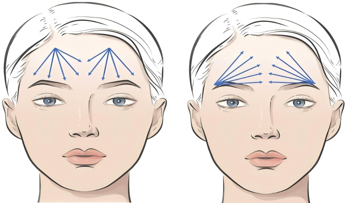
Forehead ( Figure 1 ): A blunt needle can be used for supraperiosteal injection. Depending on the degree of volume deficiency, an injection volume of 1 mL to 5 mL may be used.
Glabella ( Figure 2 ): A blunt or sharp needle can be used for supraperiosteal injection. The glabellar region can accommodate an injection volume of 0.5 mL to 2 mL.
Temporal Region ( Figure 3 ): A sharp needle can be used for supraperiosteal injection in the area approximately 1 cm below the temporal crest and 1 cm above the superior orbital rim. Alternatively, a blunt needle can be used for injection into the subcutaneous layer or the superficial aspect of the deep temporal fascia. The recommended volume per side is 0.5 mL to 5 mL.
Eyebrow Ridge ( Figure 4 ): A sharp needle can be used for supraperiosteal injection. Alternatively, a blunt needle can be used for injection into the retro-orbicularis oculi fat (ROOF) layer, subcutaneous fat layer, or the supraperiosteal layer. The recommended volume per side is 0.5 mL to 1 mL.
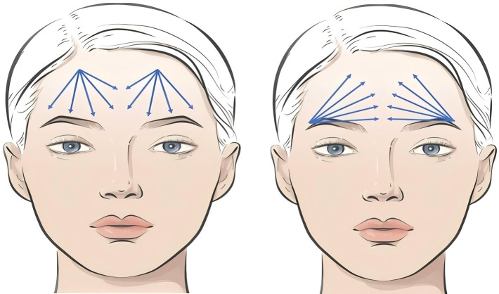
Figure 1 Upper Facial Operational Plan Design — Frontal Region. Blue indicates blunt cannula.额部(上面部)操作设计图:蓝色示钝针进针规划,体现「由上到下、由外到内、由深到浅」的进针顺序原则。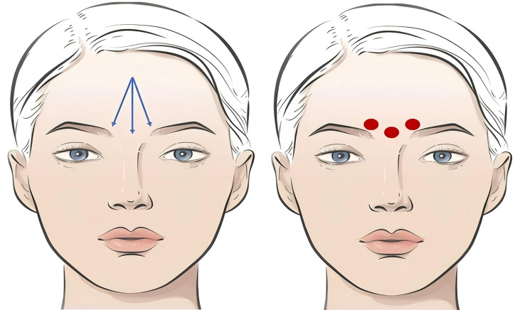
Figure 2 Upper Facial Operational Plan Design — Glabellar Region. Blue indicates blunt cannula; red indicates sharp needle.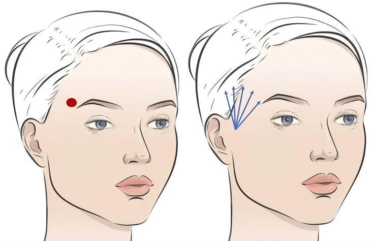
Figure 3 Upper Facial Operational Plan Design — Temporal Region. Blue indicates blunt cannula; red indicates sharp needle.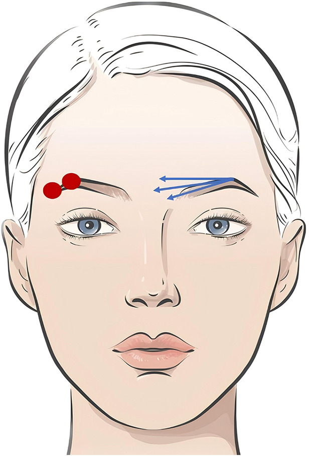
Figure 4 Upper Facial Operational Plan Design — Supraorbital Ridge. Blue indicates blunt cannula; red indicates sharp needle.
Periorbital Area ( Figure 5 ): A sharp needle can be used, contacting the periosteum for injection along the attachment zones of periorbital ligaments, such as the temporal fusion zone, lateral orbital thickening, periorbital retaining ligaments, and the tear trough ligament area, as well as into the suborbicularis oculi fat (SOOF) layer medially and laterally around the orbit. Alternatively, a blunt needle can be chosen for injection onto the periosteum, under the orbicularis oculi muscle, or into the subcutaneous layer. The recommended periorbital injection volume per side is 0.5 mL to 2 mL. For upper eyelid hollowing, a blunt or sharp needle can be used for injection into the ROOF layer or within the orbital septum. The recommended upper eyelid injection volume per side is 0.2 mL to 1 mL.
Cheeks (Malar Region) ( Figure 6 ): A blunt needle is typically chosen for injection into the subcutaneous layer. The recommended volume per side is 0.5 mL to 4 mL.
Nose ( Figure 7 ): A blunt or sharp needle can be used for injection onto the periosteum or into the subcutaneous layer. For defining the double C-curves, a sharp needle contacting the periosteum or a blunt needle for subcutaneous sculpting can be used. The recommended nasal injection volume is 0.5 mL to 2 mL.
Buccal Fat Pad Area ( Figure 8 ): A sharp needle can be used for supraperiosteal injection. When using a blunt needle, injection can be performed into the medial buccal fat compartment. The recommended volume per side is 0.5 mL to 1 mL.
Nasolabial Folds ( Figure 9 ): A sharp needle can be used for supraperiosteal injection. When using a blunt needle, injection can be performed into the nasolabial fat compartment. The recommended volume per side is 0.5 mL to 2 mL.
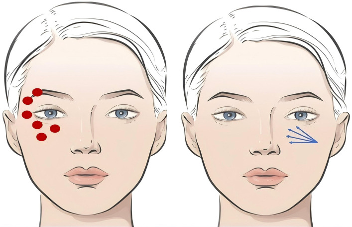
Figure 5 Mid-Facial Operational Plan Design — Periorbital Region. Blue indicates blunt cannula; red indicates sharp needle.眶周操作设计图:示锐/钝针在眶周支持韧带附着区及 SOOF 层的进针层次,为胶原用于黑眼圈与眶周年轻化的核心区域示意。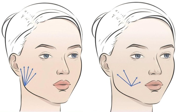
Figure 6 Mid-Facial Operational Plan Design — Cheek Region. Blue indicates blunt cannula.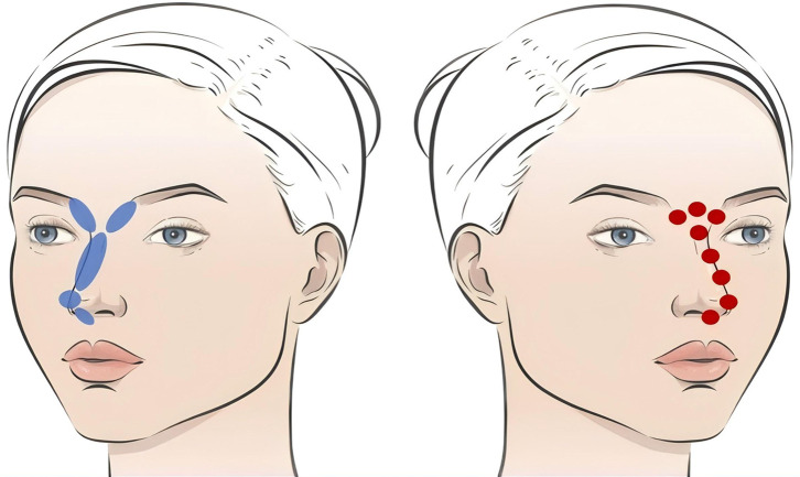
Figure 7 Mid-Facial Operational Plan Design — Nasal Region. Blue indicates blunt cannula; red indicates sharp needle.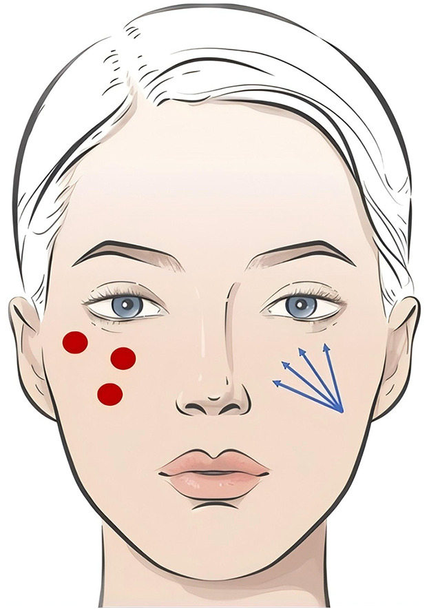
Figure 8 Mid-Facial Operational Plan Design — Malar Fat Pad Region. Blue indicates blunt cannula; red indicates sharp needle.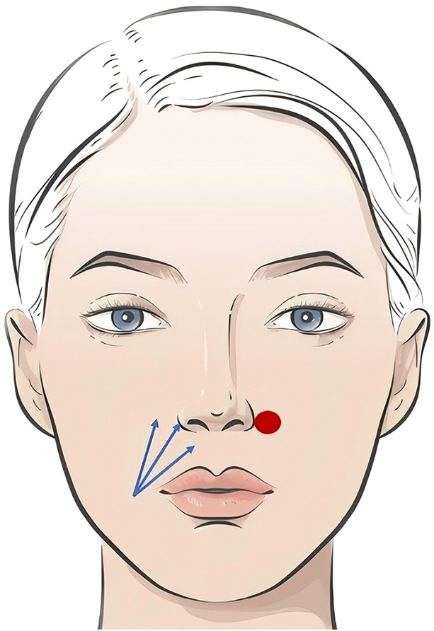
Figure 9 Mid-Facial Operational Plan Design — Nasolabial Fold. Blue indicates blunt cannula; red indicates sharp needle.鼻唇沟操作设计图:示锐针骨膜上或钝针鼻唇脂肪室进针,每侧推荐 0.5–2 mL,是胶原容量补充的代表性适应证。
Pre-jowl Sulcus ( Figure 10 ): A sharp needle can be used, contacting the periosteum in the mandibular ligament area. Alternatively, a blunt needle can be chosen for injection into the deep subcutaneous layer. The recommended volume per side is 0.5 mL to 2 mL.
Chin ( Figure 11 ): A sharp needle can be used, contacting the periosteum at the pogonion or pre-pogonion point, with blending and transition to areas approximately 1–2 cm on either side. The recommended chin injection volume is 0.5 mL to 4 mL.
Labiomental Fold (Mental Crease) ( Figure 12 ): A blunt needle is typically chosen for injection into the deep subcutaneous layer. The recommended volume per side is 0.5 mL to 1 mL.
Mandibular Line (Jawline) ( Figure 13 ): At the mandibular angle and mandibular ligament area, a sharp needle can be used for periosteal layer injection. Alternatively, a blunt needle can be chosen for subcutaneous sculpting to enhance the mandibular line contour. The recommended volume per side is 0.5 mL to 1 mL.
Lips ( Figure 14 ): Non-crosslinked collagen can be injected using a sharp needle into the labial mucosa and subdermal layer. The recommended lip injection volume is 0.5 mL to 1 mL.
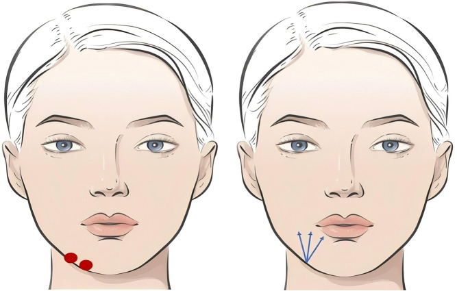
Figure 10 Lower Facial Operational Plan Design — Prejowl Sulcus. Blue indicates blunt cannula; red indicates sharp needle.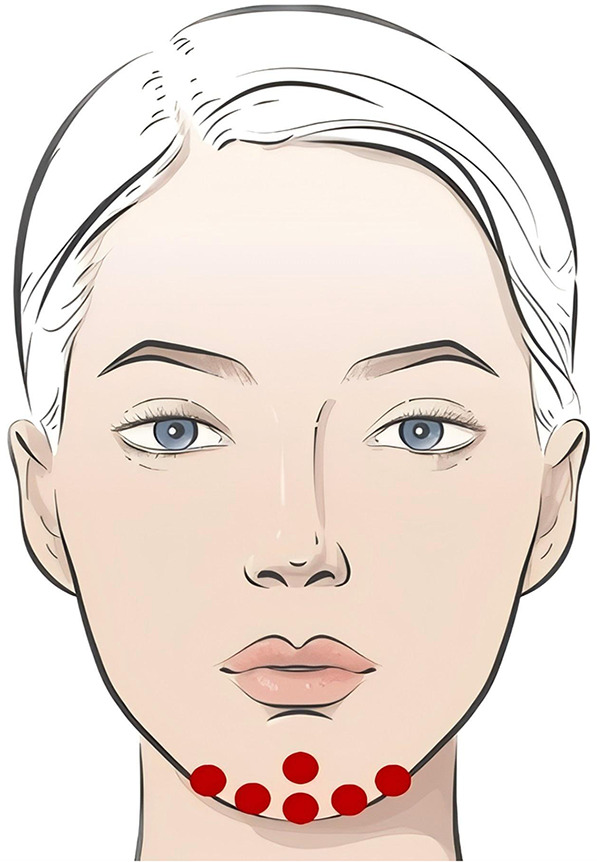
Figure 11 Lower Facial Operational Plan Design — Chin Region.red indicates sharp needle.
Figure 12 Lower Facial Operational Plan Design — Mentolabial Fold. Blue indicates blunt cannula.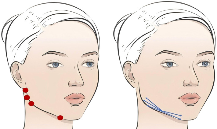
Figure 13 Lower Facial Operational Plan Design — Jawline. Blue indicates blunt cannula; red indicates sharp needle.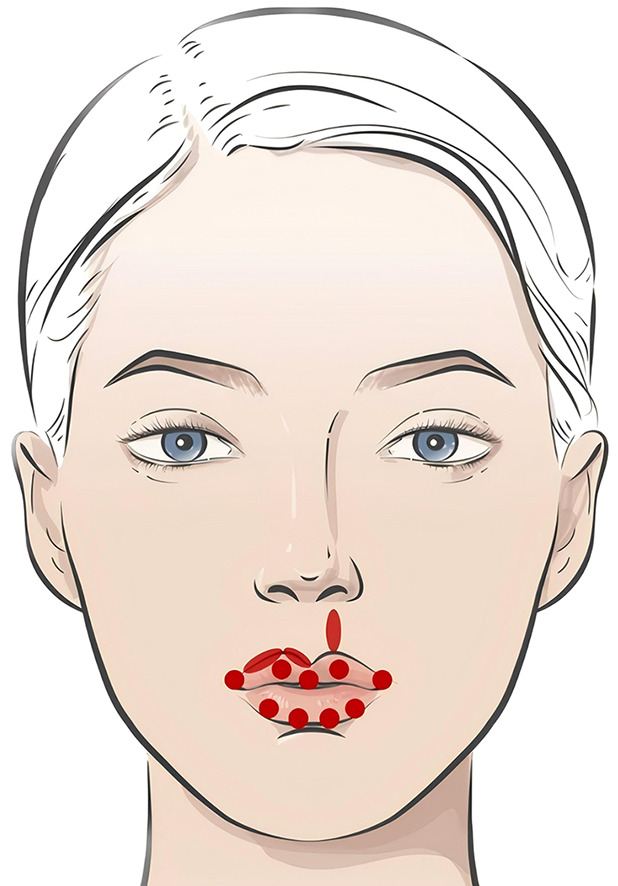
Figure 14 Lower Facial Operational Plan Design — Lip Region red indicates sharp needle.
Neck ( Figure 15 ): A sharp needle may be selected for intradermal or subcutaneous injection. When using a blunt needle, injection is performed into the superficial subcutaneous layer along the neck lines. For the treatment of neck lines alone, the recommended single-session volume is 1 mL to 3 mL.
Mesotherapy ( Figure 16 ): Collagen is diluted with a diluent (non-crosslinked or minimally crosslinked hyaluronic acid and normal saline) at a ratio of 1:2:2. The mixture is injected into the deep dermal layer, with an injection volume of 0.025 mL per point. The recommended total collagen volume for a full-face treatment is 1 mL to 2 mL per session.
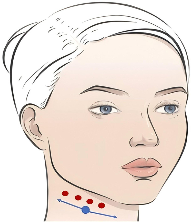
Figure 15 Other Regional Operational Plan Design — Neck Region. Blue indicates blunt cannula; red indicates sharp needle.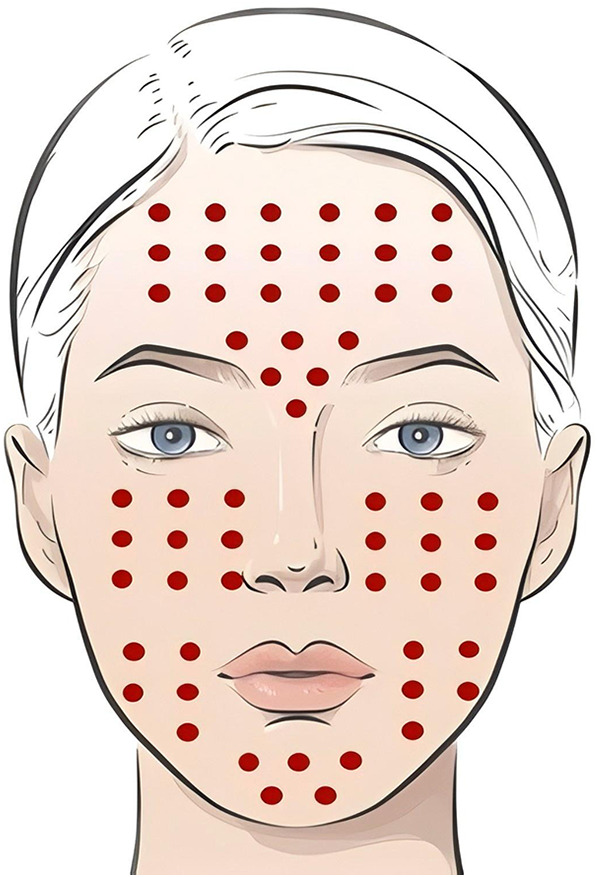
Figure 16 Other Regional Operational Plan Design — Mesotherapy. Red indicates sharp needle.中胚层疗法操作图:胶原按 1:2:2 与稀释液(非交联/低交联透明质酸+生理盐水)稀释,真皮深层每点 0.025 mL,全脸单次约 1–2 mL。
The common injection techniques involve a combination of blunt and sharp needles. If blunt needles cause mechanical trauma due to aggressive dissection, postoperative swelling is likely. 36 To prevent this, practitioners must have a thorough understanding of local anatomy, perform precise injections during the procedure, avoid repeated punctures and blunt dissection with blunt needles, and apply appropriate cold compresses postoperatively. Oral anti-edema medication may also be administered. 37
Immediate cold compress and pressure can help reduce bruising and hematoma. Topical ointment application 12 hours post-treatment may accelerate the resolution of bruising. 38
Pain is commonly associated with aggressive manipulation, such as nerve irritation or injection into sensitive areas during the procedure. Injectable products containing lidocaine can effectively reduce intraoperative injection pain. If arterial or venous embolism occurs during injection, immediate, intensified pain may be observed. 39 Prompt assessment of the injection site for skin mottling or whitening is necessary, and reference should be made to Section 10.8 for further management.
Nodules often result from overly superficial injection, excessive dose per injection point, or failure to massage and shape the area post-injection. Manifestations include abnormal protrusions or white spots at the injection site. 40 For nodules detected immediately or shortly after injection, massage may be performed to promote even dispersion. Larger nodules may require aspiration for improvement. Chronic nodules are often associated with induration at the injection site and reduced skin elasticity. 41 Management may include intralesional injection of collagenase or corticosteroids, hot compresses, massage, or radiofrequency therapy.
Hyperpigmentation may be improved with light-based therapy or mesotherapy. Precise control of injection depth and dosage can effectively prevent immediate post-injection skin whitening. If hypopigmentation occurs, management should follow the guidelines in Combination with Other Fillers .
Currently, the telopeptide removal technology in collagen production is highly advanced, making allergic reactions due to product formulation rare. Acute or delayed allergic reactions are more likely associated with individual patient variability or collagen denaturation. 42 Strict control of cold chain transportation, pre-procedure verification of product integrity, and thorough inquiry into patient allergy history and immune-related conditions can effectively prevent allergic reactions. If allergic symptoms occur, oral antihistamines may be administered. In severe cases, short-term use of corticosteroids may be added to accelerate collagen degradation and alleviate symptoms. 43
Strict adherence to aseptic technique can effectively prevent infection. If infection is confirmed, local debridement is the primary treatment. Oral or intravenous antibiotics may be administered. If necessary, bacterial culture of drained secretions should guide antibiotic selection. 44
Pre-filling the needle with lidocaine before injection and aspirating before injection can help determine if the needle is within a blood vessel. Injection should be performed slowly. If resistance is encountered or the patient reports pain, injection should be stopped immediately, and the area should be massaged while observing changes in skin color. 45 Arterial embolism may present within 10 minutes post-injection with skin whitening or mottling accompanied by severe pain. A capillary refill test may show delayed refill. 46 Management may include warm compress massage, topical application of nitroglycerin ointment, vasodilation and microcirculation improvement, along with prophylactic anti-infection treatment. Venous embolism may manifest as a deep blue discoloration near the injection site, possibly occurring within 10 minutes to several hours post-injection. Management may involve local injection of collagenase or corticosteroids, and systemic administration of antibiotics, steroids, aspirin, low-molecular-weight heparin, etc. 47 Hyperbaric oxygen therapy may also be considered to provide anticoagulation, reduce inflammation, improve microcirculation, and alleviate tissue hypoxia. Skin necrosis may develop days or weeks later. Management may include wet compresses with nitroglycerin ointment, wound cleansing and dressing changes, and topical application of growth factors to promote tissue regeneration and wound healing. If severe symptoms such as visual field defects, blindness, hearing impairment, limb movement disorders, respiratory distress, or unstable vital signs occur, immediate consultation with a multidisciplinary team including ophthalmology, otorhinolaryngology, respiratory medicine, and interventional radiology is required. Timely symptomatic treatment such as intravascular thrombolysis should be performed. 45 , 48
Due to its unique material properties and biological performance, collagen can be widely used in combination with other therapies.
Collagen can be combined with surgical procedures. Application at the incision site can assist in hemostasis and promote rapid wound healing. In periorbital rejuvenation, collagen filler can be used 3 months postoperatively to improve fine lines, depressions, and hyperpigmentation around the eyes.
Collagen can be combined with thread lifting techniques. Layering collagen along the path of the threads can assist in hemostasis, reduce swelling, aid in thread-tissue adhesion, and minimize displacement of lifted tissues. Additionally, collagen can be injected after thread lifting to improve local depressions, fine lines, enlarged pores, skin sensitivity, and periorbital hyperpigmentation.
Energy-based devices employ physical and photobiological principles to create orderly, controlled cutaneous injury, thereby initiating post-traumatic repair mechanisms to achieve regeneration and tissue repair. 49 In facial rejuvenation treatments, these devices can improve hyperpigmentation, enlarged pores, and telangiectasia, increase skin hydration, restore barrier function, and reduce fine wrinkles. Post-procedure, combination with collagen mesotherapy can enhance efficacy, shorten recovery time, and reduce adverse reactions. For comprehensive treatment of periorbital aging concerns such as dark circles and tear troughs, intense pulsed light (IPL), Q-switched lasers, picosecond lasers, radiofrequency energy, and focused ultrasound can improve skin tone and texture. 50 Following energy-based treatments, provided there is no significant exfoliation or erythema, collagen mesotherapy or filler injections can be immediately combined to yield superior therapeutic outcomes. If open wounds are present (eg., from fractional microneedling radiofrequency), a 1 to 2-week interval is recommended before combination treatment. When performing energy-based therapies after collagen filler injection, an interval of over 2 months is advised. 51
Collagen presents as an opaque, milky white substance. Subcutaneous injection can optimize skin tone and improve both pigmented and vascular dark circles. In facial soft tissue augmentation, it can be combined with harder, longer-lasting fillers such as hyaluronic acid or biostimulators for supraperiosteal and deep fascial layer injection, leveraging complementary advantages. 52 For periorbital filling, collagen offers the distinct benefit of avoiding the Tyndall effect, making its combination particularly advantageous. If the Tyndall effect occurs due to hyaluronic acid filler, hyaluronidase can be used for dissolution followed by collagen injection. When combining collagen with other facial fillers, layered injection is recommended. 53 Additionally, as an excellent carrier material, collagen can be directly mixed in specific proportions with injectable materials such as modified hydroxyapatite, polylactic acid, or acellular dermal matrix particles for injection therapy, for which both domestic and international clinical experience has been accumulated.
Botulinum toxin type A improves dynamic wrinkles by modulating facial muscle activity. Reduced muscle movement is conducive to the long-term maintenance of facial collagen. 54
As a skin nutrient, collagen can be used in mesotherapy. Intradermal injection improves dull complexion, dryness, enlarged pores, and fine wrinkles, and promotes the repair of sensitive skin and melasma. It can be combined with other mesotherapy products, typically administered via intradermal injection following mesotherapy. 55
This document establishes a comprehensive technical framework for tissue-extracted collagen facial injection, spanning fundamental principles to clinical practice. It systematically outlines the unique advantages of collagen fillers—such as low immunogenicity, biodegradability, procoagulant function, and non-hydrating properties—differentiating them from hyaluronic acid and other dermal fillers. In conclusion, this operational standard serves as a practical, safety-oriented clinical guide to enhance treatment outcomes and patient satisfaction. It also provides a foundation for professional training, quality assessment, and regulatory oversight, supporting the standardized and sustainable advancement of tissue-extracted collagen injection therapy in clinical practice.
声明:中文精读 · 仅供学术参考。内容来自公开文献检索,不代表本人观点,不构成诊疗建议。 医疗美容需在正规医疗机构由执业医师实施。
基于相同主题推荐 · 继续深挖
分层注射+注前回抽,有助于降低胶原血管栓塞风险
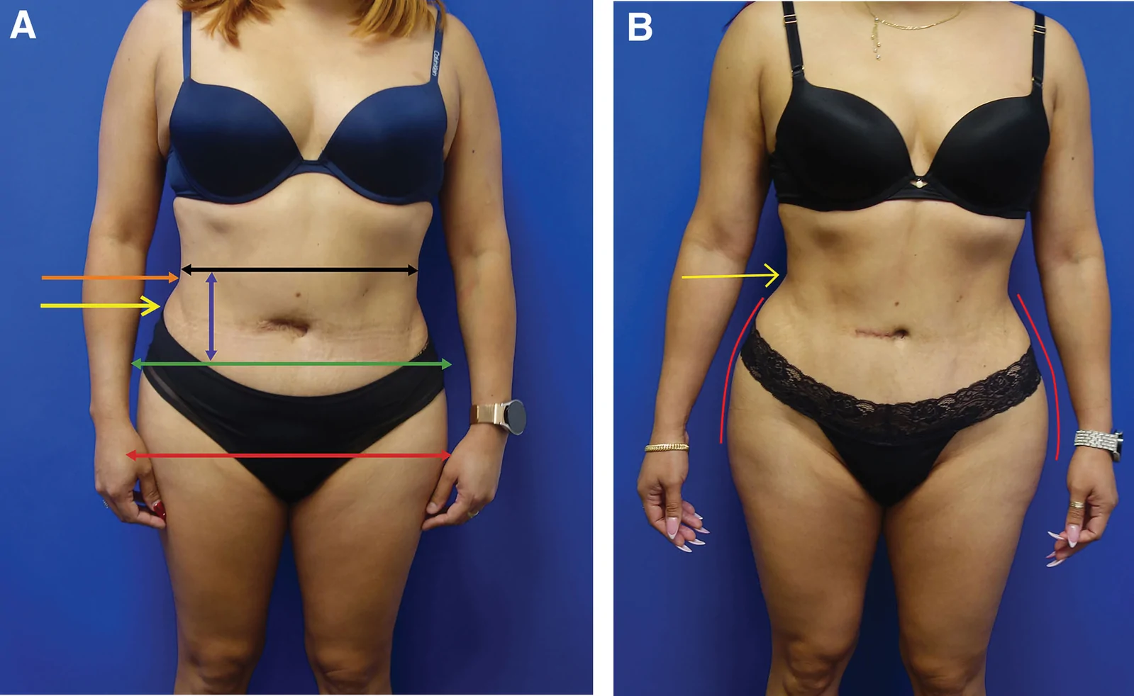
回顾性大样本病例系列(2016年11月至2025年1月),共1999例连续手术,中位年龄36岁(IQR 31至42),中位BMI 29(IQR 26至32),最短随访6个月。
鼻部塑形疗效与安全性:12个月前瞻性研究/PMC13100342_fig1.jpg)
这项单中心前瞻性研究纳入24名中国女性受试者,接受CaHA凝胶鼻部注射(骨膜上/骨膜浅层,单侧单次总量≤2.0 mL,平均0.93±0.22 mL),全部完成12个月随访,无脱落。