经结膜延长下入睑成形术长期结果:200 例连续病例前瞻性研究
2026年10月3日29 min read
Plastic and Reconstructive Surgery
结论与实践要点
研究类型:单中心前瞻性病例系列(level of evidence 偏低,非随机对照试验),由单一术者完成 200 例连续病例,旨在报告其经结膜延长下入睑成形术的长期结果。
临床落地解读:
证据边界/局限: 样本虽大但为单术者单中心、无对照与盲法,随访中位数约 22 个月,长期稳定性(如脂肪存活、复发)仍需更久随访;文中“较传统术式更具可预测性”等比较为术者自身经验性判断,非头对头随机证据,临床采纳应结合术者经验与个体解剖(如 maxilla 矢量方向影响解剖难度)。
Background:The authors first described the extended transconjunctival lower eyelid blepharoplasty in 2017. In this prospective study, the authors present their long-term outcomes with this technique.
Methods:A total of 200 consecutive patients who underwent the extended transconjunctival lower eyelid blepharoplasty between June of 2017 and December of 2024 were prospectively evaluated. Ninety-six percent of patients received concomitant midcheek fat grafting. The mean follow-up was 22 months. Two specific aspects unique to our technique were analyzed: (1) the long-term effects of releasing the tear trough ligament and orbicularis oculi origins, and (2) the use of free fat grafts, fashioned from excised orbital fat, placed along the orbital rim. Patients were evaluated for aesthetic outcomes, lower eyelid position stability, and dynamic changes with smiling. Patient satisfaction was assessed with an outcome questionnaire.
Results:This technique is both effective and safe in addressing eye bags and the tear trough deformity. No occurence of scleral show was observed. Free fat grafting, using excised orbital fat, effectively corrected the tear trough deformity and softened the lid-cheek junction. Release of the orbicularis oculi origins consistently resulted in long-lasting improvement in periorbital aesthetics during smiling, without compromising orbicularis oculi function. Patient satisfaction was high. The overall complication rate was low, with fat necrosis occurring in 3.5% of patients and a revision rate of 1.0% for residual lower eyelid bulges.
Conclusions:This technique delivers superior aesthetic outcomes compared with conventional approaches that do not release the tear trough ligament. A novel finding in this study is that the technique predictably improves the aesthetics of the smile, which is a significant benefit.
The transconjunctival lower eyelid blepharoplasty has distinct advantages over subciliary approaches, including better safety and faster recovery. 1 – 3 However, to fully harness its potential in delivering transformative lower eyelid rejuvenation, specific maneuvers must be incorporated into the transconjunctival lower blepharoplasty. These maneuvers, which include precise excision of orbital fat and release of the tear trough ligament, orbicularis oculi origins, and part of the orbicularis retaining ligament, along with free fat grafting using the excised orbital fat along the orbital rim, were incorporated into our extended transconjunctival lower blepharoplasty technique that we published in 2017. 4 – 6 The technique has since gained popularity and has been adopted by many surgeons. However, questions remain regarding its long-term efficacy and safety. 7 – 18 Specific concerns about our technique included the long-term effects of surgically releasing the tear trough ligament and the orbicularis oculi origins, and the viability of the free fat grafts used, fashioned from excised orbital fat and placed along the orbital rim. 19 – 24 In this study, we present our technical refinements, address concerns associated with this procedure, and report our long-term outcomes and complication rates.
Patients presenting for lower eyelid blepharoplasty with (1) prominent eye bags, (2) tear trough deformities, and (3) no history of lower eyelid blepharoplasty were enrolled in this study. A comprehensive periorbital, ophthalmic, and facial examination was performed for each patient. For those with a history of dermal filler injections to the tear trough, maxillary, or zygomatic regions, a detailed history was obtained, including the type of filler, quantity administered, and timeline of injections. Clinically, these patients often exhibited lumpy, palpable irregularities with sausage-like fullness along the infraorbital region. If the fillers were hyaluronic acid–based, they were removed with intralesional hyaluronidase injection at least 1 week before surgery. ( See Figure, Supplemental Digital Content 1 , which shows a [ left ] 43-year-old woman who had 4 cc of a hyaluronic acid filler injected into her tear trough/upper midcheek along the orbital rim in 3 treatments over 2 years. This was dissolved with hyaluronidase. She was seen 1 week later [ right ], and the true prominence of her eye bags and midcheek aging changes was evident, https://links.lww.com/PRS/I462.) The true extent of the eye bags and midcheek volume deficiency were then more accurately assessed and the surgery planned accordingly. Preoperative and postoperative photographs and videos were taken. Patients were reviewed at 1 week, 1 month, 3 months, 6 months, 1 year, and annually thereafter. Long-term outcomes were assessed at least 6 months after surgery using a patient satisfaction questionnaire. A targeted survey addressing 2 domains—(1) appearance and (2) quality of life—was administered. Responses were recorded using an ordinal scale with 4 categories: very satisfied, satisfied, neutral, and dissatisfied. Written informed consent was obtained from all patients, and the study was conducted in accordance with the principles of the Declaration of Helsinki.
A 15-mm transconjunctival incision is made through the conjunctiva and lower eyelid retractors. ( See Video 1 [online] , which demonstrates our technique of extended transconjunctival lower blepharoplasty.) Dissection with cutting cautery is performed until the orbital septum—a whitish, semitranslucent structure—is visualized. With an insulated Desmarres retractor, upward traction is applied to place the orbital septum under tension. The preseptal space is then gently opened using tenotomy scissors. The orbital septum is incised directly over areas of planned fat pad removal. The medial, middle, and lateral fat pads are excised as indicated. The lower eyelid contour is then assessed to determine the adequacy of fat removal. Gentle pressure on the globe reveals any residual bulging fat pads and visual comparison with the contralateral lower eyelid helps with the determination of the adequacy of the fat excision.
With the right-angle end of the cat paw retractor placed in the roof of the preseptal space, gentle sweeping with a cotton-tipped applicator is used to bluntly open the preseptal space to its inferior boundary. The key landmark to look for is the white line. This locates the arcus marginalis. The origin of the palpebral part of the orbicularis oculi is located slightly caudal to this key landmark. With the assistant providing upward retraction and the surgeon applying downward pressure on the orbital septum using the cotton tip, the orbicularis oculi origins are sharply released with cutting cautery at its bony origin. Sequential release of the palpebral part of the orbicularis oculi, the tear trough ligament, and the orbital part of the orbicularis oculi is performed along the anterior maxilla. The endpoint of the release is visualization of the muscle fibers of the levator labii superioris, which signals entrance into the premaxillary space and confirms complete release of the tear trough ligament. Laterally, the orbicularis retaining ligament is also released. The extent of surgical release is guided by preoperative markings, which delineate the area of the lid-cheek junction requiring correction.
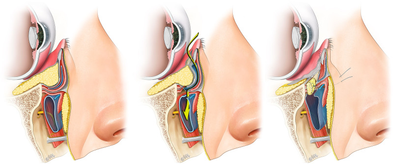
The excised orbital fat pads are carefully trimmed into small lobules for grafting along the orbital rim to smooth the lid-cheek junction. To optimize graft survival, the fat lobules are cut into small pieces, approximately 5 mm in diameter to facilitate revascularization. Cauterized portions of fat are removed to ensure that only healthy adipose tissue is used. Typically, 5 small pieces of fat are grafted under the released tear trough ligament medially and the orbicularis retaining ligament laterally. To achieve precise placement of each graft, a bite is taken through each fat lobule using 6-0 Vicryl suture, and the suture is tied onto itself. The needle is then placed transconjunctivally at the desired location and exits percutaneously. Traction on the suture allows precise positioning of the fat graft along the orbital rim. ( See Figure, Supplemental Digital Content 2 , which illustrates the placement of the free fat grafts. [ Left ] Preoperatively. [ Center ] Day 2 after surgery with the percutaneous sutures in situ before trimming. [ Right ] This diagrammatically indicates the locations of the free fat grafts placed along the orbital rim, https://links.lww.com/PRS/I463.) The sutures are left long and secured externally with Steri-Strips to maintain graft position during early healing. The dissection pocket is irrigated with gentamicin-saline, and the conjunctival incision is closed using 2 buried 6-0 plain catgut sutures. Figure 1 illustrates the key steps of the procedure. If indicated, midcheek microfat grafting is performed using the Coleman technique. Nanofat grafting is performed using the technique and instrumentation described by Tonnard et al. 25 The percutaneous fat stabilization sutures are cut at postoperative day 2, and the lower eyelids further immobilized for 5 to 7 days with Steri-Strips.
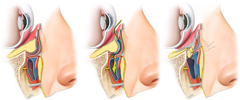
Fig. 1. Illustration of our surgical technique. The dissection is performed though the facial soft-tissue space of the midcheek, from the preseptal into the premaxillary space (left). The tear trough ligament and origins of the orbicularis oculi located between these spaces are precisely released (center). This provides a safe dissection plane and minimizes the surgical trauma associated with the surgical mobilization required for the surgery. The excised fat is then precisely grafted, in small pieces, under the tear trough ligament to complete the procedure (right). (Used with permission from Levent Efe, CMI.)补充视频 / 附件(原文未随文提供原图,可于出版方页面查看)prs-157-0975-s001.mp4Video 1. This operative video demonstrates our technique of extended transconjunctival lower blepharoplasty.
From June of 2017 to December of 2024, 200 (152 female and 48 male) patients underwent this procedure performed by the first author (C.-H.W.). The mean patient age was 41 years (range, 24 to 74 years), with a mean follow-up duration of 22 months (range, 6 to 95 months). Most patients (152 patients [76%]) were healthy, with no significant chronic medical conditions. The remaining patients (48 patients [24%]) had at least 1 of the following medical comorbidities: diabetes mellitus, hypertension, or ischemic heart disease. These were managed optimally before surgery. For patients on anticoagulant therapy, medications were stopped for an appropriate duration before surgery, with approval from the prescribing physician. Midcheek fat grafting was performed concurrently in 192 patients (96%). The mean volume of fat grafted per side was 2.4 cc over the maxilla (range, 1.0 to 3.0 cc) and 1.6 cc over the zygoma (range, 0.5 to 4.5 cc). The total mean graft volume was 4.0 cc per cheek. Eighty patients (40%) underwent the lower eyelid procedure in combination with other facial aesthetic procedures. Among these, 64 patients received concurrent upper blepharoplasty, 8 underwent medial epicanthoplasty, 18 underwent a deep plane face lift, and 3 had rhinoplasty. Fractionated carbon dioxide laser resurfacing was performed for patients with skin excess and fine lower eyelid fine wrinkles. A total of 30 patients (15%) underwent resurfacing using the carbon dioxide MIXto Pro laser system (Lasering USA, Medical Laser). None of the patients in this cohort underwent canthopexy, canthoplasty, or skin-pinch excisions.
Patient satisfaction was high, with 198 patients (99.0%) reporting being satisfied or highly satisfied with their surgical outcome. Early and late complications are summarized in Table 1 . Three patients experienced minor delayed bleeding, which presented as sudden discharge of a small amount of stale, liquefied blood from the lower eyelid between postoperative days 5 and 9. This was attributed to spontaneous discharge from liquefaction of small blood clots at the incision site and was self-limiting. Chemosis occurred in 3.5% of patients. Subset analysis revealed that all cases of chemosis occurred in patients who underwent concurrent upper blepharoplasty and/or face lift. The incidence of chemosis among patients undergoing isolated extended transconjunctival lower blepharoplasty was 0%. Chemosis resolved in all patients within 3 weeks with conservative treatments of eyelid lubrication and steroid eyedrops. No patient complained of dry eyes or epiphora after surgery.
Table 1. Our Early and Late Complications Rates
| Complications | Value (%) |
|---|---|
| Early | |
| Minor delayed bleeding | 3 (1.5) |
| Middle lamella scarring | 2 (1.0) |
| Hematoma | 0 |
| Chemosis | 6 (3.0) |
| Infection | 0 |
| Late | |
| Lower eyelid malposition | 0 |
| Ectropion | 0 |
| Residual eye bag needing revision surgery | 2 (1.0) |
| Fat necrosis | 7 (3.5) |
| Dry eyes | 0 |
| Epiphora | 0 |
| Late infection | 0 |
| PIH from laser treatment | 0 |
| Overall revision rate | 2 (1.0) |
Scarring of the middle lamella occurred in 2 patients with a genetic predisposition for scarring. Both were Asian patients and had a history of hypertrophic scar formation following previous operations. These patients were successfully treated with 2 sessions of dilute triamcinolone and 5-fluorouracil injections into the middle lamella (spaced 1 month apart), combined with daily gentle upward stretching of the lower eyelid. Both patients achieved full resolution of eyelid contracture within 2 months, without long-term sequelae. In the long-term follow-up, there were no cases of scleral show, lower eyelid retraction, or ectropion.
Fat necrosis was observed in 3.5% of patients, presenting as a palpable mass over the orbital rim. All cases were successfully managed with triamcinolone and 5-fluorouracil injections, without the need for surgical excision (see later under Discussion). The overall revision rate was 1.0%, from 2 patients presenting with small residual bulges of the lateral fat pad. These were addressed either by means of a repeated transconjunctival approach or, when an upper blepharoplasty was planned, through a lateral upper eyelid incision.
Figure 2 , above , and Supplemental Digital Content 3 and 4 show the long-term results of the patient shown in Video 1 . ( See Figure, Supplemental Digital Content 3 , which shows three-fourths views before [ left ] and 2 years after surgery [ right ], showing effective long-term correction of the eye bags and the tear trough deformity, https://links.lww.com/PRS/I464. See Figure, Supplemental Digital Content 4 , which shows comparison with eyes looking upward before [ left ] and 2 years after surgery [ right ], https://links.lww.com/PRS/I465.) The technique delivers predictable and permanent removal of the eye bags, permanent correction of the tear trough deformity, and predictable smoothing of the lid-cheek junction (Figs. 3 through 5 ). ( See Figure, Supplemental Digital Content 5 , which shows three-fourths views before surgery [ above ], 2 years after surgery [ center ], and 8 years after surgery [ below ], showing effective long-term correction of the eye bags and tear trough deformity, https://links.lww.com/PRS/I466.) The patient’s smile changes in a predictable and reproducible manner with the surgery (Fig. 2 , below ). In patients presenting with increased prominence of the eye bags and tight tethering at the tear trough ligament with smiling, the procedure is able to simultaneously remove the eye bags and permanently eliminate the tethering at the tear trough ligament with smiling (Figs. 6 and 7 ). In addition, the fine wrinkling on the lower eyelid with smiling is reduced. ( See Video 2 [online] , which shows the long-term change in the pattern of orbicularis oculi contraction of the lower eyelid after the described surgical technique is used.)
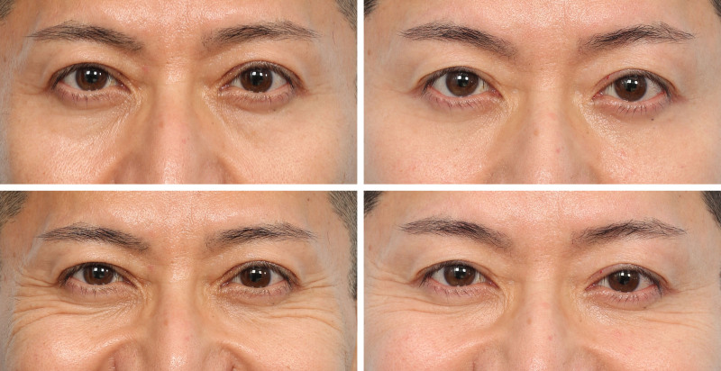
Fig. 2. (Above) A 52-year-old man presented for eye bag correction and appearing tired. His operation is shown in Video 1. Here he is shown at 2 years after surgery with good long-term correction of the eye bags and the tear trough deformity, and enhancement of the midcheek projection from the fat grafting. (Below) Smiling views before and after surgery. The procedure enhanced the aesthetics of his smile. The tethering centered at the tear trough ligament is eliminated, and the fine wrinkles of the lower eyelid are reduced.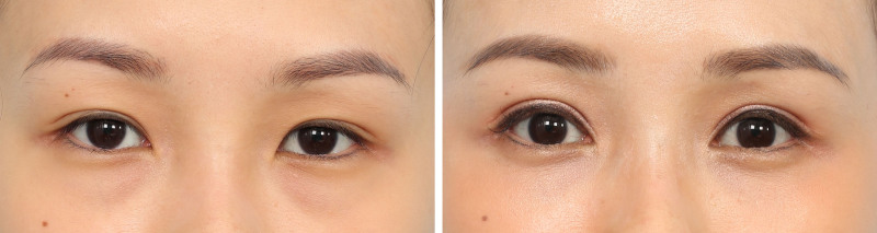
Fig. 3. A 35-year-old woman with prominent eye bags. The extended transconjunctival lower blepharoplasty was performed. No midcheek fat grafting was performed. Concurrently, she underwent upper blepharoplasty with levator advancement and medial epicanthoplasty. She is shown 2 years postoperatively. The technique was effective in permanently eliminating the eye bags and correcting the tear trough deformity.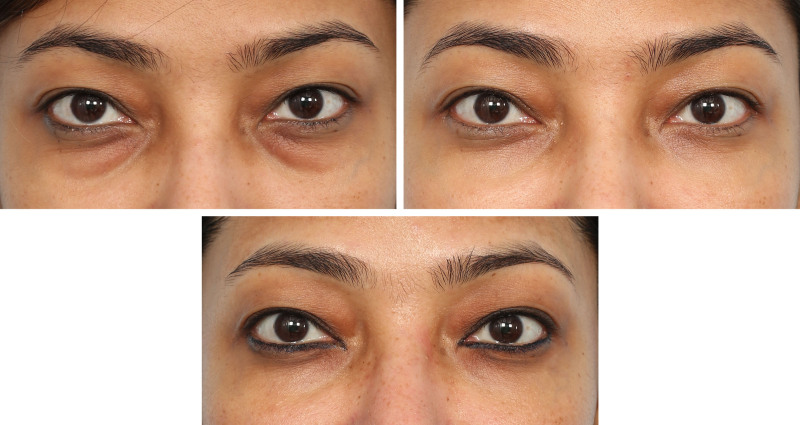
Fig. 4. (Above, left) A 39-year-old woman presented with prominent eye bags and lid-cheek junction. She underwent the extended transconjunctival lower blepharoplasty with facial fat grafting. A total of 3.5 cc of microfat was injected per side of her midcheek (1.5 cc over the zygoma and 2.0 cc over the maxilla). Nanofat was injected into her lower eyelid as well. (Above, right) She is shown at 2 years (age 41) and (below) at 8 years (age 47) after surgery. The stability of the result over the long term is clearly demonstrated.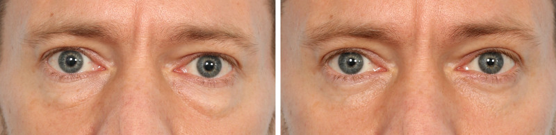
Fig. 5. A 43-year-old man presented with eye bags. He underwent the extended transconjunctival lower blepharoplasty. Midcheek fat grafting was not required. An excellent correction is shown at 6 months after surgery.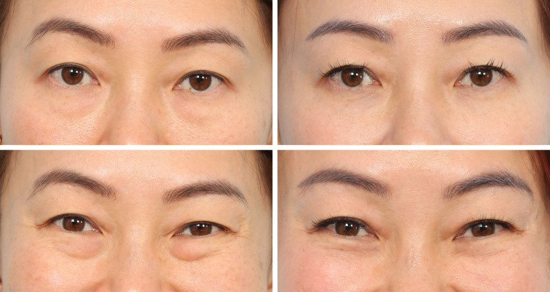
Fig. 6. (Above) A 43-year-old woman presented with eye bags. She specifically complained of a deep groove on the lower eyelid-cheek junction when smiling. She underwent the extended transconjunctival lower blepharoplasty with facial microfat and nanofat grafting. She is shown 3 years after surgery. (Below) The effect of the procedure on the aesthetics of the smile is shown. The prominent tethering at the location of the tear trough ligament and muscle origins has been permanently eliminated, allowing a more relaxed and aesthetically pleasing smile.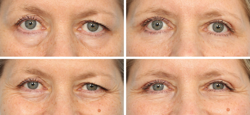
Fig. 7. (Above) A 54-year-old woman presented with periorbital aging changes with droopy eyelids and prominent eye bags. She underwent upper blepharoplasty with levator advancement, extended transconjunctival lower blepharoplasty, and midcheek fat grafting. She is shown 2 years after surgery. No skin excision or pinch blepharoplasty was performed. With the skin contraction and remodeling after her surgery, the fine wrinkles of the lower eyelid have improved. (Below) Changes of aesthetics of the smile after surgery with elimination of the tethering centered at the tear trough ligament and orbicularis oculi origins, giving a significantly more relaxed and aesthetically pleasing smile.补充视频 / 附件(原文未随文提供原图,可于出版方页面查看)prs-157-0975-s002.mp4Video 2. This video shows the long-term change in the pattern of orbicularis oculi contraction of the lower eyelid after the described surgical technique is used.
This technique is advantageous because of its simplicity, safety, and effectiveness. Key surgical steps include (1) atraumatic dissection through the facial soft-tissue spaces of the lower eyelid and midcheek; (2) precise release of the tear trough ligament, orbicularis retaining ligament, and origins of the orbicularis oculi; (3) adequate removal of the excess orbital fat pads; (4) precise orbital fat pad grafting along the orbital rim to prevent reattachment of the tear trough ligament and orbicularis retaining ligament to efface the lid-cheek junction; and (5) optimizing the orbital facial vector of the midcheek with judicious structural fat grafting. 13 , 16 , 26
A key difference of our technique, compared with most previously described methods, is the use of free fat grafts fashioned from the excised lower eyelid fat pads, rather than pedicled fat flaps transposed over the orbital rim. The primary advantage of the pedicled fat flap approach lies in the preservation of vascular supply of the transposed fat, theoretically reducing the risk of fat necrosis. 27 – 29 However, orbital fat pad transposition has some potential limitations and risks. 30 First, it is difficult to determine whether the transposed fat sufficiently reduces the excess lower eyelid fat while simultaneously providing adequate volume enhancement and caudal reach to prevent the tear trough ligament from reattaching to the anterior maxilla. This manifests as residual bulges of the lower eyelid and inadequate correction of the tear trough deformity. Furthermore, the transposition of the pedicled fat flap may exert downward traction on the orbital septum—either through direct tension or inadvertent inclusion of orbital septum during suture fixation. This increases the risks of middle lamella contracture and lower eyelid malposition. 31 In contrast, the free fat graft approach is technically simpler to execute and is safer. 23 , 24 , 32 – 35 By dividing the maneuver into 2 distinct steps—fat excision followed by graft placement—each step can more predictably be optimized. First, the excess orbital fat pads are precisely excised. The extent of removal is determined by the degree of lower eyelid fat excess, with greater excision performed for more prominent eye bags. Only the fat that readily extrudes on opening the orbital septum is removed. Final adjustment is guided intraoperatively by gently applying pressure on the globe to assess the contour of the lower eyelid, providing a visual endpoint for adequate excision. The second step is to augment the tissue deficiency along the orbital rim with free fat grafting using small lobules fashioned from the excised orbital fat to eliminate the “lid-cheek junction.” These grafts can be precisely placed at anatomically optimal locations, without the limitations imposed by pedicle length or reach. Long-term follow-up in our cohort demonstrated consistent correction of the tear trough deformity and effacement of the lid-cheek junction. Importantly, because the orbital septum remains undisturbed during graft placement, there is no risk of inferior traction on the septum—a factor that may contribute to lower eyelid malposition caused by contracture of the middle lamella seen in other transconjunctival techniques. This lack of tension on the orbital septum may explain the 0% incidence of scleral show and lower eyelid retraction observed in our long-term results. The effectiveness of our approach may be seen in the high patient satisfaction rate and very low revision rates of 1.0%.
Fat necrosis is a potential risk with free fat grafting. Since our initial publication, we have refined our technique to enhance fat graft survival and reduce this complication. Several modifications were implemented. First, the size of each fat graft was reduced to approximately 5 mm in diameter. 36 This improves the surface area–to-volume ratio, enhancing the revascularization potential. 16 We are also using more pieces of graft per eyelid to more evenly distribute the fat grafts along the orbital rim. Whereas our original technique in 2017 used 3 fat grafts per eyelid, our current approach uses 5 smaller sized fat grafts, Second, during fat preparation, any tissue that has been “burned” by the cautery is excised to ensure that only viable adipose tissue is used for grafting. Several other authors have also used free fat grafts from the excised orbital fat pads. These authors used various techniques of fat graft placements, ranging from free placement of the fat grafts without suture fixation to mincing the fat and then placing the fat into syringes and injecting the fat from percutaneously. 23 , 30 , 37 In our experience, precise placement under direct vision with fixation of each fat graft lobule along the orbital rim using percutaneous pull-through sutures is essential for predictable fat survival. 6 Finally, further postoperative immobilization of the lower eyelid with Steri-Strips for a minimum of 5 days promotes revascularization during the early healing phase. With these refinements, fat graft survival has been favorable, and the incidence of fat necrosis in our series was low at 3.5%. Importantly, when fat necrosis occurred, it was effectively managed nonsurgically. Treatment involved puncture of the fat cyst using a 30-gauge needle, followed by injection of 0.15 to 0.25 cc of a triamcinolone and 5-fluorouracil mixture (40% triamcinolone, 60% 5-fluorouracil) directly into the lesion. The puncture of the cysts disperses the necrotic fat, and the injected mixture promotes the resorption of the capsule associated with the cyst. ( See Figure, Supplemental Digital Content 6 , which shows [ above ] a 37-year-old woman who underwent the extended transconjunctival lower blepharoplasty with midcheek fat grafting procedure. [ Center ] Four weeks after surgery, she presented with a small cystic lesion on her right cheek [ arrow ]. This was a small fat cyst from fat necrosis. The cyst was managed with a single intralesional injection of 0.15 cc of a mixture of triamcinolone and 5-fluorouracil. [ Below ] Six months after surgery, the cyst has completely resolved and tissues are soft and supple with no mass palpable, https://links.lww.com/PRS/I467.) No patients in our series required surgery to remove the fat cysts.
The long-term effect of releasing the tear trough ligament and the origins of the orbicularis oculi without refixation remains a point of contention among surgeons. Anatomically, the tear trough ligament functions to anchor the medial lower eyelid soft tissues to the bone. 6 The orbicularis oculi is a sphincteric muscle that functions to voluntarily and involuntarily close the eyelids to protect the eyes. 13 With aging, a deep groove develops along the course of the tear trough ligament, accompanied by increased contraction and puckering of the associated muscular attachment. Age-related changes such as dermal thinning, increased protrusions of orbital fat pads, maxillary retrusion, and soft-tissue volume loss collectively contribute to the accentuation of the tear trough deformity. 38 , 39 Given the anatomical role of the tear trough ligament and orbicularis oculi origins in tethering the overlying soft tissues, their release is a prerequisite for effective long-term correction. 4 , 40 , 41 Our data demonstrate that release of the tear trough ligament does not compromise long-term tarsoligamentous integrity and support of the lower eyelid. Postoperative pretarsal orbicularis oculi function remained normal and there was no increased risk of scleral show. This is attributed to the preserved integrity of the medial and lateral canthal ligaments—both of which are located above the meridian of the tear trough ligament—and the unaltered pretarsal orbicularis oculi sling, which continues to provide support to the lower eyelid against the globe.
The release of the orbicularis oculi origins results in a weakening of orbicularis oculi function. In the native state, the medial suborbital region becomes gathered or constricted because of contraction of the orbicularis oculi muscle fibers both superior and inferior to the tear trough ligament tethering point. This constriction, combined with protrusion of orbital fat and periorbital skin wrinkling, produces a deep, contracted groove that contributes to a squint-like smile. By releasing the muscle origins, this focal contraction point is eliminated, reducing the formation of fine lower eyelid wrinkles and producing a more open, aesthetically pleasing smile ( Video 2 and Figs. 6 , below , and 7, below ). This contrasts with traditional transconjunctival techniques that excise fat alone without release of the tear trough ligament and orbicularis oculi origins. Tethering at the ligaments remains unchanged so that those techniques are unable to deliver the kind of transformative lower eyelid rejuvenation as demonstrated here. ( See Figure, Supplemental Digital Content 7 , which shows a 52-year-old man who had previously undergone a standard transconjunctival eye bag removal with midcheek fat grafting 2 years before, performed by another surgeon. Although his result appears to be reasonable in repose [ left ], he appears considerably more aged when smiling [ right ], because of the persistent tethering at the tear trough ligament with animation, https://links.lww.com/PRS/I468.)
Because of the limited access, variations in individual anatomy can significantly influence the technical ease of performing the procedure. This can be predicted preoperatively on clinical examination. Favorable factors include patients having a longer lower eyelid and more laxity of the tarsoligamentous sling of the lower eyelid. In contrast, it is more difficult to perform the procedure in patients with shorter and tighter lower eyelids, because of the more limited exposure provided by such anatomy and tissue quality. The angulation of the anterior maxilla is another factor that influences the ease of performing this procedure (Fig. 8 ). In patients with a more retruded maxilla (negative or neutral vector), it is easier to perform the procedure, as the dissection plane is on a downward incline from the surgeon’s line of sight. In contrast, for patients with a projected maxilla (positive vector), the procedure is more challenging to perform, as the plane of dissection is on a more upward incline from the orbital rim, making visualization and instrument handling more difficult.
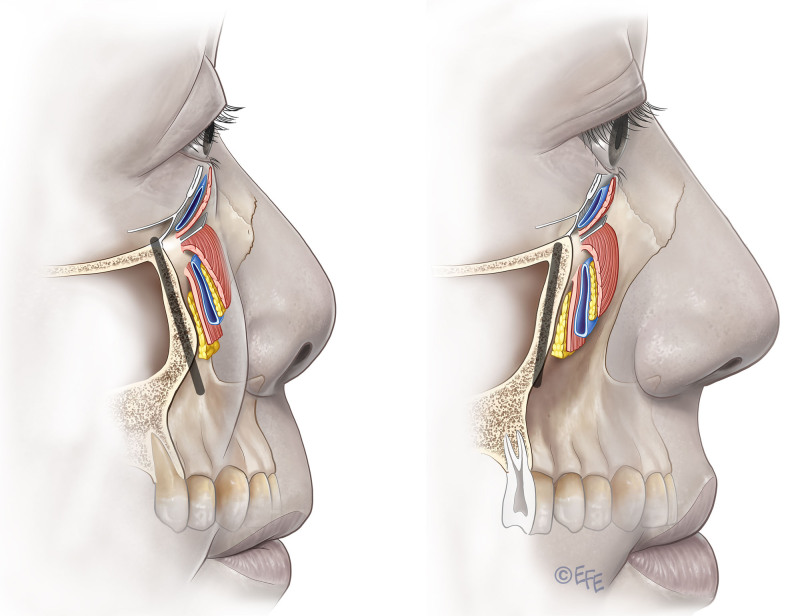
Fig. 8. In patients with a negative vector maxilla, the downward incline of the plane of dissection makes visualization and surgery easier (right). In contrast, in patients with a positive vector, the maxilla rises from the orbital rim. This upward plane of dissection makes surgery more challenging, as the surgeon has to dissect upward from the orbital rim (left). (Used with permission from Dr. Levent Efe.)Patient selection for this technique is important. The ideal patients are younger patients with prominent eye bags and tear trough deformity (Fig. 3 ). Accordingly, all younger patients (younger than 40 years of age) are excellent candidates for this procedure. In older patients undergoing a concomitant face lift, the extended transconjunctival lower blepharoplasty is also ideal for treating the eye bags, whereas the cheek is tightened by means of the face-lift approach without the need for a subciliary incision, which significantly speeds up recovery and is safer (Fig. 2 ). Older patients presenting for eye bag removal can also be good candidates for this approach. The crepey skin can be treated sufficiently with fractionated carbon dioxide laser resurfacing at the time of the surgery. This firms and smooths the skin to a sufficient degree in most patients (Fig. 7 ). None of our patients required secondary skin excision because of dissatisfaction with residual crepey skin.
The authors have no financial interest in any of the products, devices, or drugs mentioned in this article. They have no conflicts of interest to declare in regard to the present work.
Patients provided written informed consent for the use of their images.
声明:中文精读 · 仅供学术参考。内容来自公开文献检索,不代表本人观点,不构成诊疗建议。 医疗美容需在正规医疗机构由执业医师实施。
基于相同主题推荐 · 继续深挖
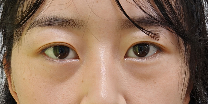
回顾性无对照研究(深圳市宝安区人民医院,2023-05 至 2025-05):32 例(17 女 15 男,平均 28.9 岁)下睑成形(LLB)后黑眼圈加重,接受自体微脂肪移植矫
本研究为单中心前瞻性病例系列(200 例,2017—2024),平均随访 22 个月(范围 6—95 个月),评估经结膜延长下入睑成形术的长期美学与安全性结果。
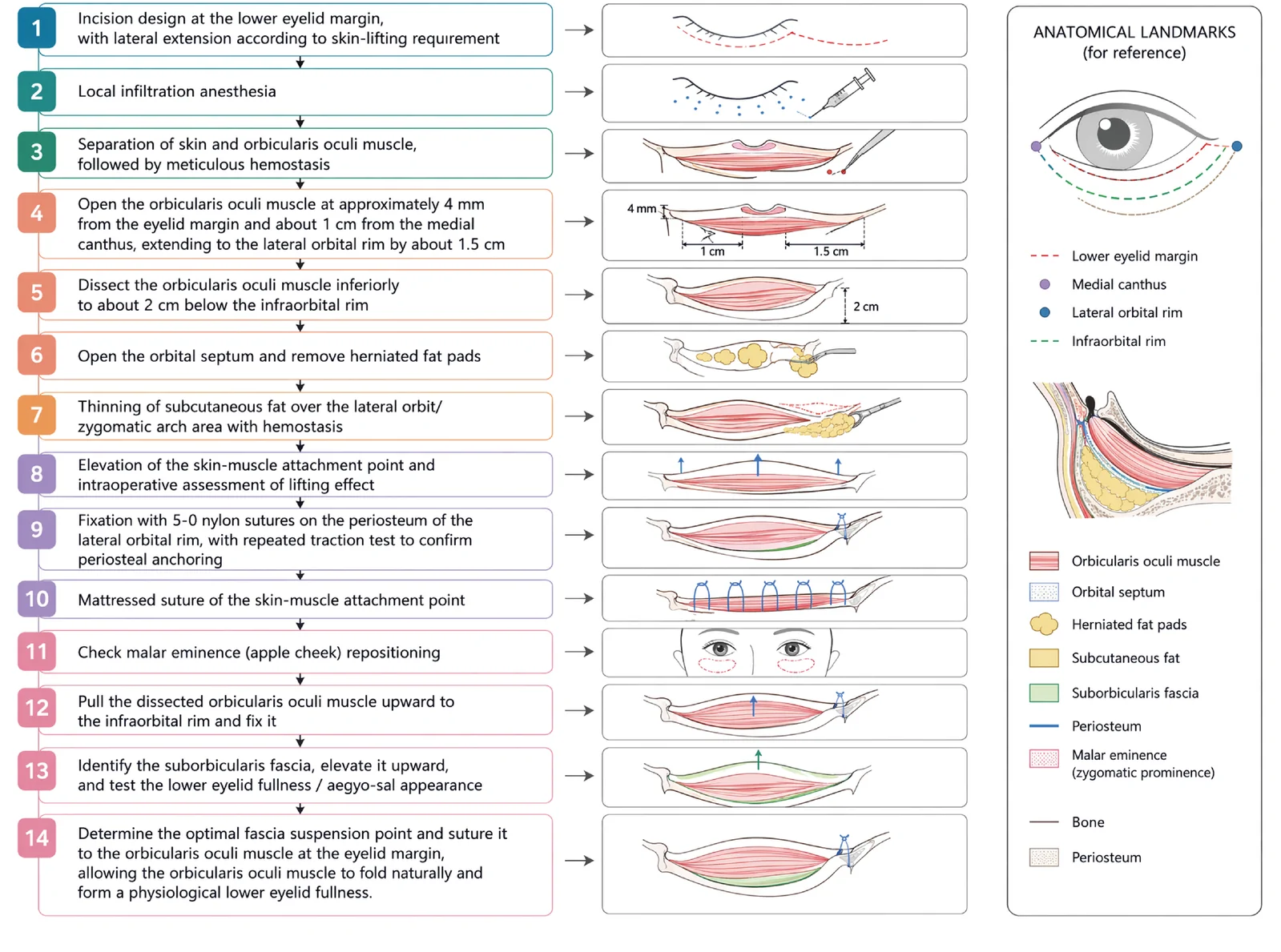
V3中面部提升经睫毛下切口,沿内眦线、外眦线、颧脂肪垫下界三个向量悬吊,可同时改善中面部下垂、下睑袋及睑颊轮廓不连续。