经下睑睫毛下切口V3中面部提升:三向量生理性复位技术
2026年10月18日15 min read
Frontiers in Surgery
结论与实践要点
本文报道了一种经睫毛下切口、以三向量(内眦线、外眦线、颧脂肪垫下界)进行骨膜/筋膜多点悬吊的中面部提升技术,并回顾性评估了42例患者的量化结果。其特点是用较局限的下睑入路同时处理中面部下垂、下睑袋与睑颊沟,且随访数据显示睑颊距离缩小、泪槽平滑,患者满意度较高。
并发症以短暂结膜水肿与轻度水肿为主,未见持久性睑外翻或面神经损伤,提示在掌握适应证与操作要点的前提下,该技术的安全性较可控。但需注意:本研究为单中心回顾性、样本量有限且缺乏对照组,结论的外推应审慎;是否较其他中面部提升术式更优,仍需更大样本与对照研究验证。临床选用时应结合患者解剖基础与对下睑支撑的需求综合判断。
BackgroundConventional midface rejuvenation through temporal or preauricular incisions often fails to adequately restore the central midface and tear trough vector. We describe the “V3 midface lift"—a transcutaneous subciliary technique utilizing a three-vector physiologic repositioning system to address midface ptosis, lower eyelid bags, and lower lid-malar contour disharmony.
MethodsA consecutive cohort of 42 patients (38 females, 4 males; mean age: 46.2 ± 6.8 years) who underwent the V3 midface lift between January 2022 and December 2024 was retrospectively analyzed. The technique incorporates an extended subciliary incision, selective orbicularis oculi release, conservative septal fat management, and multi-point periosteal/fascial suspension anchored along three specific vectors: the medial canthal line, lateral canthal line, and the inferior boundary of the malar fat pad (adjacent to the melolabial fold). Quantitative outcomes were assessed using pre- and postoperative lower lid-malar distance (LMD), tear trough rating scales, and the Global Aesthetic Improvement Scale (GAIS).
ResultsAll 42 patients completed follow-ups ranging from 12 to 36 months (mean: 22.4 months). Significant reductions in lower lid-malar distance (mean reduction: 3.4 ± 0.8 mm, P < 0.001) and marked smoothing of the tear trough/malar groove were achieved. High patient satisfaction was recorded at 12 months (92.8% rated as “Very Much Improved” or “Much Improved” on GAIS). Postoperative complications were limited to transient chemosis ( n = 3, 7.1%) and minor prolonged edema ( n = 2, 4.8%), both resolving without secondary intervention. No permanent ectropion, hematoma, or facial nerve injury occurred.
ConclusionsThe V3 midface lift provides safe, stable, and durable midface elevation and lower lid-malar continuity through a minimalist subciliary access, representing an efficient option for central facial rejuvenation.
Keywords: lower blepharoplasty、malar fat pad、midface lift、orbicularis suspension、subciliary approach、tear trough
Aging of the central face involves a complex interplay of skin laxity, orbicularis oculi attenuation, orbital fat pseudoherniation, deep fat compartment atrophy, and downward migration of the malar fat pad ( 1 , 2 ). These structural shifts disrupt the smooth contour connecting the lower eyelid to the cheek, creating a distinct tear trough deformity, accentuation of the lid-cheek junction, and flattening of the anterior malar eminence ( 3 ).
Traditional rhytidectomy procedures primarily deliver lateral traction, which frequently leaves central midface hollows and tear trough deformities under-corrected ( 4 ). Conversely, deep-plane or endoscopic midface lifts offer powerful repositioning but carry longer learning curves, higher risk to facial nerve branches, and prolonged recovery times ( 5 ). Extended transcutaneous lower blepharoplasty techniques have addressed these limitations, yet maintaining secure, multi-vector vertical suspension without increasing lower lid downward tension remains a surgical challenge ( 6 ).
To achieve direct, physiologic restoration of the lower lid-malar complex, we developed a three-vector suspension technique performed through a subciliary approach: the V3 midface lift. This method targets the superficial skin envelope, orbicularis oculi muscle, deep fascia, and descended malar fat pad simultaneously. In this report, we outline the surgical concepts, operative steps, and a consecutive clinical series evaluating its quantitative efficacy and long-term durability.
Between January 2022 and December 2024, a total of 42 consecutive patients (38 females [90.5%] and 4 males [9.5%]) underwent the V3 midface lift and were included in this study. The mean age of the cohort was 46.2 ± 6.8 years (range: 34 to 62 years). Primary procedures accounted for 88.1% of cases ( n = 37), while 11.9% ( n = 5) were revision surgeries. Concomitant upper blepharoplasty was performed in 12 patients (28.6%). All patients completed clinical evaluations with a mean follow-up duration of 22.4 ± 7.2 months (range: 12 to 36 months).
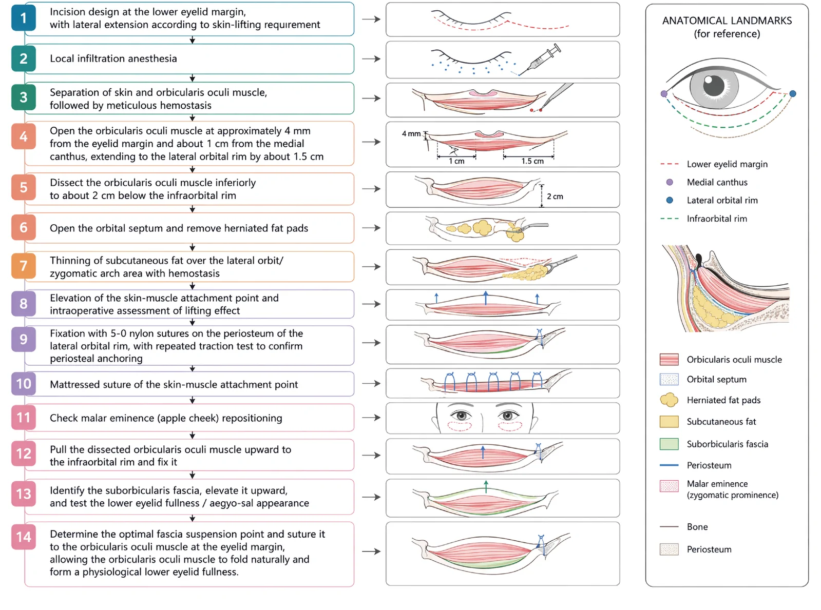
The operational protocol for the V3 midface lift is hierarchically structured into a 14-step surgical sequence ( Figure 1 ). Following local infiltration anesthesia along the subciliary line, a skin-muscle flap is dissected with meticulous release of the orbicularis retaining ligament (ORL) to approximately 2 cm below the infraorbital rim. After conservative management of herniated orbital fat and thinning of the lateral orbital subcutaneous fat pad, structural resuspension is executed. Multi-point periosteal fixation along the three primary vectors is confirmed via intraoperative traction testing using 5-0 nylon sutures. Crucially, the suborbicularis fascia is elevated superiorly and anchored to the pretarsal orbicularis muscle near the eyelid margin, promoting natural muscle folding to recreate physiologic pretarsal fullness (aegyo-sal contour) without creating tension on the lower eyelid margin ( Figure 1 ).
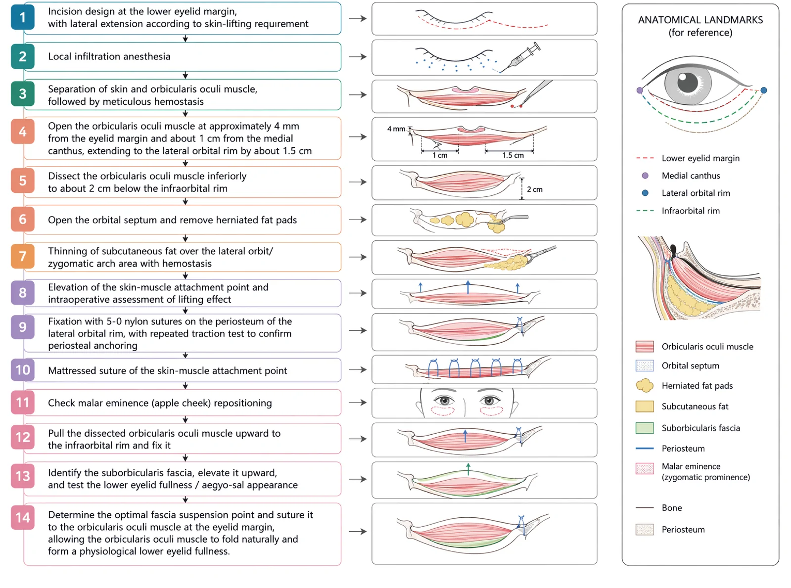
Figure 1 Step-by-step schematic surgical flowchart of the V3 midface lift procedure. The diagram illustrates the 14-step surgical sequence alongside corresponding anatomical illustrations and legends, detailing incision design, skin-muscle flap dissection, orbicularis release, septal fat management, multi-point vector elevation with periosteal anchoring at the lateral orbital rim, and superior resuspension of the orbicularis oculi and deep suborbicularis fascia to re-establish natural pretarsal roll (aegyo-sal contour) fullness.
With the patient in an upright sitting position, three key vertical vectors (V3) are marked on the midface ( Figure 2 ): Vector 1 (Medial Vector): Extends vertically from the medial canthal region down through the tear trough toward the superior border of the nasolabial fold. Vector 2 (Lateral Vector): Extends from the lateral canthus toward the zygomatic arch. Vector 3 (Malar Vector): Passes through the point of maximum malar projection down to the inferior boundary of the malar fat pad (adjacent to the melolabial groove).
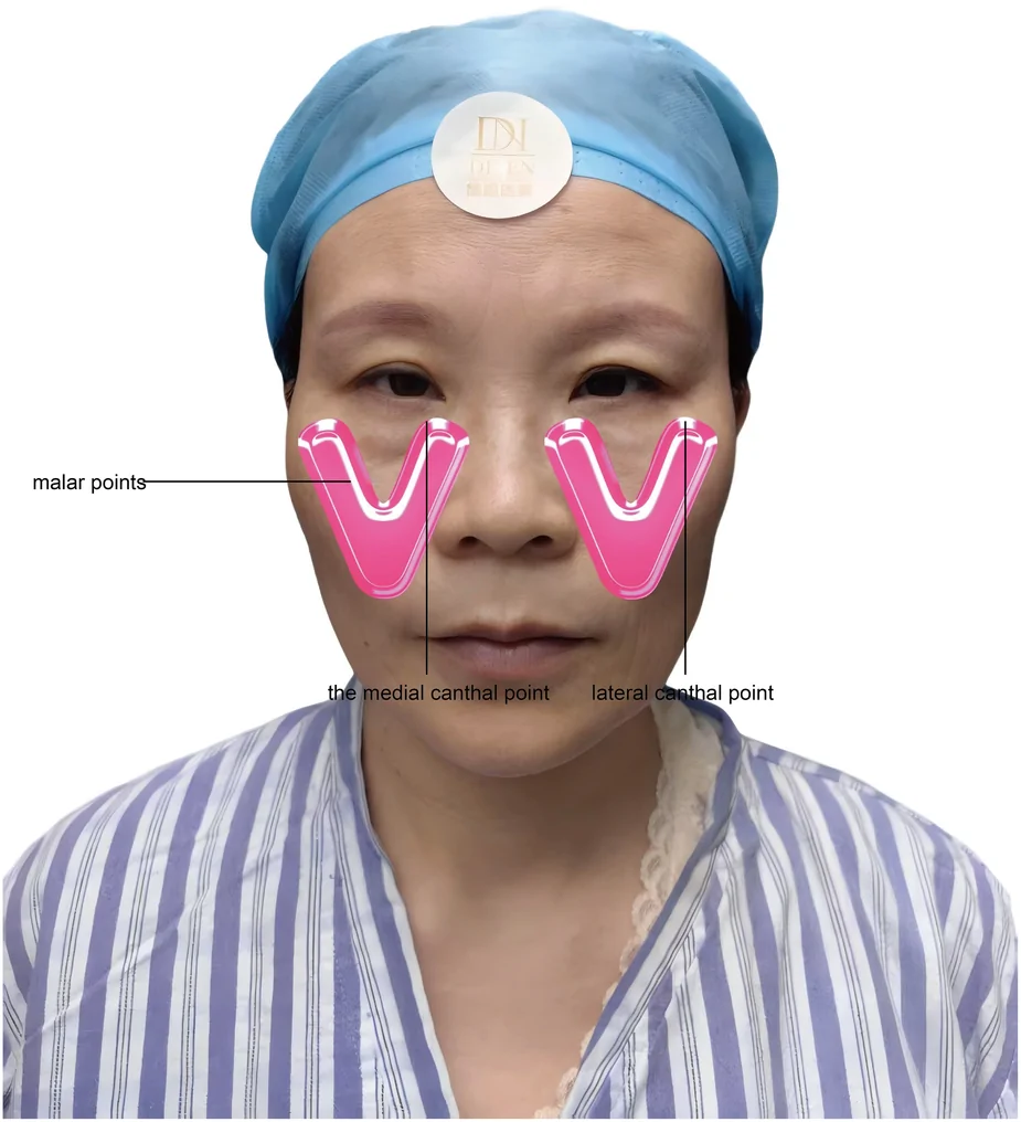
Figure 2 Preoperative design demonstrating the three-vector (V3) lifting system. Preoperative surface markings indicating the three principal vertical lifting vectors: Vector 1 (Medial Vector) extending from the medial canthal region down through the tear trough; Vector 2 (Lateral Vector) extending from the lateral canthus toward the zygomatic arch; and Vector 3 (Malar Vector) passing through the point of maximum malar projection down to the inferior boundary of the malar fat pad (adjacent to the melolabial groove).2.
A subciliary incision is used and extended laterally beyond the orbital rim according to the amount of redundant skin requiring redraping. After local infiltration anesthesia, the skin is separated from the orbicularis oculi muscle. Meticulous hemostasis is obtained before proceeding deeper. Approximately 1 cm lateral to the medial canthus and 4 mm below the ciliary margin, the orbicularis is opened and the dissection is carried laterally toward the orbital rim. The orbicularis is then released inferiorly to approximately 2 cm below the infraorbital rim, releasing the orbicularis retaining ligament (ORL) and zygomaticocutaneous ligaments (ZCL) as needed to fully mobilize the malar soft tissue complex. The orbital septum is opened when indicated, and herniated fat is conservatively removed or transposed into the tear trough defect.
The subcutaneous fat of the lateral orbital region (above the zygomatic arch) is judiciously thinned as needed. Tissue elevation is performed along the three pre-designed vectors: Deep Anchoring: The sub-orbicularis fascia and deep malar soft tissues along Vector 3 are recruited upward and secured to the lateral orbital periosteum using 5-0 clear nylon or PDS sutures. Muscle Resuspension: The lateral orbicularis oculi muscle flap (Vectors 1 and 2) is advanced superolaterally and anchored to the internal rim of the lateral orbital periosteum above the lateral canthal level. Pretarsal Fullness Creation: The deep fascial layer inferior to the orbicularis is anchored to the pretarsal orbicularis, allowing the muscle to fold naturally, restoring a physiologic pretarsal roll rather than an artificial bulge.
Redundant skin is redraped tension-free in a superior-lateral direction. Excess skin (typically 2 to 4 mm) is trimmed conservatively. Skin closure is performed with 6-0 polypropylene sutures, which are removed on postoperative day 5 to 7.
All 42 surgeries were completed without intraoperative complications. Patients were evaluated preoperatively and at 1, 6, 12, and 24 months postoperatively under standardized clinical studio conditions.
To illustrate the acute recovery trajectory of the V3 midface lift, serial photographs were documented across the early perioperative phase. Mild to moderate subcutaneous edema and local swelling reached their peak at 24 h post-operation, accompanied by visible supportive steristrip taping over the midface vector anchoring regions. By 72 h post-operation, peri-incisional edema and tissue swelling had visibly subsided without significant ecchymosis or severe chemosis. Immediately following suture removal (postoperative day 5 to 7), the subciliary skin incision demonstrated excellent primary healing with minimal tension, significant improvement in tear trough contour, and early restoration of a smooth lower lid-malar junction. ( Figure 3 ).
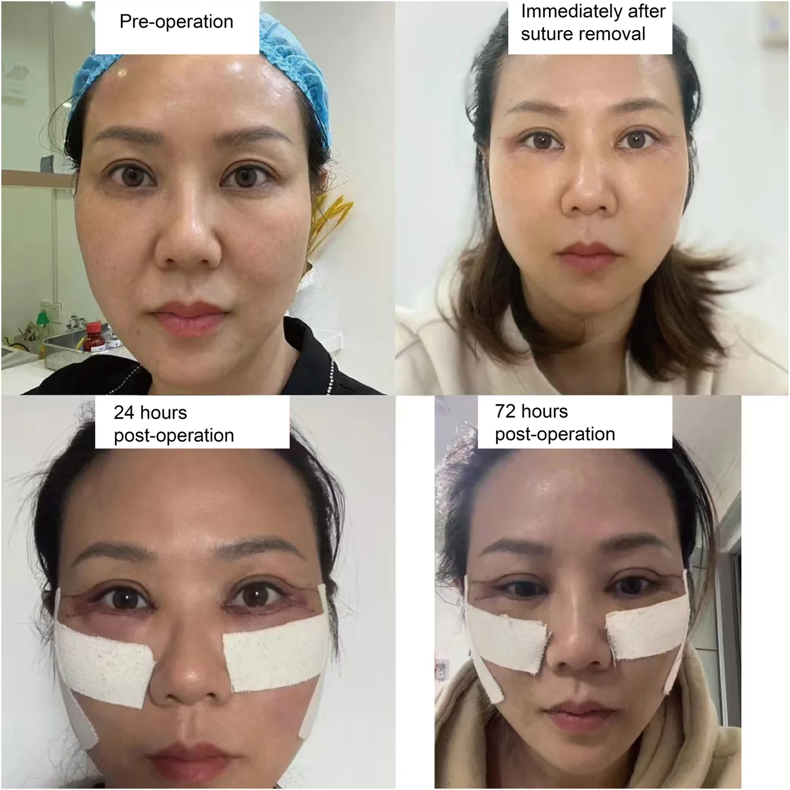
Figure 3 Representative perioperative recovery course of a patient undergoing the V3 midface lift. Sequential clinical photographs document the surgical trajectory at baseline (Pre-operation), 24 h post-operation (showing peak mild edema and supportive taping along vector tension lines), 72 h post-operation (demonstrating progressive swelling resolution), and immediately after suture removal (showing primary incision healing, minimal erythema, and early smoothing of the cheek-lid continuum).
The distance between the pupil center and the lowest point of the lid-cheek junction (Lower Lid-Malar Distance, LMD) was measured using standardized medical photographs. The mean LMD decreased significantly from 18.6 ± 1.4 mm preoperatively to 15.2 ± 1.1 mm at 12 months postoperatively ( P < 0.001), reflecting significant midface elevation and shortening of the lower eyelid vertical height.
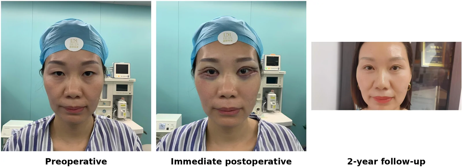
Figure 4 Representative case 1. Preoperative baseline, immediate postoperative appearance, and 2-year follow-up of a 45-year-old female, demonstrating sustained lower lid-malar continuity, elimination of infraorbital hollowing, and durable midface elevation.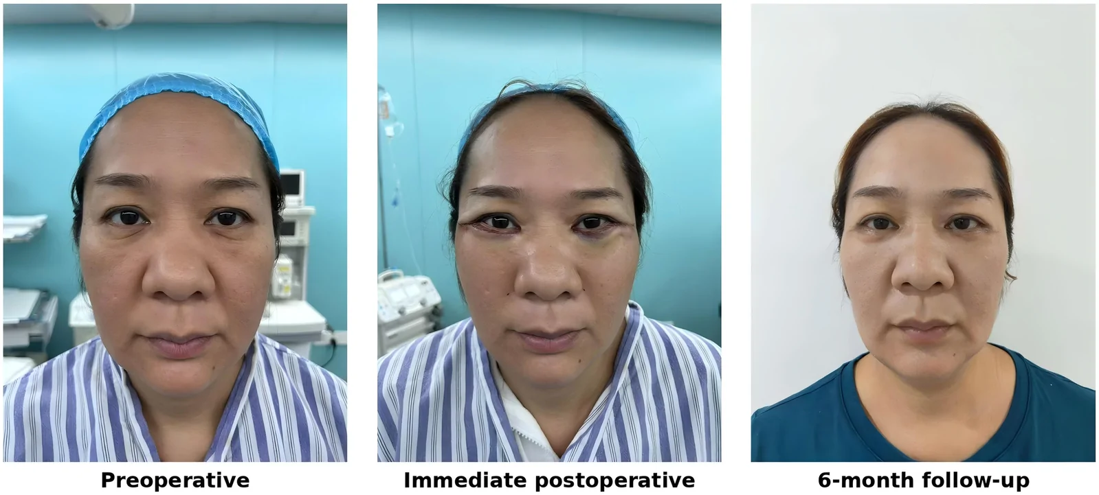
Figure 5 Representative case 2. Preoperative baseline, immediate postoperative appearance, and 6-month follow-up of a 48-year-old female, showing correction of orbital fat pseudoherniation, smoothing of the tear trough, and natural pretarsal roll restoration without scleral show or lid margin distortion.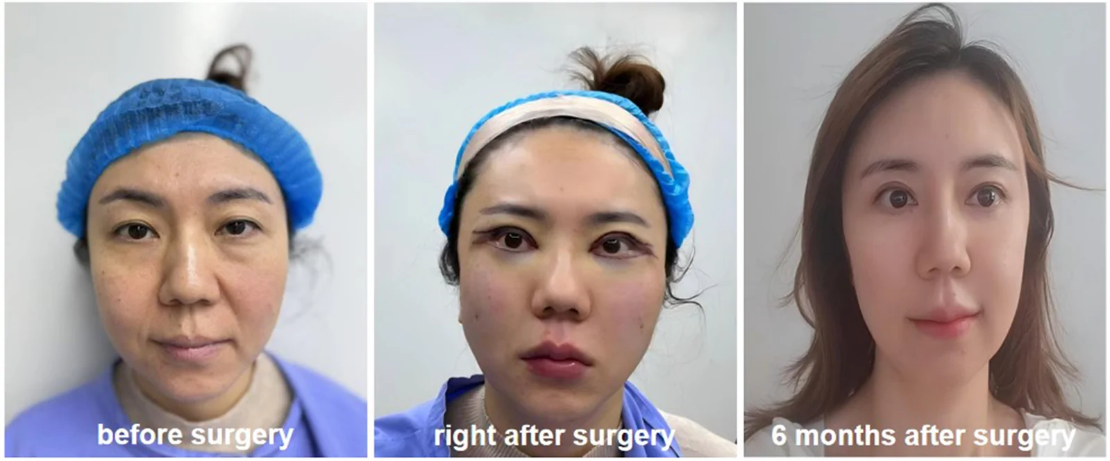
Figure 6 Representative case 3. Standardized clinical photographs of a 42-year-old female treated with the V3 midface lift: preoperative baseline (left), immediate postoperative appearance (middle), and 6-month postoperative follow-up (right). The outcome demonstrates stable midface elevation, seamless rejuvenation of the lid-cheek junction, complete resolution of tear trough grooving, and well-matured subciliary healing without residual deformity or eyelid malposition.
No cases of permanent ectropion, retrobulbar hematoma, infection, or facial nerve injury were observed. Transient Chemosis: 3 cases (7.1%), resolved within 2 weeks with topical anti-inflammatory drops. Prolonged Subcutaneous Edema: 2 cases (4.8%), resolved by 6 weeks with warm compresses and physical therapy. Minor Asymmetry: 1 case (2.4%), required no secondary intervention as the patient was satisfied with the overall outcome.
The V3 technique is conceptually distinct from lateral skin traction alone. Instead, it emphasizes direct restoration of the lower eyelid-malar unit through a subciliary route, with combined treatment of the superficial skin envelope, orbicularis oculi, deeper fascial layer, and descended malar fat ( 2 , 3 ).
Single-vector vertical or lateral traction often results in an unnatural “swept” appearance or recurrent midface sagging due to focal stress concentrations ( 7 ). The V3 system distributes lifting tension across three functional anatomical points: Medial Vector: Corrects the tear trough and redrapes the thick skin-muscle layer over the inner infraorbital margin. Lateral Vector: Re-establishes lateral orbital rim support and counteracts downward eyelid pull. Malar Vector (Inferior Boundary of the Malar Fat Pad): Lifts the heavy descent of the midface from its true inferior margin near the melolabial fold, restoring youthful anterior malar projection.
Compared to classical procedures like Rohrich's multi-step lower blepharoplasty or subperiosteal midface lifts, the V3 technique offers key advantages ( 5 , 6 ): (1) Lower neurological risk due to maintaining dissection in the pre-zygomatic sub-orbicularis plane above the periosteum, minimizing risk to the zygomatic and buccal branches of the facial nerve; (2) Natural aesthetic restoration by combining orbicularis flap advancement with deep fascial suspension to recreate a youthful pretarsal roll while smoothing the lid-cheek junction without creating a hollowed or skeletonized lower lid appearance ( 8 ).
The V3 midface lift is a reliable, three-vector subciliary technique that achieves physiologic repositioning of the midface soft tissue complex. By restoring lower eyelid support, smoothing the tear trough, and re-establishing malar convexity with low complication rates, this technique represents a practical and effective approach for central facial rejuvenation.
声明:中文精读 · 仅供学术参考。内容来自公开文献检索,不代表本人观点,不构成诊疗建议。 医疗美容需在正规医疗机构由执业医师实施。
基于相同主题推荐 · 继续深挖
V3中面部提升经睫毛下切口,沿内眦线、外眦线、颧脂肪垫下界三个向量悬吊,可同时改善中面部下垂、下睑袋及睑颊轮廓不连续。
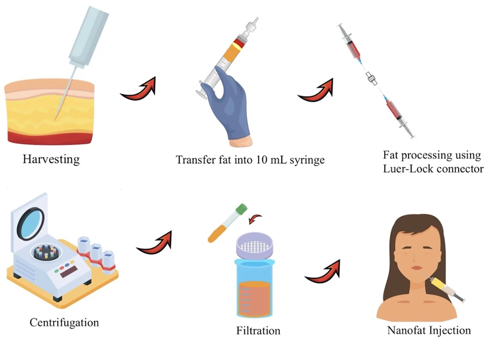
PRISMA 系统综述(2024-12),纳入 18 项研究、768 例患者;纳米脂肪为机械乳化/过滤的自体脂肪,富集 SVF 与脂肪来源干细胞,侧重再生而非单纯容积充填。
背景:面部老化包含组织下垂、容积缺失与皮肤老化三方面;面部提升术是面部年轻化的核心术式,但仅靠提升难以纠正容积缺失与皮肤质地的改变,需联合辅助术式。