接触冷却式单极射频对小型猪皮肤胶原及弹性纤维的增强作用:临床前研究
2026年10月3日33 min read
Clinical, Cosmetic and Investigational Dermatology
Clinical, Cosmetic and Investigational Dermatology
结论与实践要点
本文在 小型猪模型 中评估了一种基于珀尔帖(Peltier)直接接触冷却(DCC)的单极射频系统(频率 6.78 MHz、最大输出 140 W、电极 20×20 mm)。其核心机制是:射频能量向真皮及皮下层递送均匀热刺激,同时电极尖端通过接触式冷却实时降低表皮温度,使热损伤风险偏向更深层的胶原与弹性纤维重塑。研究设置最小、默认、最大三档能量,于术后即刻及 30 天取材观察。
安全性边界 方面,表皮温度基线约 35.1°C,治疗后降至 18.1–20.2°C,且全程监测未超过 43°C 的热损伤阈值;真皮峰值温度分别达 55.6°C(最小)、61.7°C(默认)、69.2°C(最大)。在 3 只 6 月龄雄性小型猪、单次治疗、随访 30 天的设定下,肉眼、血液生化及组织学(H&E、NBTC)均未发现红斑、水肿、坏死或炎症浸润等不良反应,提示在该参数范围内较可控。
有效性信号 :Masson 染色定量显示,治疗 30 天时胶原面积分数较 Day 0(设为 100)在默认组升至 197.7、最大组升至 213.7(**p<0.01),最小组为 167.9;Victoria blue 染色显示弹性纤维阳性面积在最大能量组增至 188.4(较 Day 0 呈增高趋势,p<0.1)。结果支持该设备在 非侵入性皮肤年轻化 方向的潜力。
证据等级与局限 :本研究为临床前动物实验,样本量小(n=3)、仅为单次治疗、随访 30 天,尚不能直接外推至人体临床疗效与适宜能量方案。对临床的参考价值在于:证实了「接触冷却 + 单极射频」在保护表皮的同时实现真皮加热与细胞外基质重塑的思路可行;后续仍需人体随机对照研究来验证适用人群、治疗频次与长期安全性。
PurposeTo investigate the thermal effects, dermal remodeling, and safety of a novel direct contact cooling (DCC) based monopolar radiofrequency (RF) system using a minipig model.
MethodsMonopolar RF was applied at three levels. Temperature changes, visual skin assessment, hematological analysis, and histological and histochemical evaluations were performed at baseline and up to 30 day post-treatment.
ResultsDCC-based monopolar RF achieved effective dermal heating without excessive epidermal temperature elevation. No adverse visual, systemic, or histological reactions were observed. Collagen density and elastic fiber remodeling were significantly enhanced at 30 days.
ConclusionDCC-based monopolar RF safely induces controlled dermal thermal stimulation and promotes extracellular matrix remodeling, supporting its potential as an effective non-invasive skin rejuvenation modality.
Keywords: radiofrequency system、skin rejuvenation、collagen remodeling、elastic fiber regeneration
Over the past several years, remarkable advances in laser- and energy-based medical devices have significantly influenced the field of aesthetic dermatology. Among these technologies, radiofrequency (RF) devices have gained widespread attention due to their ability to provide relatively simple and cost-effective treatment options for both clinicians and patients, while minimizing pain, adverse effects, and downtime, thereby achieving favorable clinical outcomes. 1 , 2 In aesthetic medicine, RF technology has been applied to address a broad range of medical and cosmetic indications, including skin laxity, photoaging, cellulite, and localized adiposity. 3 , 4 Currently available RF medical devices are classified according to electrode configuration and energy delivery mode, including monopolar, bipolar, multipolar, and fractional systems. Except for fractional RF devices, which may induce coagulative effects and tissue ablation, most RF systems are considered non-invasive. The size and geometry of the electrodes play a critical role in determining RF penetration depth and energy distribution. 5 Monopolar RF technology has emerged as an effective modality for skin rejuvenation due to its ability to deliver uniform thermal energy to deeper dermal layers with minimal adverse effects and short recovery periods. Monopolar RF induces homogeneous heating of the dermis, resulting in immediate collagen denaturation and contraction, which leads to improvement in skin tightness. Over time, neocollagenesis and reorganization of collagen fiber bundles further enhance these effects, with newly formed collagen becoming more densely arranged and sustained for several months following treatment. 6 , 7 In Vivo studies have demonstrated that monopolar RF devices effectively promote collagen and elastic fiber synthesis and increase dermal thickness. Kinney et al 8 reported that monopolar RF treatment administered for one month in a minipig model resulted in a significant increase in dermal collagen and elastic fiber expression. Minipigs are widely used as preclinical models in dermatologic research due to the close anatomical and physiological similarities between porcine and human skin. Notably, the skin of approximately 2-month-old minipigs has been reported to closely resemble adult human skin in terms of dermal cellular composition as well as the physical and chemical properties of collagen, making this model highly suitable for evaluating the effects of energy-based dermatologic therapies. The therapeutic efficacy of monopolar RF is highly dependent on achieving an optimal thermal effect within the dermal layer. Previous studies have indicated that collagen remodeling occurs within a temperature range of approximately 42–65 °C, whereas temperatures exceeding 85 °C may increase the risk of neural injury and other adverse effects. 9 Accordingly, most RF systems incorporate contact cooling mechanisms to protect the epidermis and ensure procedural safety. 10 Cooling allows the epidermis to remain at a controlled and safe temperature while enabling deeper penetration of RF, thereby elevating dermal temperatures to levels sufficient to induce collagen remodeling. However, conventional monopolar RF systems commonly employ cryogen- or liquid nitrogen (LN 2 )-based cooling techniques, which are associated with several limitations, including procedural complexity, limited cooling capacity, increased treatment-related pain, and a higher risk of burns or other complications. To address these limitations, a novel monopolar RF device incorporating a direct contact cooling (DCC) system was developed for the present study. The DCC-based cooling approach directly cools the skin surface through contact, effectively reducing treatment-related pain. Reduced pain perception may allow the application of higher levels, thereby enhancing therapeutic efficacy. The purpose of this study was to investigate the temperature changes induced within the dermal layer by DCC-based monopolar RF treatment and to evaluate subsequent changes in collagen and elastic fibers, as well as overall tissue responses, using a minipig model. In addition, safety-related parameters associated with both the thermal and cooling effects of monopolar RF were systematically assessed to establish the clinical feasibility and safety of this novel device.
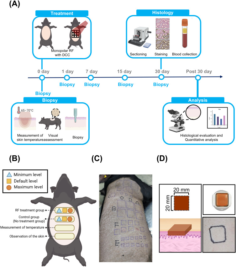
Three 6-month-old male minipigs (Sus scrofa domestica) were purchased from a certified breeder (M-pig ® ; CRONEX Inc., Cheongju, Republic of Korea). After a 7-day quarantine and acclimatization period, animals with a body weight ranging from 50 to 55 kg were used for the experiments. All experimental procedures were reviewed and approved by the Institutional Animal Care and Use Committee of CRONEX Inc., Cheongju, Republic of Korea (CRONEX-IACUC approval no. 202506005). All animal experiments were conducted in accordance with the National Institutes of Health (NIH) Guide for the Care and Use of Laboratory Animals and the guidelines of the Institutional Animal Care and Use Committee. For general anesthesia, tiletamine/zolazepam (5 mg/kg) and xylazine (2 mg/kg) were administered via intramuscular injection. Subsequently, endotracheal intubation was performed, and the pig was ventilated with oxygen and 2% isoflurane. Intravenous hydration through the superficial auricular vein with 0.9% normal saline at 25 mL/hour was maintained during the experiments. The lateral flank region of each minipig was selected as the treatment area. Hair was carefully removed from the skin surface using gentle clipping, and a grid corresponding to each experimental condition was marked on the skin using black ink prior to treatment. At the end of the experimental procedures, animals were humanely euthanized by intravenous administration of an overdose of pentobarbital sodium (≥100 mg/kg), in accordance with the American Veterinary Medical Association (AVMA) Guidelines for the Euthanasia of Animals. Death was confirmed by the absence of heartbeat and respiration ( Figure 1 ).
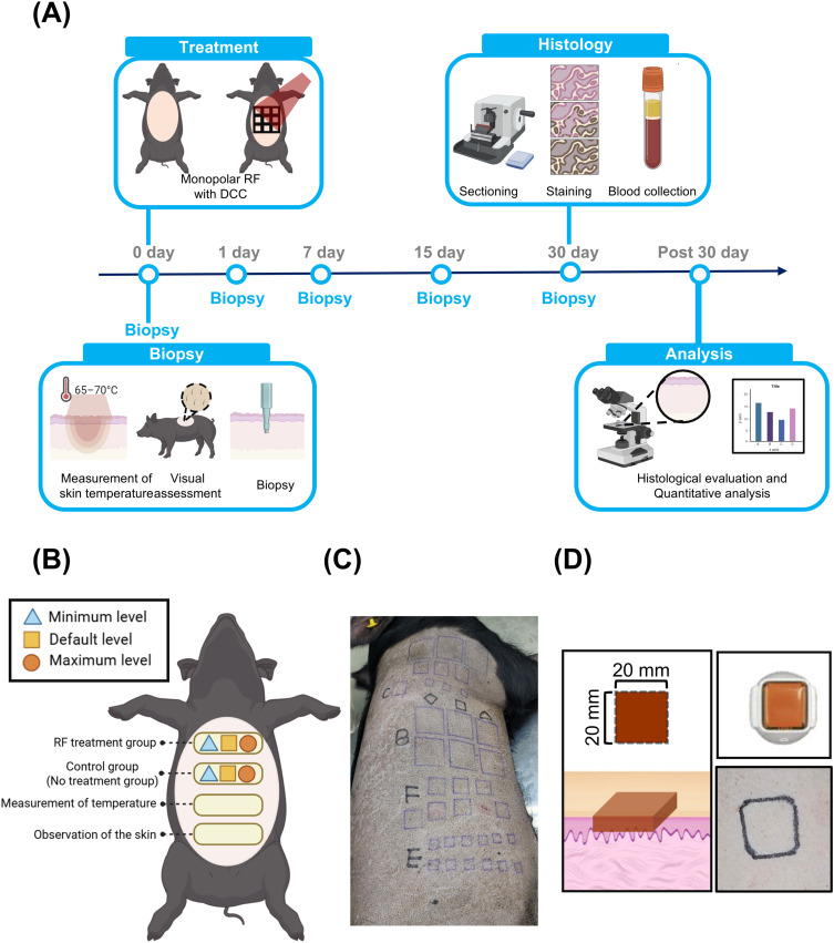
Figure 1 Experimental design and treatment configuration of DCC-Based monopolar RF in a minipig model. (A) Visual observation and skin tissue biopsy were performed at 0, 1, 7, 15, and 30 days after treatment to assess temporal tissue responses. (B) Schematic overview of the in vivo experimental design in minipigs. Treatment sites were categorized into minimum (blue triangles), default (Orange squares), and maximum (red circles) levels according to the applied monopolar RF parameters. (C) Representative photograph of the dorsal skin illustrating the delineation of RF-treated areas. (D) Schematic illustration of the electrode tip (CF-CT20) and its corresponding dimensions, together with a representative image of the actual electrode tip and examples of the associated dorsal treatment areas.
In this study, a non-invasive monopolar radiofrequency (RF) system incorporating a direct contact cooling (DCC) mechanism was used (Coolfase, ASTERASYS CO., Ltd., Seoul, Republic of Korea) ( Table 1 ). This device was designed to deliver thermal energy beyond the epidermis into the dermal and subdermal layers while simultaneously cooling the skin surface through direct contact ( Figure 2 and Supplementary Figure S1). To evaluate temperature changes within the dermal layer during DCC-based monopolar RF treatment, a fiber optic temperature sensor (EA10, Extech Instruments, Nashua, NH, USA) was employed. Prior to RF application, the fiber optic probe was inserted approximately 3 mm below the skin surface within the dermal layer of the target treatment area. RF was then applied at three predefined levels (Minimum, Default, and Maximum), and dermal temperatures were recorded in real time using the fiber optic thermometer. Skin surface and dermal temperatures were measured immediately before and immediately after RF treatment. In addition, a thermal imaging camera (A20pro, Test Tools, China) was used to monitor skin surface temperature and to confirm that the device maintained safe operating conditions without exceeding critical temperatures that could induce thermal injury. Surface temperature was continuously monitored throughout the treatment procedure to ensure that it did not exceed 43 °C, thereby verifying the safety of the DCC-based monopolar RF system.
Table 1 Characteristics of DCC-Based Monopolar RF
| Product Type | DCC-Based Monopolar RF |
|---|---|
| Manufacturing company | ASTERASYS Co. Ltd., ROK |
| Electrode type | Monopolar RF |
| Rated voltage | AC 100–240 V |
| Rated frequency | 6.78 MHz |
| Impedance range | 75 to 400 Ω |
| Maximum output | 140 W |
Skin biopsies were performed on days 0, 1, 7, 15, and 30 following treatments. At each time point, tissue samples were obtained exclusively from the treated areas using an 8-mm biopsy punch to enable temporal evaluation of treatment-induced histological changes.
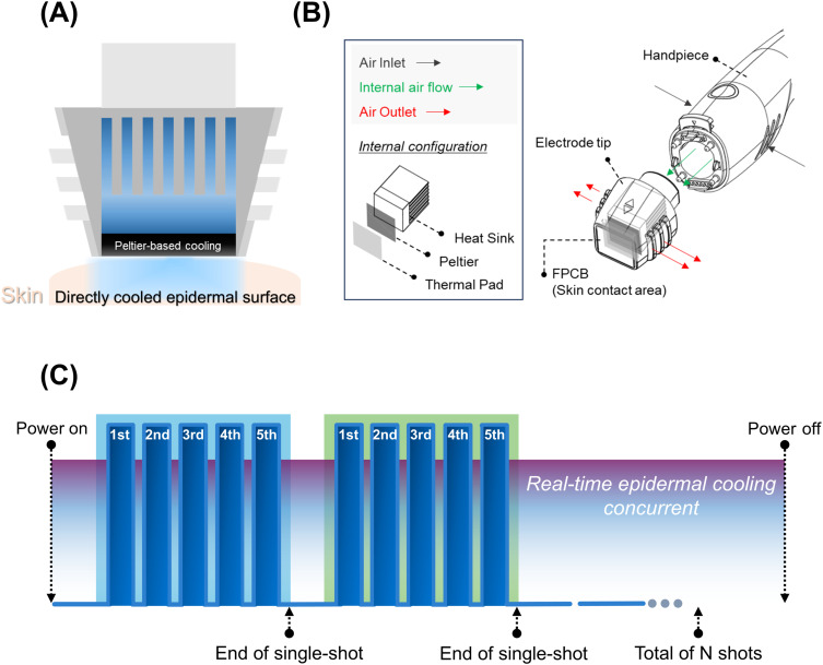
Figure 2 The mechanism of Peltier-based direct contacting cooling (DCC). (A) The schematic diagram shows the Peltier-based cooling system. Peltier provides direct-contacting cooling (DCC) and reduces the temperature of the epidermal surface. (B) Engineering drawings indicated the handpiece, electrode tip and illustrated the air-flow pathway during the treatment. Also, peltier operates in combination with the heat sink and the thermal pad and they are integrated in the tips not in the handpiece. (C) A single shot of DCC-Based monopolar RF consists of 5 pulses and each pulse duration of 100 ms. During RF treatment, the peltier continuously cools the surface of the skin and the cooling effect is maintained before, between, and after the pulses throughout the whole procedure in real time.展示珀尔帖直接接触冷却(DCC)原理:冷却模块集成于电极尖端,在射频脉冲前、中、后实时降低表皮温度。
To evaluate skin irritation and local cutaneous responses induced by DCC-based monopolar RF treatment, a skin magnification imaging device (Folliscope ® , Lead M, Seoul, Republic of Korea) was used to examine both treated and untreated skin areas. The treatment sites were imaged on days 0, 1, 7, 15, and 30 following RF treatment. Skin surface conditions were assessed according to predefined skin reaction evaluation criteria, and the presence of abnormal responses, including erythema and surface damage, were systematically examined.
For systemic safety evaluation, blood samples (5 mL) were collected via the jugular vein from each of the three minipigs before treatment and at 2, 4, 8, 12, 24, 48, and 72 hours after RF treatment. Collected blood samples were allowed to clot at room temperature and subsequently centrifuged at 3,000 rpm for 10 minutes to separate the serum. Serum biochemical analyses were performed to assess liver function, kidney function, and electrolyte balance. Liver function parameters included aspartate aminotransferase (AST), alanine aminotransferase (ALT), alkaline phosphatase (ALP), total bilirubin (T-BIL), albumin (ALB), and the albumin/globulin (A/G) ratio. Renal function was evaluated by measuring phosphorus (P), blood urea nitrogen (BUN), and creatinine (CREA). Electrolyte levels, including calcium (Ca) and inorganic phosphorus (IP), were also analyzed ( Table 2 ).
Table 2 Blood Parameters
| Test Category | Parameter | Description |
|---|---|---|
| Liver Function Test | AST (Aspartate aminotransferase | Indicator of hepatocellular injury |
| ALT (Alkaline aminotransferase) | Marker of liver-specific cellular damage | |
| ALP (Alkaline phosphatase) | Indicator of bile flow abnormality or bone disease | |
| T- BIL (Total bilirubin) | Assessment of jaundice | |
| ALB (Albumin) | Major protein synthesized in the liver | |
| A/G Ratio (Albumin/Globulin ratio) | Indirect assessment of liver function and immune status | |
| Kidney Function Test | P (Total protein) | Total protein concentration in serum |
| BUN (Blood urea nitrogen) | Evaluation of renal function related to protein metabolism | |
| CREA (Creatine) | Indicator of renal function associated with muscle metabolism | |
| Electrolyte Test | Ca (Calcium) | Regulation of bone health, nerve, and muscle function |
| IP (Inorganic phosphorus) | Related to bone metabolism and renal function |
At the end of the experiment, skin tissues were harvested from the sacrificed minipigs and immediately fixed in 4% formaldehyde at 4 °C overnight to preserve tissue morphology. Following fixation, the tissues were processed through routine histological procedures, including dehydration, clearing, and paraffin embedding. Paraffin-embedded tissue blocks were sectioned at a thickness of 4 µm using a microtome (Leica RM2235, Leica Biosystems, Germany). The sections were mounted on silicone-coated glass slides and dried in a 60 °C oven for 1 hour. Prior to staining, the slides were deparaffinized in xylene and rehydrated through a graded ethanol series (100%, 95%, and 70%). The sections were stained with hematoxylin solution for 5 minutes, rinsed with distilled water, and counterstained with eosin solution for 2 minutes. Stained sections were examined and imaged using a light microscope (BX53; Olympus, Tokyo, Japan).
Nitro blue tetrazolium chloride (NBTC) staining was performed to identify non-viable (necrotic) regions in porcine skin tissue following DCC-based monopolar RF treatment. After completion of the treatment, skin tissues were excised and embedded in optimal cutting temperature (OCT) compound. Cryosections with a thickness of 20 μm were prepared using a cryostat (Leica Biosystems, Germany). The sections were air-dried at room temperature for approximately 10 minutes and then incubated in 0.1% NBTC solution dissolved in 0.1 M phosphate buffer (pH 7.4) at 37 °C for 30 minutes in the dark. After incubation, the slides were gently rinsed with phosphate-buffered saline (PBS) to remove excess staining solution and subsequently mounted with coverslips. NBTC staining serves as an indicator of dehydrogenase activity within the tissue. In viable cells, NBTC is reduced to blue formazan crystals, resulting in intense blue staining, whereas necrotic or non-viable regions lacking dehydrogenase activity remain unstained and appear as colorless areas. Stained sections were examined and imaged under a light microscope to evaluate the extent and depth of necrotic regions within the tissue.
Masson’s Trichrome (MT) staining was performed to visualize collagen fibers within connective tissue, as well as cytoplasm and nuclei. Like the H&E staining procedure, deparaffinized tissue sections were rinsed under running tap water for 5 minutes and mordanted in Bouin’s solution preheated to 60 °C. The sections were then washed under running water for an additional 5 minutes. Nuclear staining was carried out by immersing the sections in a freshly prepared mixture of Weigert’s iron hematoxylin A and B solutions (1:1) for 10 minutes, followed by rinsing in 3% acetic acid to remove excess dye. At this stage, cell nuclei were stained dark purple to black. The sections were subsequently stained with Biebrich Scarlet–acid fuchsin solution for 10 minutes to stain cytoplasm and muscle fibers. Thereafter, the sections were stained with aniline blue for 5 minutes to visualize collagen fibers. Following staining, the slides were briefly rinsed with distilled water and treated with 1% acetic acid for 1 minute to stabilize color differentiation. The stained sections were then dehydrated, cleared, and mounted. Histological images were obtained using a light microscope. In MT-stained sections, nuclei appeared dark brown to black, cytoplasm and muscle fibers were stained bright red, and collagen fibers were distinctly visualized in blue. Quantitative analysis of collagen fibers was performed using ImageJ software (National Institutes of Health, Bethesda, MD, USA).
Victoria Blue staining was performed to evaluate the distribution and morphology of elastic fibers in skin tissue. The staining solution was prepared by dissolving 0.1 g of Victoria Blue R (Sigma-Aldrich, St. Louis, MO, USA) in 100 mL of 70% ethanol. Hydrated tissue sections were immersed in the prepared Victoria Blue staining solution and incubated at room temperature for approximately 30 minutes. After staining, the slides were rinsed thoroughly with running distilled water to remove excess dye. Differentiation was then performed for 2–5 seconds using an acetic acid solution containing 1% hydrochloric acid. During this step, elastic fibers appeared distinctly blue, whereas the background tissue became lightly stained. Following differentiation, the sections were immediately rinsed thoroughly to prevent further destaining. Counterstaining was performed using Van Gieson’s solution (1% acid fuchsin and 1% picric acid) for 1–2 minutes to stain collagen fibers red, thereby providing clear contrast with elastic fibers. After counterstaining, the slides were rinsed with distilled water, dehydrated through graded ethanol solutions (95% and 100%), cleared twice in xylene, and mounted using a mounting medium. The stained sections were examined and imaged under a light microscope.
Quantitative histological analysis was performed using ImageJ software (National Institutes of Health, Bethesda, MD, USA). For each treatment site, tissue samples were collected from three randomly selected locations within the RF-treated area. The entire dermal region was defined as the region of interest (ROI) for quantitative assessment of collagen and elastic fibers. Measurements obtained from multiple sections were averaged to generate a single representative value for each animal prior to statistical analysis.
All quantitative data are presented as the mean ± standard error of the mean (SEM). Statistical analyses were performed using GraphPad Prism software (GraphPad Software, San Diego, CA, USA). Comparisons between groups were performed using two-way analysis of variance (ANOVA), followed by Bonferroni’s post hoc correction for multiple comparisons. A p-value of less than 0.05 was considered statistically significant. For histological quantification, multiple tissue sections were obtained and analyzed from each animal. Measurements from individual sections were averaged to generate a single representative value for each animal prior to statistical analysis. Therefore, all statistical comparisons were performed using animal-level data (n = 3 animals).
Prior to in Vivo temperature assessment, the reproducibility of the device focal configuration was verified under ex vivo conditions.
Monopolar RF was applied to excised porcine skin at each level, and the resulting coagulation length, depth, and width were consistently reproduced across conditions (Supplementary Figure S2 and Supplementary Table S1).
These findings confirmed the stability and reliability of the focal delivery, supporting subsequent in vivo thermal evaluation. Skin surface temperature was continuously monitored in real time using a thermal imaging camera during DCC-based monopolar RF treatment. Maintaining epidermal temperature below the threshold for thermal injury is essential to prevent epidermal damage. 11 As shown in Figure 3A , the DCC cooling system effectively maintained epidermal temperature within a safe range following RF treatment. Quantitative analysis demonstrated that, under all tested levels, the skin surface temperature remained well below 42 °C and did not exceed approximately 20 °C throughout the treatment period ( Figure 3B ), indicating effective epidermal protection by the direct contact cooling system. The relatively low post-treatment epidermal temperatures reflect the active cooling effect of the DCC system, which continuously extracts heat from the skin surface during RF delivery. In contrast, the thermal generated by monopolar RF was efficiently delivered from the skin surface to the dermal layer. The dermal temperature range considered optimal for collagen denaturation, fibroblast activation, and subsequent tissue remodeling is selectively maintained between 50 and 70 °C. 12 Dermal temperature measurements obtained using a fiber optic thermometer revealed that, at the Minimum level, dermal temperature rapidly increased to 55.6 °C, followed by a gradual decline. At the Default level, dermal temperature reached 61.7 °C before slowly decreasing. At the Maximum level, dermal temperature increased to a peak of 69.2 °C and subsequently decreased in a controlled manner ( Figure 3C ).
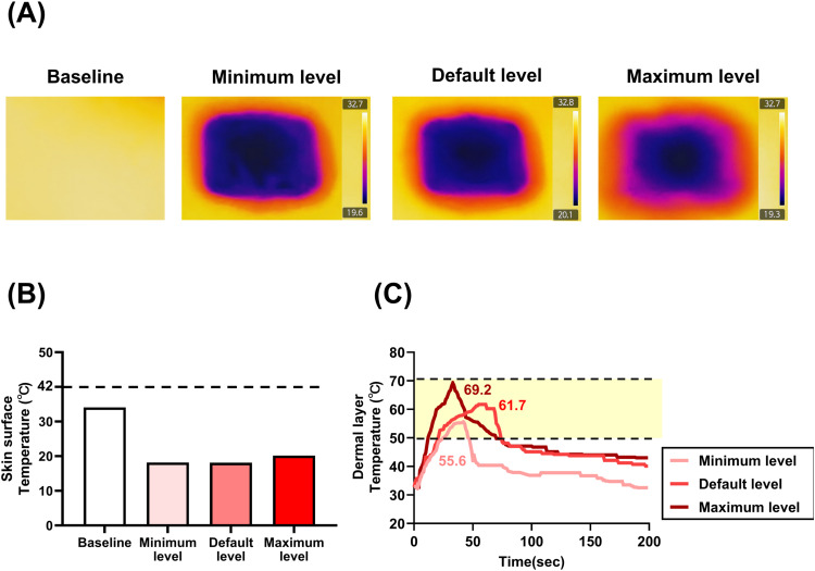
Figure 3 Skin surface temperature control and dermal thermal response after DCC-based monopolar RF treatment. (A) Representative thermal images of the treatment site before RF application and immediately after treatment at the minimum, default, and maximum energy levels. Skin surface temperatures were measured using a thermal imaging camera. (B) Comparison of skin surface temperatures before and immediately after treatment. Baseline skin surface temperature was 35.1 °C and decreased to 18.2 °C (minimum), 18.1 °C (default), and 20.2 °C (maximum) following treatment. The dashed line indicates the thermal injury threshold. (C) Dermal temperature profiles measured during RF treatment at different energy levels. Peak dermal temperatures reached 55.6 °C (minimum), 61.7 °C (default), and 69.2 °C (maximum). The shaded region indicates the dermal temperature range associated with collagen remodeling.显示治疗后表皮温度降至约 18–20°C(全程≤43°C),而真皮峰值温度达 55.6–69.2°C,直观体现表皮保护、真皮加热的温度分隔。
Visual assessment of skin surface changes following monopolar RF treatment was performed using a skin magnification imaging device. After treatment at different levels on the abdominal skin of minipigs, skin surface appearances were compared at Day 0 and Day 30, while observations at pretreatment, Days 1, 7, and 15 are provided in Supplementary Figures S3. Across all observation time points, including immediately after treatment (Day 0), no visible adverse skin reactions such as erythema or edema were observed at any treatment level ( Figure 4 ). These findings indicate that monopolar RF treatment within the tested range did not induce observable inflammatory responses or surface damage throughout the observation period. Overall, the results demonstrate that the applied DCC-Based monopolar RF treatment parameters maintained epidermal surface safety under the experimental conditions used in this study.
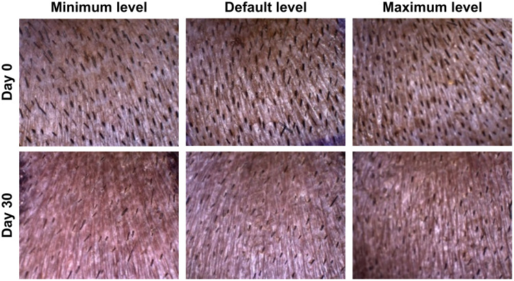
Figure 4 Visual Assessment of Skin Surface Following DCC-based Monopolar RF Treatment. Representative clinical photographs of treatment sites obtained immediately after treatment (Day 0, upper panels) and 30 days after treatment (Day 30, lower panels) at the minimum (left panels), default (middle panels), and maximum (right panels) energy settings. Visual assessment was performed to evaluate treatment-related adverse skin reactions, including erythema and edema. No noticeable erythema, edema, burns, or other visible adverse skin reactions were observed at any energy level throughout the observation period.
To evaluate systemic physiological responses following monopolar RF treatment, hematological and serum biochemical analyses were performed. Blood samples were collected before treatment and at 2, 4, 8, 12, 24, 48, and 72 hours post-treatment to assess key biochemical parameters. Overall, serum ALP levels exhibited relatively higher inter-individual variability in some animals; however, all values remained within the normal physiological range. A transient increase in AST levels was observed between 4 and 48 hours post-treatment, which is associated with anesthesia-related physiological responses rather than treatment-induced effects. No notable or consistent alterations were observed in other biochemical parameters when compared with baseline values. Across all minipigs, hematological parameters remained stable throughout the 72-hour observation period, and no biologically meaningful abnormalities or signs of systemic toxicity were detected ( Figure 5 ). These findings indicate that DCC-Based monopolar RF treatment did not induce adverse systemic physiological effects across the tested levels.
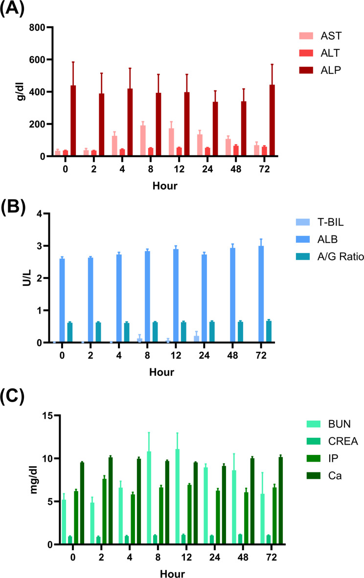
Figure 5 Hematological and biochemical safety evaluation following DCC-based monopolar RF treatment. (A–C) Serial blood analyses revealed no statistically or clinically meaningful changes in hematological or biochemical parameters in minipigs (n = 3) before and after DCC-Based monopolar RF treatment across all tested levels. No treatment-related systemic adverse reactions were observed during the observation period.
Histological evaluation using H&E staining was conducted to assess both acute and long-term tissue responses following monopolar RF treatment. Skin samples obtained immediately after treatment (Day 0) and at Day 30 were analyzed, while pretreatment and intermediate time points (Day 1, 7, and 15) are presented in Supplementary Figure S4.
Across all treatment levels and time points examined, the epidermal and dermal architecture remained well preserved, with no evidence of epidermal disruption, coagulative necrosis, inflammatory cell infiltration, or abnormal structural alterations. The treated skin at Day 0 showed intact epidermal layers and normal dermal organization, indicating the absence of acute tissue injury. Consistently, histological features observed on Day 30 were comparable to those at Day 0, with no signs of chronic inflammation, fibrosis, delayed tissue damage, or pathological remodeling ( Figure 6 ). Overall, the absence of histopathological abnormalities at both early and late point demonstrates that DCC-Based monopolar RF treatment did not induce acute or long-term tissue damage, supporting the histological safety of the procedure within the tested ranges.
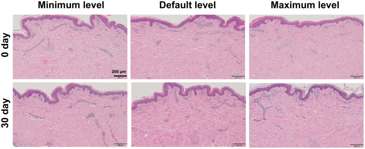
Figure 6 Histological assessment of skin tissue after DCC-Based monopolar RF treatment. Representative hematoxylin and eosin (H&E)-stained sections of minipig skin obtained immediately after treatment (Day 0, upper panels) and 30 days after treatment (Day 30, lower panels) at the minimum (left panels), default (middle panels), and maximum (right panels) energy settings. No histopathological abnormalities, including coagulation necrosis, inflammatory cell infiltration, epidermal disruption, or structural damage, were observed at either time point across all tested energy levels. Scale bar = 200 μm.
To evaluate tissue viability and identify potential necrotic changes following monopolar RF treatment, nitroblue tetrazolium chloride (NBTC) staining was performed. NBTC staining distinguishes viable tissue from necrotic tissue based on cellular dehydrogenase activity, with metabolically active cells exhibiting blue staining, whereas necrotic areas remain unstained or appear faint. Across all treatment levels (Minimum, Default, and Maximum), no distinct epidermal necrosis or structural damage was observed at either Day 0 or Day 30 following treatment ( Figure 7 ). NBTC staining demonstrated preserved cellular viability within the epidermal layer, with uniformly positive blue staining patterns. Evaluation of all time points, including pre-treatment and Days 1, 7, and 15, is provided in the Supplementary Figure S5. In addition, no evidence of extensive thermal-induced necrosis or degenerative changes was detected within the dermal layer. Dermal tissue architecture remained intact, without signs of widespread cell death or heat-related structural disruption. Overall, these findings indicate that DCC-Based monopolar RF treatment, within the applied ranges, does not induce significant epidermal or dermal necrosis and preserves tissue viability, supporting the histological safety of the procedure.
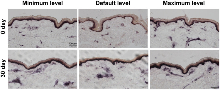
Figure 7 Evaluation of Epidermal Injury in Skin tissue Following DCC-based Monopolar RF Treatment. Representative NBTC-stained images of mini-pig skin obtained immediately after treatment (Day 0) and 30 days after DCC-based monopolar RF treatment at minimum, default, and maximum energy levels. NBTC staining was used to assess epidermal viability and thermal injury. No evidence of epidermal damage was observed at any treatment level or time point. Scale bar = 100 μm.
Masson’s trichrome staining was performed to assess dermal collagen remodeling following monopolar RF treatment at three different levels (Minimum, Default, and Maximum). Dermal collagen fibers were visualized as blue-stained structures. Quantitative analysis was conducted at Day 0 and Day 30, while histological findings at pretreatment and intermediate time points (Day 1, 7, and 15) are provided in Supplementary Figure S6.
At Day 0, a modest reduction in collagen staining intensity was observed across all treatment groups compared with the pretreatment, suggesting transient collagen alteration immediately after RF exposure, possibly due to thermal denaturation or compaction within the dermis. By Day 30, collagen staining intensity was markedly increased, indicating enhanced collagen deposition and improved dermal matrix organization. Notably, the Default and Maximum level groups exhibited a statistically significant increase in collagen density at Day 30 compared with Day 0. These findings suggest that DCC-Based monopolar RF treatment facilitates collagen regeneration and fiber reorganization during the dermal remodeling process ( Figure 8 ).
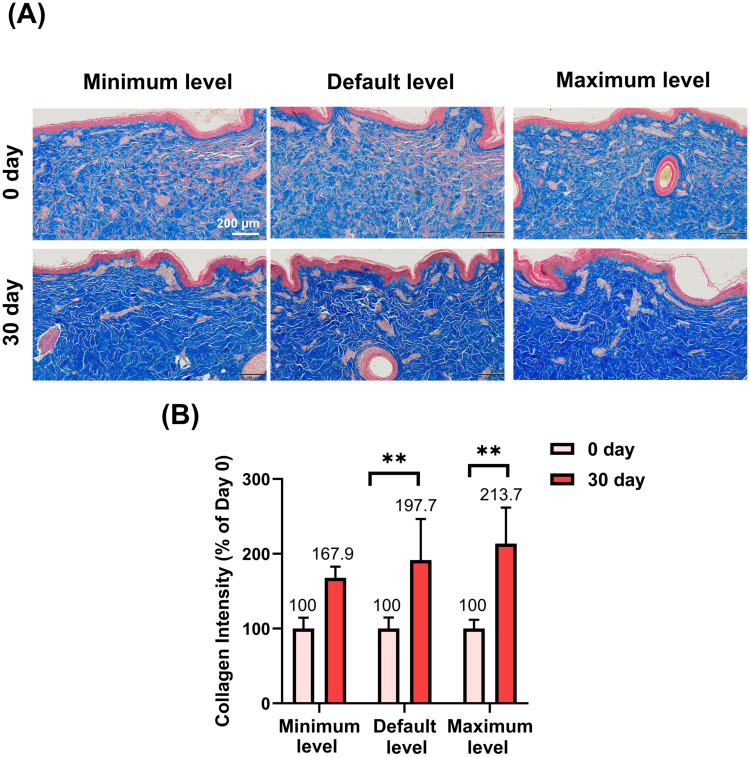
Figure 8 Collagen Fiber Remodeling in Skin Tissue Following DCC-based Monopolar RF Treatment. (A) Representative Masson’s trichrome-stained histological sections obtained immediately after treatment (Day 0, upper panels) and 30 days after treatment (Day 30, lower panels) at the minimum (left panels), default (middle panels), and maximum (right panels) energy settings. Increased collagen density and dermal collagen organization were observed at Day 30, particularly at the default and maximum energy levels. Scale bar = 200 μm. (B) Quantitative analysis of collagen area fraction in the dermis. Compared with Day 0, collagen area fraction was significantly increased at Day 30 in the default and maximum energy groups (**p < 0.01 versus Day 0). Data are presented as mean ± SEM.Masson 染色显示治疗 30 天胶原面积分数在默认组(197.7)与最大组(213.7)显著升高(**p<0.01)。
Victoria blue staining was performed to evaluate time-dependent changes in dermal elastic fibers following monopolar RF treatment at three different levels (Minimum, Default, and Maximum). Elastic fibers within the dermis were visualized as blue-stained structures. Quantitative analysis was conducted at Day 0 and Day 30, while histological findings on pretreatment and intermediate time points (Day 1, 7, and 15) are presented in Supplementary Figure S7. Across all treatment levels, the density and staining intensity of elastic fibers at Day 30 increased compared with those observed immediately after treatment (Day 0). Notably, a trend toward increased elastic fiber density was observed in the Maximum-level treatment group. These findings indicate progressive restoration and enhancement of dermal elastic fibers over time. The observed increase in elastic fiber density and organization is consistent with elastic fiber regeneration and remodeling during the dermal repair process following monopolar RF treatment. Overall, these results suggest that monopolar RF may contribute to elastic fiber remodeling in the dermis in a time-dependent manner ( Figure 9 ).
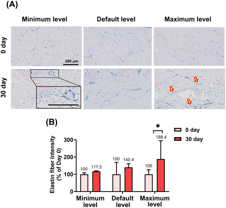
Figure 9 Elastic Fiber Remodeling Following DCC-based Monopolar RF Treatment. (A) Representative Victoria blue-stained histological sections of dermal tissue obtained immediately after treatment (Day 0, upper panels) and 30 days after treatment (Day 30, lower panels) at the minimum (left panels), default (middle panels), and maximum (right panels) energy settings. Elastic fibers were visualized as blue-stained structures within the dermis. Increased elastic fiber density and altered distribution were observed at Day 30, particularly in the maximum energy group. The inset shows a higher-magnification view of the boxed region. Red arrows indicate representative areas of increased elastic fiber accumulation. Scale bar = 200 μm. (B) Quantitative analysis of elastic fiber-positive area following monopolar RF treatment at different energy levels. Compared with Day 0, the maximum energy group showed a trend toward increased elastic fiber-positive area at Day 30 (*p < 0.1). Data are presented as mean ± SEM.Victoria blue 染色示最大能量组弹性纤维阳性面积增至 188.4,呈增高趋势(p<0.1)。
This study evaluated the thermal characteristics, tissue responses, and safety of a novel DCC-based monopolar RF system using a minipig model. The results demonstrate that this device effectively elevates dermal temperature to a range associated with collagen remodeling while maintaining epidermal integrity and overall tissue safety. Temperature measurements confirmed that DCC-based monopolar RF induced sufficient dermal heating without excessive epidermal temperature elevation. This finding is clinically relevant, as effective collagen denaturation and subsequent remodeling are known to occur within a controlled thermal window, whereas excessive temperatures may increase the risk of neural or thermal injury. 13 The direct contact cooling system appeared to stabilize epidermal temperature while permitting efficient energy delivery to deeper dermal layers, supporting its role in improving both treatment safety and energy efficiency. 10 Visual evaluation revealed no visible adverse skin reactions following treatment, and systemic safety was further supported by the absence of significant changes in hematological and biochemical parameters. Histological analysis corroborated these findings, demonstrating preservation of normal skin architecture without evidence of sustained inflammation, necrosis, or structural injury. These results indicate that the DCC-based monopolar RF system is well tolerated at the tested energy settings. Importantly, histochemical analyses revealed increased collagen density and elastic fiber remodeling within the dermis following treatment. Masson’s trichrome and Victoria blue staining demonstrated qualitative enhancement of collagen fiber organization and elastic fiber expression, consistent with previously reported RF induced dermal remodeling processes. 14 , 15 These findings align with prior in vivo and clinical studies show that monopolar RF promotes neocollagenesis and elastic fiber synthesis through controlled thermal stimulation of the dermis. 16 , 17 Minipigs were selected as an experimental model due to their well-established anatomical and physiological similarity to human skin, particularly with respect to dermal thickness and extracellular matrix composition. 18 Conventional monopolar RF systems often rely on cryogen- or LN 2 -based cooling mechanisms, which may be associated with procedural complexity, discomfort, and inconsistent cooling performance. 19 In contrast, the DCC-based cooling approach used in this study provides continuous and uniform epidermal cooling through direct contact while enabling consistent dermal heating. These characteristics may contribute to efficient energy delivery and favorable tissue responses; however, further comparative studies are required to determine whether these findings translate into superior clinical outcomes. Several limitations should be acknowledged. First, this study was conducted in a limited number of animals and should therefore be considered a preliminary preclinical feasibility study. Although multiple tissue sections were analyzed from each animal, statistical comparisons were performed using animal-level data, and larger studies are needed to further validate the observed effects. Second, direct comparison with conventional cooling systems was not included in the study design, limiting definitive conclusions regarding the relative advantages of the DCC-based cooling approach. In addition, although pretreatment histological samples were collected and analyzed, longitudinal evaluation of an untreated control site was not performed. Therefore, the extent to which the observed tissue remodeling can be attributed exclusively to RF treatment should be interpreted with caution. Third, this study was conducted in an animal model, and direct extrapolation to human clinical outcomes should be approached with caution. In addition, potential nerve injury, long-term fibrosis, and delayed tissue responses beyond the current observation period were not specifically evaluated. Future studies incorporating larger sample sizes, direct comparative analyses, extended follow-up durations, and controlled clinical trials will be necessary to further validate the efficacy, durability, and long-term safety profile of DCC-based monopolar RF treatment in human subjects. These findings provide preliminary preclinical evidence supporting the potential utility of this novel RF system for non-invasive skin rejuvenation and warrant further clinical investigation.
In conclusion, the DCC-based monopolar RF system demonstrated controlled dermal heating and was associated with collagen and elastic fiber remodeling without apparent adverse histological or systemic findings in a minipig model. The device enabled thermal stimulation of the dermis while maintaining epidermal integrity throughout the study period. These findings provide preliminary preclinical evidence supporting the potential utility of DCC-based monopolar RF for non-invasive skin rejuvenation. Further studies with larger sample sizes and clinical investigations are warranted to confirm its safety, efficacy, and long-term outcomes in human subjects.
声明:中文精读 · 仅供学术参考。内容来自公开文献检索,不代表本人观点,不构成诊疗建议。 医疗美容需在正规医疗机构由执业医师实施。
基于相同主题推荐 · 继续深挖
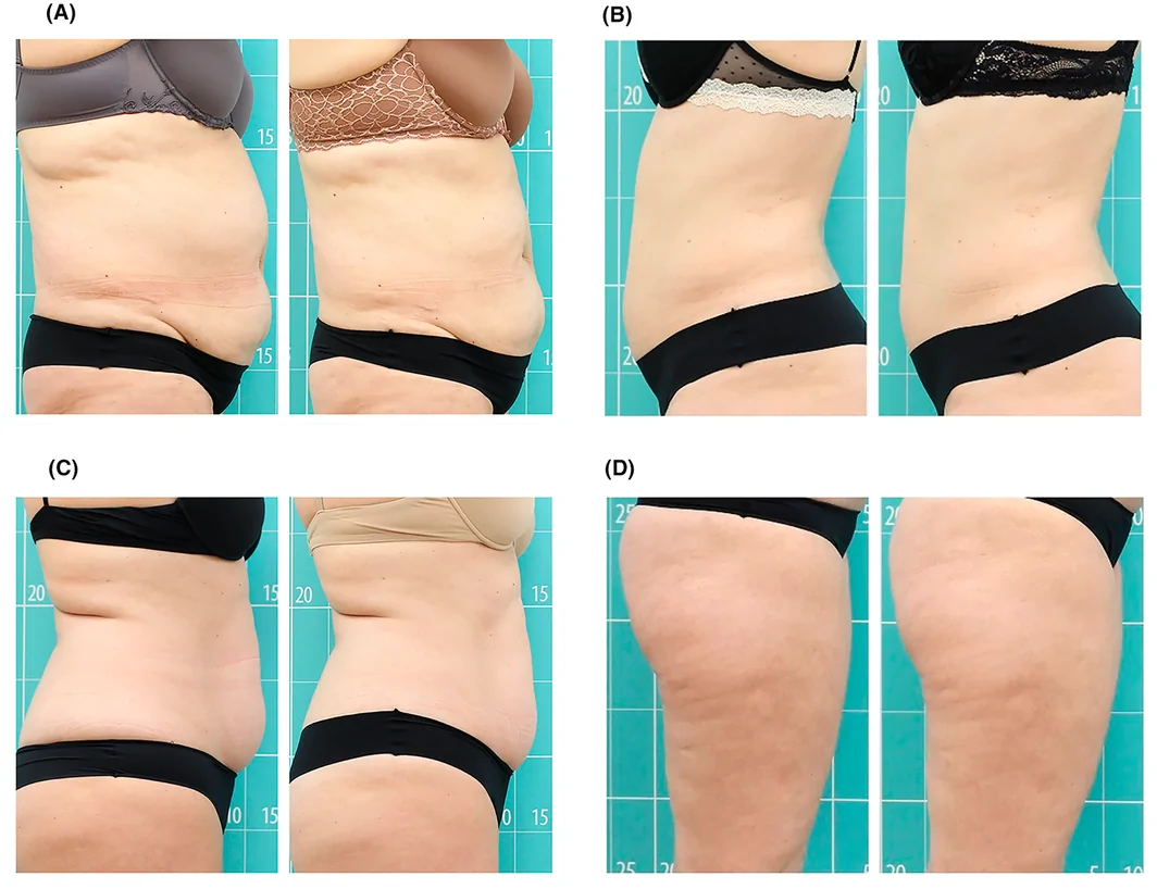
前瞻性单臂研究(西班牙 Asturias 伦理委员会批准),39 例健康女性志愿者(23–68 岁,平均 47 岁,Fitzpatrick II–III),腹部/腰侧 25 例、大
接触冷却单极射频可在小型猪安全诱导真皮热刺激
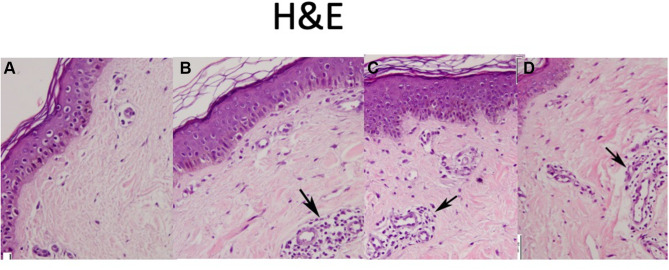
纳入 10 例 Fitzpatrick IV 型女性白纹(striae albae)患者;每人在三个部位分别接受 PRP、点阵 CO₂/射频(CO₂/RF)、联合治疗,每月 1 次