睑板囊筋膜切开下睑成形术 340例前瞻性研究
2026年10月4日25 min read
Plastic and Reconstructive Surgery
结论与实践要点
证据等级: 单中心前瞻性连续病例系列(prospective case series),无对照组,在循证体系中属低至中等证据(病例系列约为 Ⅳ 级证据)。
适用人群: 拟行经皮下睑成形术、以改善泪槽、睑颊沟、眼袋及细纹为主要诉求者;特别强调下睑松弛不明显、snapback 试验正常者。snapback 试验不佳者不适用本术式(应改用外眦固定等术式)。
关键结果: 340 例连续手术中未观察到不可逆(持续性)下睑外翻;暂时性睑外翻症状 15 例(4.4%)均在 1 个月内自行消退。其余并发症轻微:感染 2 例(0.6%)、血清肿 1 例(0.3%)、增生性瘢痕 3 例(0.9%)、血肿 6 例(1.8%)。最近 50 例由 3 名独立医师按 5 分制盲评,4 项美学指标(泪槽、睑颊沟、眼袋、细纹)术后 3、6 个月均较术前显著改善(P<0.001)。
证据边界与局限:
Background:Lower-eyelid surgery by means of the transcutaneous approach is challenging because of common complications such as ectropion and scleral show.
Methods:The authors devised a transcutaneous lower-eyelid technique that incorporates 4 modifications that greatly reduce these complications—namely, tension-reducing incision of the capsulopalpebral fascia to prevent ectropion and preserve the pretarsal fullness; selective omission of lateral canthoplasty; universal application of arcuate expansion cuts to prevent lateral compartment bulging; and reattachment of the tear-trough and orbital-retaining ligaments to the orbital septum. A prospective cohort study was conducted on all consecutive patients who underwent the technique in January of 2020 to March of 2023. All complications were reviewed. The 50 most recent patients were assessed objectively by 3 independent plastic surgeons with regard to patient preoperative and 3- and 6-month postoperative tear trough, lid-cheek groove, eye bag, and fine lines. Each variable was scored on a 5-point scale.
Results:In total, 340 patients underwent the procedure. No major complications, including ectropion, were observed. All 4 aesthetic variables improved significantly at 3 and 6 months relative to preoperative baseline.
Conclusions:This new approach significantly mitigates common complications of transcutaneous lower-eyelid operations, including persistent postoperative ectropion. Thus, it may be a promising advance in surgical correction of lower-eyelid aesthetics.
Lower-eyelid surgery is among the most challenging procedures in cosmetic surgery. classic methods primarily involve excising skin, orbicularis oculi muscle, and orbital fat. However, these methods can cause long-term complications, including malposition of the lower eyelid, scleral show, and sunken eyes. 1 – 3 In particular, scleral show and ectropion are common and troubling complications of transcutaneous lower-eyelid surgery. 4 – 6 Common preventative measures include routine lateral canthoplasty 7 and avoiding transcutaneous methods in favor of combining the transconjunctival approach with skin-pinch removal. 8
However, in our experience, it is possible to virtually eliminate the risk of ectropion and other complications after transcutaneous lower-eyelid surgery by avoiding excessive skin removal and using surgical processes that do not overly pull downward during the repositioning of fat and septa. In particular, we use a tension-reducing incision of the capsulopalpebral fascia (CPF) before definitively suturing the cut ends of the septum and the orbital fat to the periosteum at the lower orbital edge. This CPF incision is termed the Nomoto incision. Here, we detail our approach, describe its outcomes in a large series of patients, and discuss our findings in the context of a literature review.
This prospective cross-sectional study was conducted in the BR Clinic Ginza (Tokyo, Japan) in accordance with the Declaration of Helsinki. All patients approved in writing the use of their medical information for research purposes.
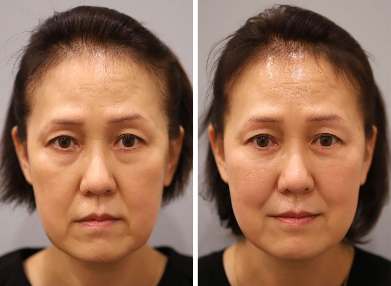
Our surgical procedure is not appropriate for patients with unfavorable snapback test results; such patients are instead managed with alternative techniques, such as canthopexy. The procedure is illustrated in case 1 (Fig. 1 ). This 60-year-old patient had tear-trough deformity, a lid-cheek groove, malar fat descent, and grade 0 on the snapback test (Fig. 1 , left ).

Fig. 1. A 60-year-old woman with tear-trough deformity, lid-cheek groove, and malar fat descent (case 1). Before surgery (left) and 6-month postoperative result (right).First, to prevent postoperative ectropion, skin excision is always performed conservatively. It is crucial that there is no vertical tension when the eyes are closed. 9 After skin excision, the pretarsal orbicularis oculi muscle is left somewhat wider in band form, and the muscle fibers are split along their course toward the deeper layer. To preserve the nerve supply of the pretarsal orbicularis oculi muscle, the fibers on the medial (nasal) side are not completely detached from the orbital part of the muscle. 10 Moreover, a minimum approximately 6.5-mm width is preserved to maintain the motor nerves of the pretarsal orbicularis oculi muscle. 11 Preserving the strength of this portion of the muscle is also crucial for preventing postoperative ectropion. Dissection above the orbital septum is then advanced to manage the arcus marginaris (Fig. 2 , above, left ). The orbicularis-retaining ligament below the orbital rim 12 , 13 is severed to enter the prezygomatic space, and the origin of the orbital part of the orbicularis oculi muscle is carefully incised. Although this part of the procedure provides a lifting effect, its primary purpose is to contour the cheek. It is unlikely to contribute significantly to tension offloading. Therefore, it is not required in all cases—particularly in younger patients without malar ptosis. The decision to perform this step should be guided by the patient’s skeletal anatomy and individual needs. As the dissection progresses, the tear-trough ligament is identified. This is a genuine ligament that is derived from the periosteum. It is thin but quite firm, and strongly connects the maxilla to the skin. 14 Sharp and thorough dissection improves the deep groove beside the nose. The orbital rim is exposed widely to approximately 10 mm in width. Broad dissection of the zygomatic cutaneous ligament, whose range starts at the origin of the levator labii superioris muscle, 15 can improve the midcheek groove. Because it is not as tough as the tear-trough ligament, blunt dissection is also possible.
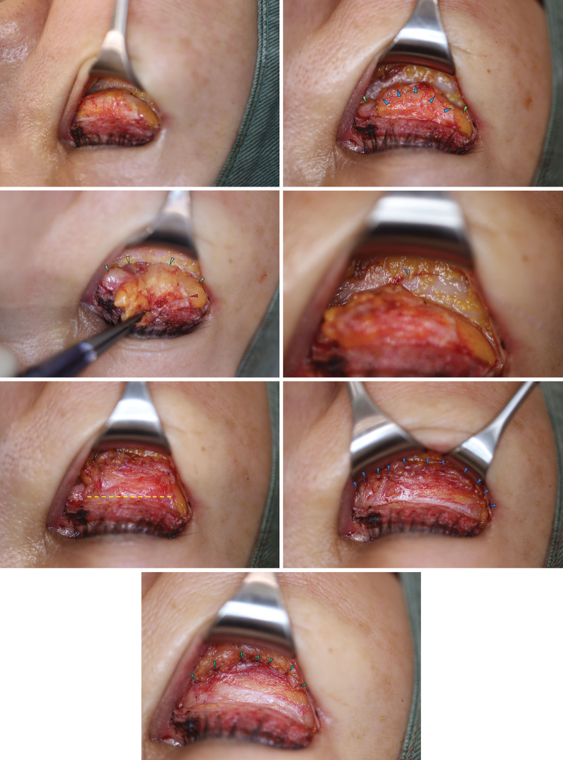
Fig. 2. Details of lower-lid surgery for the patient in case 1. (Above) Orbital-retaining and tear-trough ligaments were released; the septum was incised, and the compartments were exposed. (Second row and third row, left) Periosteal sutures were placed, followed by a tension-reducing CPF incision (Nomoto incision). (Third row, right) Septal reset with 11 sutures. (Below) Sutures lifting the malar fat.An alternative method involves entering beneath the periosteum 16 ; this results in less bleeding and simplifies the operation, thus reducing the surgical time. Once beneath the periosteum, the risk of damaging infraorbital nerves or angular vessels is lowered. However, to eliminate the tear trough, it is preferable to partially sever the origin of the orbicularis oculi muscle above the periosteum.
Next, the orbital septum is incised at the orbital rim to release the orbital fat (Fig. 2 , above , right ). At this time, both the lateral and medial angles are sharply incised to fully release the orbital fat. Severing the arcuate expansion of the Lockwood ligament treats the central and lateral fat as 1 mass, thus preventing unnatural bulging. The arcuate expansion is also said to function as a pulley that prevents the protrusion of the eyeball and orbital fat and suppresses overcontraction of the inferior oblique muscle 17 ; thus, it is always reattached to the periosteum during the subsequent septal reset. The cut ends of the septum are then flipped forward, which reveals the entirety of the orbital fat (Fig. 2 , second row , left ). The medial orbital fat appears white and has thick nutritive vessels running on its surface. The orbital fat, the cut ends of the incised septum, and the periosteum at the lower orbital edge are then sutured together in a temporary fashion with approximately 3 sutures to create tension on the CPF (Fig. 2 , second row , right ). A horizontal tension-reducing incision of the CPF is then made in its tense area, namely, approximately 5 mm below the tarsal plate (Fig. 2 , third row , left ). This incision, termed the Nomoto incision, significantly reduces the risk of postoperative ectropion by freeing the tissue surrounding the tarsal plate from the CPF and orbital septum.
A broad septal reset is then performed: a 6-0 absorbable suture (polydioxanone) is used to comprehensively suture the cut ends of the septum and the orbital fat to the periosteum (Fig. 2 , third row , right ). 18 The malar fat is gently lifted, and the cut ends of the tear-trough and orbicularis-retaining ligaments are sutured above the orbital septum (Fig. 2 . below ). Because the CPF incision sufficiently prevents ectropion, routine lateral canthoplasty is not performed. Lateral canthoplasty should be considered as an independent treatment. The operation is completed with epidermal suturing using 7-0 nonabsorbable sutures (nylon): knot sutures are placed at the corner of the eye and continuous sutures below the eyelashes.
The 6-month postoperative outcomes of our method with case 1 are shown in Figure 1 , right . The condition of the lower-eyelid area has improved and ectropion was not observed.
In January of 2020 to March of 2023, we treated 340 consecutive cases with our transcutaneous lower-eyelid surgery method. Postoperative complications such as ectropion, deformation, and infections were recorded. The most recent 50 cases were evaluated objectively before and 3 and 6 months after surgery by 3 independent plastic surgeons. For this, 4 aesthetic criteria—namely, tear trough, lid-cheek groove, eye bag, and fine wrinkles—were scored using 5-point scales, where 5 was the most aesthetically pleasing.
The changes in tear trough, lid-cheek groove, eye bag, and fine wrinkle scores at 3 and 6 months relative to baseline were assessed for statistical significance with the Friedman test followed by the Wilcoxon signed rank test with the Bonferroni correction to adjust the alpha error. The level of statistical significance was set at 0.05. Analyses were performed with SPSS software (version 16; IBM Japan, Tokyo, Japan).
Over the 27-month study period, 340 patients underwent our transcutaneous lower-eyelid surgery method. All were Japanese women aged between 30 and 63 years (average age, 46 years). The average follow-up duration was 227 days. The postoperative complications included 2 infections (0.6%), 1 seroma that lasted for longer than 1 month (0.3%), 3 cases of hypertrophic scarring at the suture site near the corner of the eye that required minor revision (0.9%), and 6 cases of hematoma removal (1.8%). Recurrence of the 3 hypertrophic scars was not observed during the 6-month follow-up period. Of the 6 hematomas, 4 were removed the day after surgery. All hematomas initially associated with mild scleral show that lasted approximately 1 month, but all cases resolved without sequelae. Temporary ectropion symptoms were reported in 15 cases (4.4%), but all resolved naturally within 1 month without the need for interventions, such as triamcinolone acetonide injections or taping. This condition is caused by temporary postoperative paralysis of the orbicularis oculi muscle. No noticeable conjunctival edema was observed in any of the cases. At 6 months postoperatively, no significant complications remained.
Figure 3 shows the tear trough, lid-cheek groove, eye bag, and fine wrinkle scores of the most recent 50 patients before and 3 and 6 months after the surgery, as determined by 3 independent plastic surgeons. The average preoperative scores for tear trough, lid-cheek groove, eye bag, and fine wrinkles were 2.0, 2.2, 2.6, and 2.8, respectively. At 3 postoperative months, the scores had improved significantly to 4.1, 4.7, 4.6, and 4.4, respectively (all P < 0.001). The 6-month scores were similar to the 3-month scores.
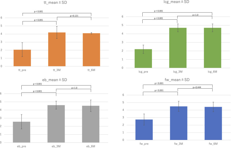
Fig. 3. The tear trough (tt), lid-cheek groove (lcg), eye bag (eb), and fine wrinkles (fw) of the patients were evaluated by 3 independent certified plastic surgeons before and 3 and 6 months after surgery by using 5-point scales. The changes relative to baseline were assessed for significance with the Friedman test followed by the Wilcoxon signed rank test with the Bonferroni correction.
Figures 1 and 4 through 7 show the postoperative outcomes of 5 representative cases (cases 1, 7, 17, 28, and 42). Case 1 is the 60-year-old female patient whose case was discussed at length under Patients and Methods (Figs. 1 and 2 ). ( See Video [online] , which demonstrates the septal reset procedure to prevent ectropion. After subperiosteal dissection and septum release, temporary fixation is made at 3 points. A subtarsal CPF incision [Nomoto incision; video time 1:30 to 2:00] with a horizontal cut 7 mm below the pretarsal orbicularis releases tension and prevents ectropion. Final sutures are placed evenly at the 3- to 9-o’clock position, with approximately 10 fixation points, paying attention to the lateral bulge.) Her tear trough score rose from 2 before surgery to 3 at 3 months postoperatively and then to 4 at 6 months postoperatively (ie, 2→3→4). Similarly, her lid-cheek groove and eye bag scores were 2→4→4, whereas her fine wrinkle scores were 3→4→4.
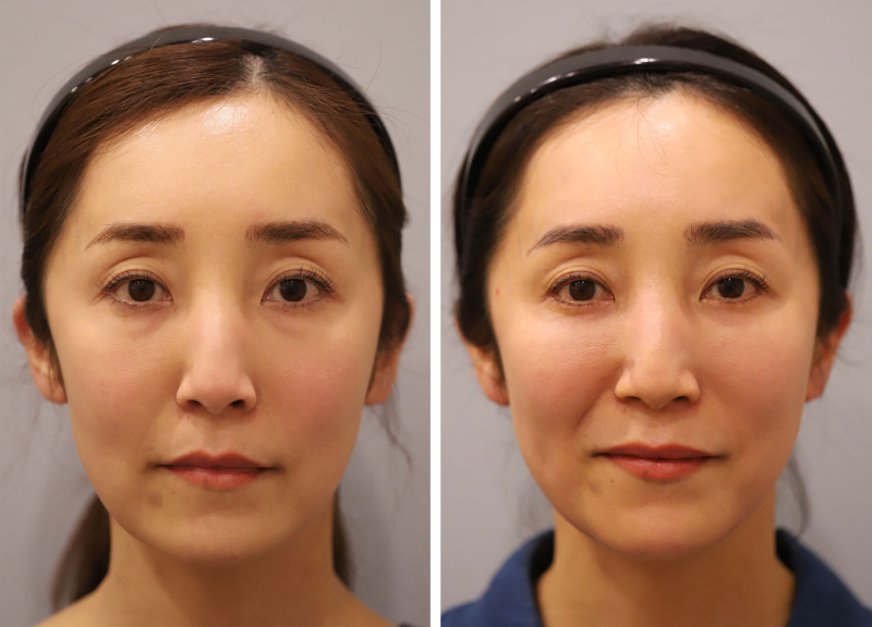
Fig. 4. Lower-lid surgery on the patient in case 7. This 44-year-old woman was concerned about mild eye bags and fine wrinkles. (Left) Before surgery. (Right) Six months after surgery.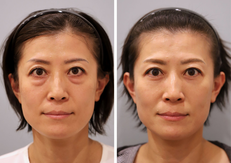
Fig. 5. Lower-lid surgery on the patient in case 17. This 48-year-old woman had notable large eye bags and a flat midface. (Left) Before surgery. (Right) Six months after surgery.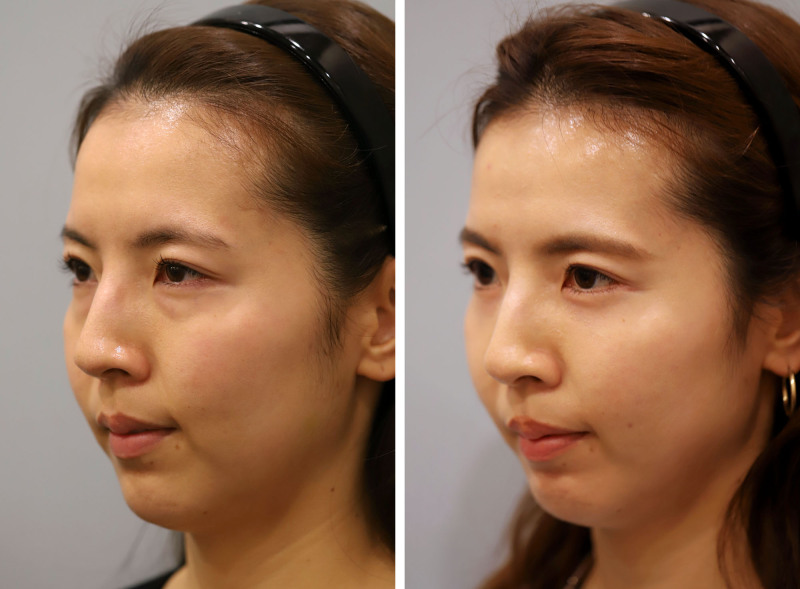
Fig. 6. Lower-lid surgery on the patient in case 28. This 37-year-old woman was troubled by large eye bags that were disproportionate to her age. (Left) Before surgery. (Right) Seven months after surgery.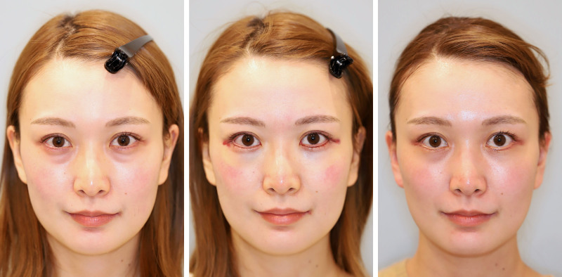
Fig. 7. Lower-lid surgery on the patient in case 42. Despite being aged 30 years, this patient had a consistently tired appearance because of eye bags. She was also concerned about fine wrinkles and requested surgery that included skin excision. (Left) Before surgery. (Center) Immediately after surgery. (Right) 6 months after surgery.Case 7 was a 44-year-old woman. Her tear trough scores were 2→4→4, whereas her lid-cheek groove and eye bag scores were both 3→5→5 and her fine wrinkle scores were 3→5→4 (Fig. 4 ).
Case 17 was a 48-year-old woman who had severe preoperative tear trough, lid-cheek groove, and eye bag scores (all 1). All improved markedly: the tear trough, lid-cheek groove, and eye bag scores were 1→4→4, 1→5→4, and 1→5→5, respectively. The mild fine wrinkles of the patient (score 4) improved to 5 at 3 months and then regressed to 4 at 6 months (ie, 4→5→4) (Fig. 5 ).
Case 28 was a 37-year-old woman whose tear trough, lid-cheek groove, and eye bag scores all improved from 2 to 5 at both postoperative time points (ie, 2→5→5). Her mild fine wrinkle scores were 4→5→4 (Fig. 6 ).
Case 42 was a 30-year-old woman whose tear trough, lid-cheek groove, and eye bag scores were 2→5→4, 2→5→5, and 2→5→5, respectively. Her fine wrinkle scores were 4→5→5 (Fig. 7 ). Figure 7 , center shows her appearance immediately after surgery: the eye bags and fine wrinkles had already improved markedly. Figure 7 , right shows the patient’s appearance at 6 months postoperatively.
补充视频 / 附件(原文未随文提供原图,可于出版方页面查看)prs-157-486e-s001.mp4Video. This video demonstrates the septal reset procedure to prevent ectropion. After subperiosteal dissection and septum release, temporary fixation is made at three points. A subtarsal CPF incision (Nomoto incision; video time 1:30 to 2:00) with a horizontal cut 7 mm below the pretarsal orbicularis releases tension and prevents ectropion. Final sutures are placed evenly at the 3- to 9-o’clock position, with about 10 fixation points, paying attention to the lateral bulge.
This study describes our transcutaneous lower-lid surgery method. It significantly improved the aesthetic outcomes, and these improvements were sustained for at least 6 months. Importantly, permanent ectropion or other complications were never observed in the 340 patients.
Our lower-eyelid surgery method involves 4 new refinements. First, the CPF is subjected to a tension-reducing incision to prevent ectropion and preserve the tear bag. Second, lateral canthoplasty is not conducted routinely. Third, the arcuate expansion is routinely sectioned to prevent bulging of the lateral compartment. Fourth, malar fat lifting is combined with reattachment of the dissected ends of the tear-trough ligament and the orbital-retaining ligament to the orbital septum.
Some of the techniques and concepts in our approach are based on several seminal studies. Thus, in 1981, Loeb 19 introduced “fat-pad sliding and fat grafting,” a technique that involves relocating the inner compartment of the orbital fat to the nasojugal groove, and removing and grafting the central and lateral compartments. However, although this method was groundbreaking, concerns were raised about the possibility of fibrosis, necrosis, and fat granuloma in the transplanted fat. Hamra later improved this concept by preserving the blood flow of all fat pads. 20 He also reported a method that involved cutting and releasing the orbital septum at its lower edge to release fibrous connections. 21 Arcus marginalis release 21 and septal reset 22 have also become foundational techniques in lower-eyelid surgery. Our transcutaneous method builds on these critical concepts.
With regard to the tension-reducing incision in the CPF below the tarsal plate (Nomoto incision), it means that the pretarsal area is unaffected by the downward pull of the septal reset. This both reduces the risk of eyelid ectropion and forms a contour below the tarsal plate that enhances the prominence of the pretarsal fullness, thus creating an aesthetically pleasing lateral contour. The inspiration for this technique came from asking 2 questions about transconjunctival lower-eyelid surgery: Why does it cause the pretarsal fullness to become beautifully prominent? Why does ectropion not occur? We hypothesized that both might be the result of the complete transection of the CPF in transconjunctival lower-eyelid surgery. The inspiration for our approach also came from the strong demand from Japanese female patients to preserve their pretarsal fullness. As we showed in our representative cases, completely detaching the CPF from the surface with the Nomoto incision resulted in prominent tear bags and no ectropion, similar to when the transconjunctival approach is used.
The CPF is reported to merge with the orbital septum approximately 5 mm below the tarsal plate, 23 which means it can be separated from the orbital septum at this location. It should be noted that Huang et al. have also reported severing the CPF during transcutaneous lower-eyelid surgery: they create a flap composed of the CPF, orbital septum, and fat complex and rotate it downward to correct orbitomalar sulcus depression. Sometimes, they release the CPF if the rotation places tension on the lower eyelid. Although this release was not conducted routinely in their cases, they reported good correction of the lid-cheek groove and tear-trough deformity. 24 We also noted that folding the upper end of the CPF downward significantly improves the lid-cheek groove. However, we do not do this because we have found that this can induce the orbital fat to move downward, thus causing excessive concavity below the tarsal plate. Instead, we make the tension-reducing CPF incision near the tarsal plate and suture the thicker and stiffer CPF membrane near the orbital rim to the septum to reinforce the anterior wall of the septum. With regard to the long-term effects of incising the CPF, it should be noted that transconjunctival lower-eyelid surgery routinely involves this incision, and it is typically not resutured after cutting. Thus, it is unlikely to pose a problem.
In their Video 6, Alghoul et al. 25 demonstrate the steps of extended skin–muscle flap lower blepharoplasty, achieving excellent results by incising the orbital septum. At first glance, their approach may appear similar to the Nomoto incision; however, there is a key difference: whereas they incise the orbital septum to expose the orbital fat, we preserve it. In our technique, a tension-releasing incision is made to prevent downward traction on the eyelid margin when the trimmed edge of the orbital fat is anchored to the periosteum. We believe this achieves a comparable effect. Although we have great respect for their technique, we consider our approach particularly effective in preventing postoperative bulging. The necessity of lateral canthoplasty is strongly debated. Some view it as an essential component of transcutaneous blepharoptosis surgery that helps tighten the lower eyelid, thus preventing ectropion. 3 Others consider it to be unnecessary if the lower eyelid has sufficient tension. 2 We avoid routine lateral canthoplasty for 2 reasons. First, the subtarsal CPF incision eliminates the risk of ectropion. Second, the aesthetic preferences of Asians differ significantly from those of Westerners: there is little aversion to round eyes, 26 whereas, conversely, there is a tendency to dislike eyes that appear to be too pulled-up or smaller. Moreover, many patients dislike the disappearance of the tear bag that occurs when the lateral angle of the eye is lifted. In addition, Asians often find a fuller volume in front of the tarsal plate attractive and youthful, whereas many Westerners deem it unnecessary. 10 , 27 , 28 Therefore, unless there is laxity in the lower eyelid that necessitates treatment, there is no need to routinely perform lateral canthoplasty.
The arcuate expansion separates the central and lateral fat compartments. Bulging of the lateral fat can be problematic in lower-eyelid surgery, but we avoid this by sectioning the arcuate expansion: the central and lateral fat compartments are then treated as a single unit that is uniformly sutured to the periosteum. The severing of the arcuate expansion is unlikely to have marked anatomical consequences, because this structure is a thin, fan-shaped membrane whose anatomy varies markedly between individuals: its angle at the circumference of the lower orbital rim relative to the eye center ranges from 5 degrees to 80 degrees, and it is narrow and string-like in some individuals. 17 Thus, unlike the inferior oblique muscle, which definitively separates the nasal and central fat pads, the arcuate expansion often does not solidly separate the lateral and central fat. Nonetheless, to ensure the long-term stability of the anterior septal wall, it is advisable to also suture and reinforce the ends of the arcuate expansion.
Releasing the tear-trough ligament and the origin of the orbicularis muscle is a critical procedure in many lower-eyelid operations. Although there are concerns about the long-term adverse effects of releasing this ligament, Wong and Mendelson 29 show that it does not associate with scleral show and ectropion in 99% of patients. It also effectively rejuvenates smiles and reduces crow’s feet. Nonetheless, to prevent long-term sagging, we reattach the released ligaments and incorporate a malar-fat lift. The fixation points for this suturing are on the septum, which may be less effective than on the periosteum. However, suturing finely at 7 to 8 points will allow broad adherence to the surface, thus promoting long-term fixation. When performing the malar-fat lift, it is essential to gently open the avascular glide planes of the midcheek facial soft-tissue spaces, such as the preseptal and prezygomatic spaces 12 : this ensures a safe and effective lift. 30 , 31
This study has several limitations. First, the postoperative follow-up period is at most 3 years. Thus, the very long-term outcomes are not known. To address this, we will report the long-term results in the future. Second, all subjects were Japanese women, meaning the applicability of the method to other races remains to be determined. We will report cases involving non-Japanese individuals in the future. Third, to avoid postoperative ectropion, we abandoned from the outset the goal of completely eliminating wrinkles. Thus, the skin excision is conservative and wrinkles may remain. This may not satisfy all patients. In such cases, secondary skin excision may be required. Lastly, many Asians have a negative vector or flat face, which means a malar lift cannot fully form the midfacial contour in some cases. In such cases, augmentation of cheek volume with fat injection may be needed. We will report these cases in a separate study.
We report on a new transcutaneous lower-eyelid surgery technique. All cases lacked any major complications, including ectropion, which is a major challenge in transcutaneous approaches. Moreover, objective evaluations of the most recent 50 cases showed that our method significantly improved 4 aesthetic variables. Thus, this technique could revolutionize the way transcutaneous surgery is conducted. This is significant because the transcutaneous approach enables meticulous internal work on a broader scale and, thus, is suitable for a wider surgical field than the transconjunctival approach. Because our method alleviates fear of postoperative ectropion symptoms, we believe that experienced surgeons will find it a useful addition to the surgical techniques available for lower-eyelid aesthetics.
The authors declare that there are no financial conflicts of interest or relationships relevant to this article.
Patients provided written informed consent for the use of their images.
声明:中文精读 · 仅供学术参考。内容来自公开文献检索,不代表本人观点,不构成诊疗建议。 医疗美容需在正规医疗机构由执业医师实施。
基于相同主题推荐 · 继续深挖
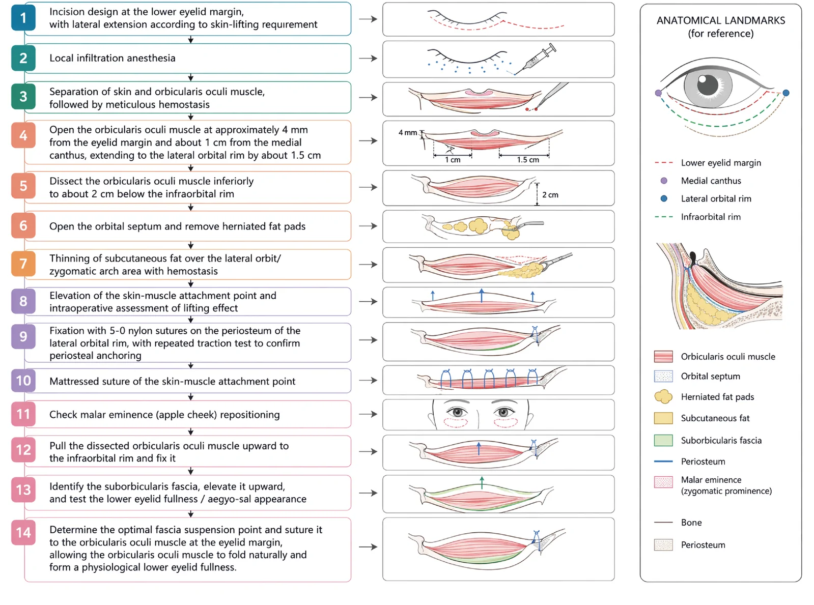
V3中面部提升经睫毛下切口,沿内眦线、外眦线、颧脂肪垫下界三个向量悬吊,可同时改善中面部下垂、下睑袋及睑颊轮廓不连续。
由6名意大利多学科专家(皮肤科、整形外科、神经科)基于文献回顾(PubMed 1973–2025)与临床经验形成的专家共识,针对incobotulinumtoxin A(Bocou
回顾性队列研究(循证等级IV),50例轴向偏曲(C形28例、反C形22例)歪鼻患者,由单一术者于2021至2023年行开放鼻整形,平均随访2年(1至3年)。