无针喷射对比有针注射递送聚核苷酸填充剂的分脸试验
2026年10月4日20 min read
Journal of Cosmetic Dermatology
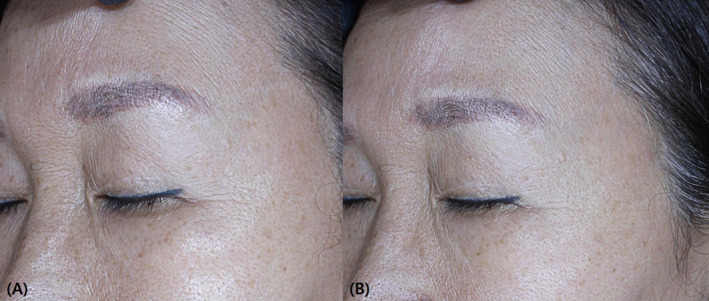
结论与实践要点
证据等级 :本研究为研究者盲法、随机、分脸(split‐face)对照临床试验,同一受试者两侧面部互为对照,可较好控制个体差异;但样本量仅 10 例,属单中心、单一种族的小样本探索性研究,证据强度有限,结论宜视为初步(hypothesis‐generating)。
适用人群 :面部皮肤老化(毛孔粗大、细纹、弹性下降)且寻求微创美容改善的健康成人(本组均 >19 岁,平均约 50 岁);对注射疼痛敏感或有针头恐惧(needle phobia)者可能更具吸引力。排除近 2 个月内接受过面部美容治疗(外用、口服、激光、化学焕肤)或近 6 个月内接受过 PN 治疗者。
关键结果(带数字)
证据边界与局限
临床落地提示 :在面部老化 PN 注射场景中,无针喷射在减轻疼痛与提升主观/客观毛孔、皱纹指标方面显示了初步优势,可作为疼痛敏感人群的可选方案;但其疗效优势尚需更大样本、更长随访与多设备对照研究验证,不宜据此宣称其优于有针注射。
BackgroundPolynucleotide (PN) filler often causes pain and can lead to delivery inaccuracies when applied via intradermal injection using a traditional needle.
AimsTo evaluate the efficacy of treatment and the pain during the procedure using conventional needle injection versus a needle‐free jet system for intradermal PN filler application.
MethodsIn this split‐face clinical trial, 10 Korean subjects were enrolled. Each subject received an intradermal injection of PN filler on one side of the face and a needle‐free jet injection using CureJet on the other side. Assessments included global and 3D skin imaging at each visit. Pain intensity was evaluated using visual analogue scale (VAS) scores during the injection. Additionally, patient satisfaction and adverse events were documented.
ResultsFindings revealed that Global Aesthetic Improvement Scale scores and patient satisfaction were significantly higher with the CureJet compared to the needle injection method. VAS scores were notably lower on the CureJet side. Improvements in both pore and wrinkle indices were observed from baseline, with a more pronounced improvement rate on the CureJet side compared to the needle injection side.
ConclusionsNeedle‐free injection of PN for aging skin was found to be effective in enhancing pore and wrinkle improvement, while reducing associated discomfort.
Keywords: needle‐free jet system、polynucleotide、pore、rejuvenation、wrinkle
1
Aged skin presents various aging indicators, including loss of elasticity, epidermal thinning, diminished collagen levels, increased wrinkles, and enlarged pores [ 1 ]. Skin aging typically involves reduced collagen production, structural alterations in the collagen fiber network, and accumulation of degenerated elastic fibers in the papillary and mid‐dermis [ 2 ]. Due to the significant social implications of skin aging, there is an increasing demand for minimally invasive dermal rejuvenation procedures with short downtime. Numerous facial fillers and skin boosters have been developed to meet this need [ 3 ].
Purified polynucleotide (PN) derived from the germ cells of salmon and other fish is widely utilized for this purpose [ 4 ]. Research indicates that PN aids in the proliferation of dermal fibroblasts and stimulates angiogenesis, contributing to effective wound healing [ 5 , 6 ]. Additionally, previous studies have shown that intradermal PN filler injections enhance skin elasticity and collagen synthesis by activating fibroblasts [ 7 ]. However, a significant limitation of intradermal PN injections is the substantial pain experienced by patients, even with local anesthesia. This discomfort can lead to involuntary facial movements or pressure application during the procedure, potentially increasing bleeding and impeding precise filler delivery.
In this investigator‐blind, randomized, split face study, we performed local PN injections using a conventional injection needle on one side of the aged face and a needle‐free jet system (CureJet; Bazbiomedic CO., LTD, Seoul, Korea) on the other side. Our objective was to compare the effectiveness of these two methods and the level of pain experienced during the procedure.
2
2.1
The Institutional Review Board granted approval for this study (IRB approval number: 2303‐001‐545), ensuring adherence to the Declaration of Helsinki guidelines. Informed consent was obtained from all participants prior to their involvement in the study.
2.2
We conducted this study as an investigator‐blind, randomized, split‐face study. Our cohort comprised 10 healthy Korean volunteers, all over 19 years old, seeking cosmetic enhancement of their facial skin. Exclusion criteria included individuals who had received any facial skin cosmetic treatments in the preceding 2 months. These treatments included topical agents, oral medications, laser procedures, and peels, as well as those who had undergone PN treatments on their facial skin in the past 6 months.
2.3
This study utilized the CureJet, a needle‐free, low‐pressure jet injector with adjustable volume and pressure settings. The device operates by generating an electro‐magnetic field via a coil, which propels fluid into the skin. It offers three modes of operation: single, auto, and burst. The burst mode is subdivided into 2‐burst, 3‐burst, and 4‐burst options, based on activation frequency. Power levels range from 1 to 5 units, incrementally increasing with voltage. The operational speed, measured in hertz (Hz), varies across five settings: 1, 3, 5, 15, and 20 Hz. The volume setting, indicating fluid dispensed, increments by 0.1, with a range from 1.0 to 2.0. Each cycle delivers between 0.5 and 0.8 μL. The jet pressure, emanating from the nozzle, scales with voltage and varies between 2.21 MPa and 6.57 MPa.
Rejuran (Pharmaresearch Products, Inc., Seoul, Korea) is a transparent liquid consisting of 20 mg/mL of PN.
2.4
To achieve local anesthesia prior to injection, EMLA 5% cream (lidocaine 25 mg/g, prilocaine 25 mg/g) was applied to both sides of the face for 30 min. The skin was then cleansed with chlorhexidine. Each participant were randomly assigned to receive intradermal needle injections of PN on one side of the face and microjet injections on the opposite side.
The intradermal injections utilized a 33‐gauge nano‐needle, employing a serial puncture technique. The injections were spaced about 1 cm apart. Each side of the face received 2.0 mL of filler, with 1.0 mL administered per side. For the clinical application of CureJet with PN, initial settings were Power level 5, Speed 10, and Volume 1.5 to create the jet. Subsequent settings during the procedure were Power level 2, Speed 5, and Volume 5. A 2 mL Rejuran syringe was attached as‐is to the CureJet for use.
2.5
During each session, a comprehensive evaluation of the facial skin was performed by the dermatologist by face‐to‐face with all participants. The degree of improvement in facial skin aging was assessed using the Global Aesthetic Improvement Scale (GAIS). This involved two independent dermatologists who were not involved in performing the treatment assigning a GAIS score, utilizing a 5‐point grading system to categorize the level of improvement (1 = worsened, 2 = unchanged, 3 = improved, 4 = much improved, 5 = very much improved). The GAIS score was presented as the average of the two evaluators' scores.
Additionally, the efficacy of the treatment was objectively measured using a noninvasive imaging technique. Various skin rejuvenation parameters, including dilated pores, brightness, sebum levels, wrinkles, melanin content, erythema, and overall skin tone, were quantified using the Mark–Vu system.
Pain intensity was also evaluated by the participants using a visual analogue scale (VAS), which ranged from 0 (no pain) to 10 (extreme pain) during the treatments. Participants were requested to report any discomfort or adverse effects, such as erythema, edema, or bruising, both during and after the procedure. Regular physical examinations were conducted as part of safety monitoring.
Participant satisfaction was assessed immediately following the treatment and again after 2 weeks. Satisfaction levels were classified as: 1 for unsatisfied, 2 for slightly unsatisfied, 3 for neutral, 4 for slightly satisfied, and 5 for satisfied.
2.6
Data analysis was conducted using SPSS version 26.0 (IBM Corp., Armonk, NY, USA). The results are presented as mean ± standard deviation. Descriptive statistics were applied to demographic data (age and sex). Due to the small sample size and the lack of normal distribution, the Wilcoxon signed‐rank test was utilized to compare differences in GAIS scores, patient satisfaction, and VAS pain scores and was employed for the analysis of skin parameters such as dilated pores, brightness, sebum, wrinkles, melanin, and erythema, as measured by the Mark–Vu system. p < 0.05 was considered statistically significant.
3
3.1
This study enrolled 10 patients with an average age of 49.9 ± 9.47 years. The GAIS score, indicating improvement, was significantly higher in the CureJet‐treated areas (3.85 ± 0.63) compared to the manually injected areas (3.6 ± 0.52) ( p = 0.037) (Table 1 ). The VAS score, reflecting pain intensity, was notably lower in the CureJet‐treated areas (2.9 ± 1.52) compared to the manually injected areas (5.4 ± 1.42) ( p = 0.017). The overall patient satisfaction scores were also greater in the CureJet‐treated areas (4.5 ± 0.53) compared to the manually injected areas (3.9 ± 0.73) ( p = 0.019). Figure 1 presents a representative case.
TABLE 1 GAIS score evaluated by two dermatologists after 2 weeks.
| Needle‐free jet injection | Conventional injection | |
|---|---|---|
| Dermatologist 1 | Dermatologist 2 | |
| Subject 1 | 5 | 4 |
| Subject 2 | 5 | 5 |
| Subject 3 | 3 | 4 |
| Subject 4 | 4 | 4 |
| Subject 5 | 4 | 3 |
| Subject 6 | 4 | 3 |
| Subject 7 | 3 | 4 |
| Subject 8 | 3 | 3 |
| Subject 9 | 4 | 3 |
| Subject 10 | 5 | 4 |
| Average | 3.85 | 3.6 |
| SD | 0.6258 | 0.5164 |
| p value | 0.037 |
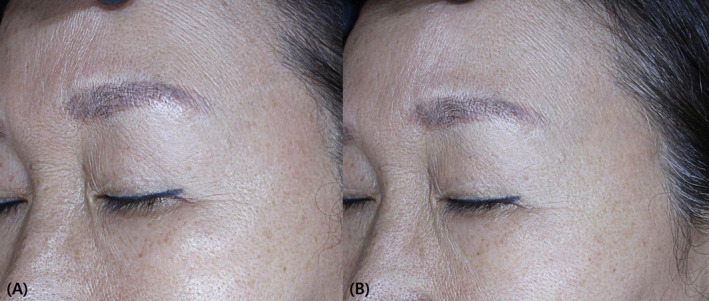
FIGURE 1 Representative case demonstrating noticeable enhancement in overall skin tone, wrinkle reduction, and smaller pore size following a 2‐weeks treatment period. (A) Before and (B) after 2 weeks of Curejet® injection.3.2
Skin parameters, including dilated pores, sebum levels, wrinkles, pigmentation, and skin brightness indices were evaluated using the three‐dimensional skin imaging tool Mark‐Vu. Notably, the pore and wrinkle indices showed significant improvement post‐treatment with CureJet, whereas manually injected areas, there was no pre‐and post‐treatment difference in these indices. In addition, while there was no difference between the two areas before treatment, the CureJet‐treated areas showed significant improvement compared to the manually injected areas for the pore index ( p = 0.025) and wrinkle index ( p = 0.006) as illustrated in Figure 2 .
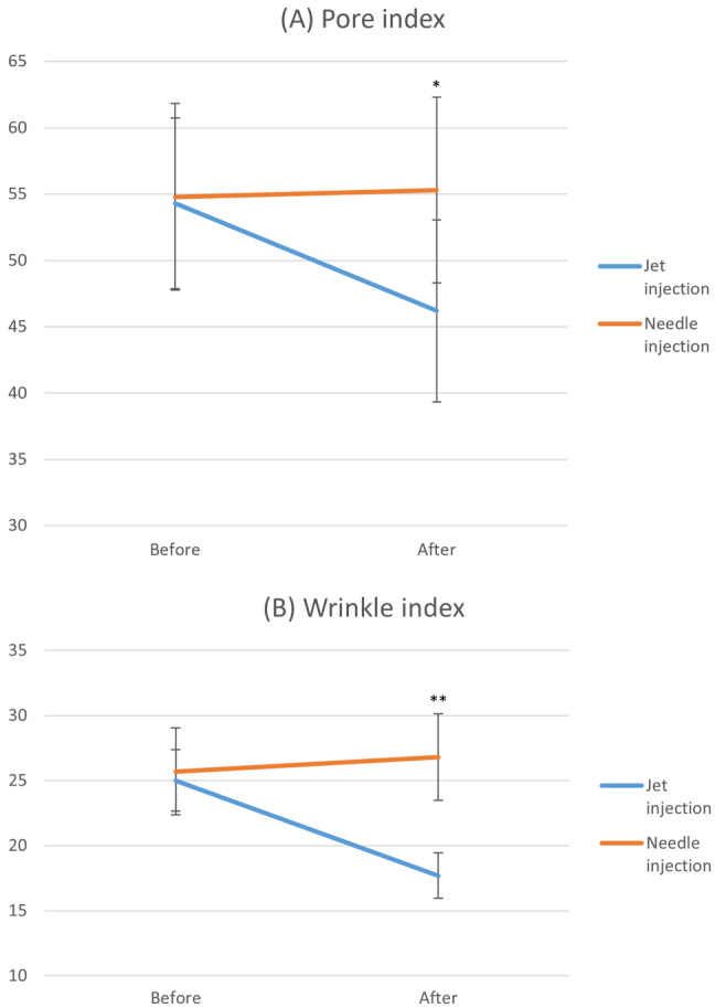
FIGURE 2 Improvement of (A) dilated pores and (B) wrinkles in the conventional injection and the needle‐free jet areas (*p < 0.05, **p < 0.01, and ***p < 0.001).Table 2 details the Mark‐Vu Indices corresponding to these findings.
TABLE 2 Measurement of the Mark–Vu indices (mean ± SD) in the needle‐free jet areas and conventional injection area.
| Baseline | 2 weeks after treatment | p ‐value | |
|---|---|---|---|
| A. Needle‐free jet areas | |||
| Pore index | 54.3 ± 6.44 | 46.2 ± 6.87 | 0.009 ** |
| Sebum index | 10.1 ± 5.45 | 9.4 ± 4.59 | 0.39 |
| Wrinkle index | 25 ± 2.37 | 17.7 ± 1.73 | < 0.001 *** |
| Pigmentation index | 27.4 ± 9.69 | 28.1 ± 10.03 | 0.44 |
| Skin brightness index | 30.6 ± 8.13 | 28.8 ± 8.91 | 0.33 |
| B. Conventional injection area | |||
| Pore index | 54.8 ± 7.04 | 55.3 ± 7.00 | 0.44 |
| Sebum index | 8.8 ± 4.49 | 8.5 ± 4.74 | 0.46 |
| Wrinkle index | 25.7 ± 3.35 | 26.8 ± 3.31 | 0.25 |
| Pigmentation index | 28.4 ± 9.34 | 27.6 ± 9.64 | 0.43 |
| Skin brightness index | 29.8 ± 10.98 | 28 ± 9.52 | 0.36 |
3.3
Treatment‐related local adverse effects included mild, transient erythema in all patients, and focal bruising in the manual injection sites of two participants. These symptoms resolved spontaneously within a week post‐treatment. All patients tolerated the treatment well, with no serious side effects reported.
4
Needle‐free injection systems are state‐of‐the‐art medical devices designed for delivering various medications, such as insulin and vaccines, into the skin without the need for traditional needle puncture [ 8 ]. The primary advantage of these systems is their ability to eliminate risks associated with skin puncture and disruption of the skin barrier [ 9 ]. This technology utilizes a needleless jet device, which administers a solution at high velocity by using pressurized air, facilitating its entry into the dermal layer. Although the procedure was performed on cadaver with a different type of filler, our previous experiments data (not shown) align with these findings, revealing that under our experimental settings, the drug was injected into and retained in the upper dermis (Figure 3 ) which suggest that a needleless jet device may be an effective way to target the upper dermis for filler injection. Since papillary fibroblasts exhibit greater proliferative and synthetic activities than reticular fibroblasts, but also show age‐related functional declines, targeted stimulation of the upper dermis using a needleless jet device may effectively enhance skin rejuvenation [ 10 ].
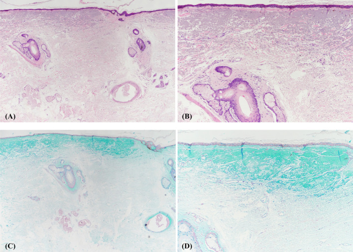
FIGURE 3 Histological samples showing poly D,L‐lactide and HA delivered through Curejet injection retained in the upper dermis. (A) H&E, ×40 (B) H&E, ×100 (C) Alcian blue, ×40 (D) Alcian blue, ×100.In our research, we observed that both the jet injection area and the conventional needle injection area exhibited improvement in GAIS, with the improvement being more significant in the jet injection area. In addition, the significant reduction in procedural pain in CureJet group resulted in overall patient satisfaction. This can help improving patient compliance to treatment and lowering psychological barrier toward injection. Also, remarkable improvement was observed in jet injection group, showing reduction of pore and wrinkles observed via magnification of MarkVu. This may be due to delicate positioning of injected materials by jet. Upon reaching the dermis, the liquid disperses evenly into the skin in a nearly spherical pattern, owing to the controlled deceleration of the jet at the exit point [ 11 ]. Over time, the orifice gradually narrows, simultaneously promoting dermal remodeling. This effect is achieved by stimulating fibroblasts through micro‐trauma, thus initiating neocollagenesis [ 12 ].
The limited sample size may have hindered the demonstration of a significant effect in the conventional needle injection area, while significant improvements can be observed in the jet injection area. This enhancement in the jet injection area is likely due to a synergistic effect resulting from the neocollagenesis induced by the jet injection.
Furthermore, there are also advantages over injection using needle such as the absence of side effects, including needle marks, bruising or bleeding, a lower risk of infection, and, most importantly, reduced pain during the procedure [ 13 , 14 ]. Since the majority of pain occurs during the initial needle insertion, using a small opening in conjunction with rapid PN delivery through a pressurized device leads to a diminished sensation of pain during administration [ 14 ]. These benefits enhance patient compliance and make the treatment more comfortable to receive, as demonstrated by high levels of patient satisfaction scores.
The results of this study should be interpreted with an acknowledgment of some limitations. First, the small sample size limits the generalizability of the results. We could not determine potential differences according to skin types. Additionally, the experiment provided only short‐term follow‐up data, spanning a duration of 2 weeks, which may not be sufficient for a comprehensive assessment of the long‐term impacts. To validate the effectiveness and establish the most appropriate parameters, further controlled clinical studies involving a larger and more diverse patient population are imperative; acknowledging the limitation of the small sample size ( N = 10), our future work will involve a more extensive clinical study with a significantly larger sample size. This will allow us to validate the preliminary results obtained from this pilot study and to perform more robust statistical analyses.
In conclusion, the needle‐free injection of PN for aging skin has demonstrated effectiveness in enhancing pore and wrinkle improvement while minimizing administration‐related discomfort. This approach may serve as a valuable alternative for patients with needle phobia, potentially working synergistically to promote neocollagenesis through the jet injection process.
The authors declare no conflicts of interest.
声明:中文精读 · 仅供学术参考。内容来自公开文献检索,不代表本人观点,不构成诊疗建议。 医疗美容需在正规医疗机构由执业医师实施。
基于相同主题推荐 · 继续深挖
前瞻性单中心上市后临床研究,28 名受试者(26 女 / 2 男,平均 43.8 岁)于面、颈、胸前区接受 PN HPT 聚核苷酸填充剂(Plinest,20 mg/mL,源自虹鳟
研究类型:研究者盲法、随机、分脸(split‐face)临床试验,2024 年在线发表于《Journal of Cosmetic Dermatology》,属小样本探索性研究。
研究类型:前瞻性自身对照split-face研究,40例(37例完成疗效评估,平均31岁、92.5%女性)接受3次、间隔3周的聚核苷酸(Rejuran,多聚核苷酸)注射,左颊皮内、