纳米脂肪在整形外科中的临床应用与安全性系统综述
2026年10月5日23 min read
Plastic and Reconstructive Surgery Global Open
结论与实践要点
临床可操作性: 纳入研究中腹部为采用频次居首的供区,机械乳化(Tonnard 法:两注射器间反复推注)为主流(83%),离心常见 3000 rpm×3 min。临床可据现有证据优先选用腹部供区+机械乳化的低成本、易操作路径。对瘢痕、脱发、烧伤、放疗后软组织损伤的改善证据较一致,可作为再生辅助手段纳入方案。
非在体边界(重要): 本综述以人体临床研究为主体(768 例),但“面部年轻化”亚组含 4 项研究、296 例患者及 68 只小鼠的在体动物实验数据,其抗皱、胶原合成等结论部分来自小鼠模型,不能外推为人体确定性证据;面部年轻化作为单一疗法的人体疗效仍不充分,实践中多与其他治疗联用。纳米脂肪制备缺乏统一标准,证据等级偏低(少 RCT、随访不一),结论应视为初步、需进一步高质量研究确认。
安全性提示: 综述层面总体耐受良好、无重大并发症报道,但属文献汇总,个体风险仍须结合具体术式、供区与制备条件评估。本篇为文献综述,所涉及的动物数据已在上文明确标注其非在体边界。
Background:The beauty industry has seen a shift in standards, with patients increasingly favoring natural and subtle enhancements over dramatic cosmetic changes. In this evolving landscape, nanofat has emerged as a promising tool to plastic surgeons due to its regenerative potential and biocompatibility. Despite its growing popularity, hesitancy regarding its efficacy and side effects persists.
Methods:A systematic literature review was conducted in December 2024 following PRISMA guidelines. Databases searched included PubMed, Embase, MEDLINE, Scopus, Web of Science, and Cochrane Library databases.
Results:A total of 18 studies met the inclusion criteria, encompassing 768 patients. Nine studies (n = 512 patients) assessed patient satisfaction, with most patients reporting above-average satisfaction. Eight studies (n = 188) reported outcomes using the Vancouver Scar Scale and Patient and Observer Scar Assessment Scale, all showing significant improvements in scar characteristics. Additional outcomes included collagen synthesis and transforming growth factor beta 1 expression (n = 68), hair density and thickness (n = 9), and histological analysis of skin samples ( n = 24). Regarding safety, 12 studies reported no significant complications such as granulomas or cysts, whereas 8 studies noted mild side effects, including erythema, hyperpigmentation, and edema.
Conclusions:Nanofat demonstrates significant potential as a cost-effective, well-tolerated regenerative therapy in both aesthetic and reconstructive contexts. Although preliminary findings are promising, further high-quality studies are necessary to establish standardized methodologies and optimize preparation techniques.
Takeaways
Question: What are the clinical outcomes, safety profile, and applications of nanofat in plastic and reconstructive surgery?
Findings: In this systematic review of 18 studies, nanofat demonstrated high patient satisfaction (88.9%); significant improvements in scar quality; and favorable outcomes in facial rejuvenation, burns, alopecia, scarring, and radiation-induced tissue damage. Complications were uncommon and limited primarily to transient erythema, edema, bruising, and pain.
Meaning: Nanofat seems to be a safe, cost-effective, and regenerative treatment modality with broad applications in aesthetic and reconstructive surgery. However, standardized processing techniques and higher-quality comparative studies are needed before widespread adoption of uniform clinical protocols.
Since Coleman’s pioneering work on structural fat grafting, the field has evolved significantly. 1 Structural fat grafting, including microfat, has long been used to restore volume and correct contour deformities in conditions associated with soft tissue loss, including congenital craniofacial anomalies, such as hemifacial microsomia and Parry-Romberg syndrome, and acquired etiologies including trauma, oncologic resection, and autoimmune-related lipoatrophy. 2 – 4 Building on this foundation, the field has increasingly incorporated regenerative medicine principles.
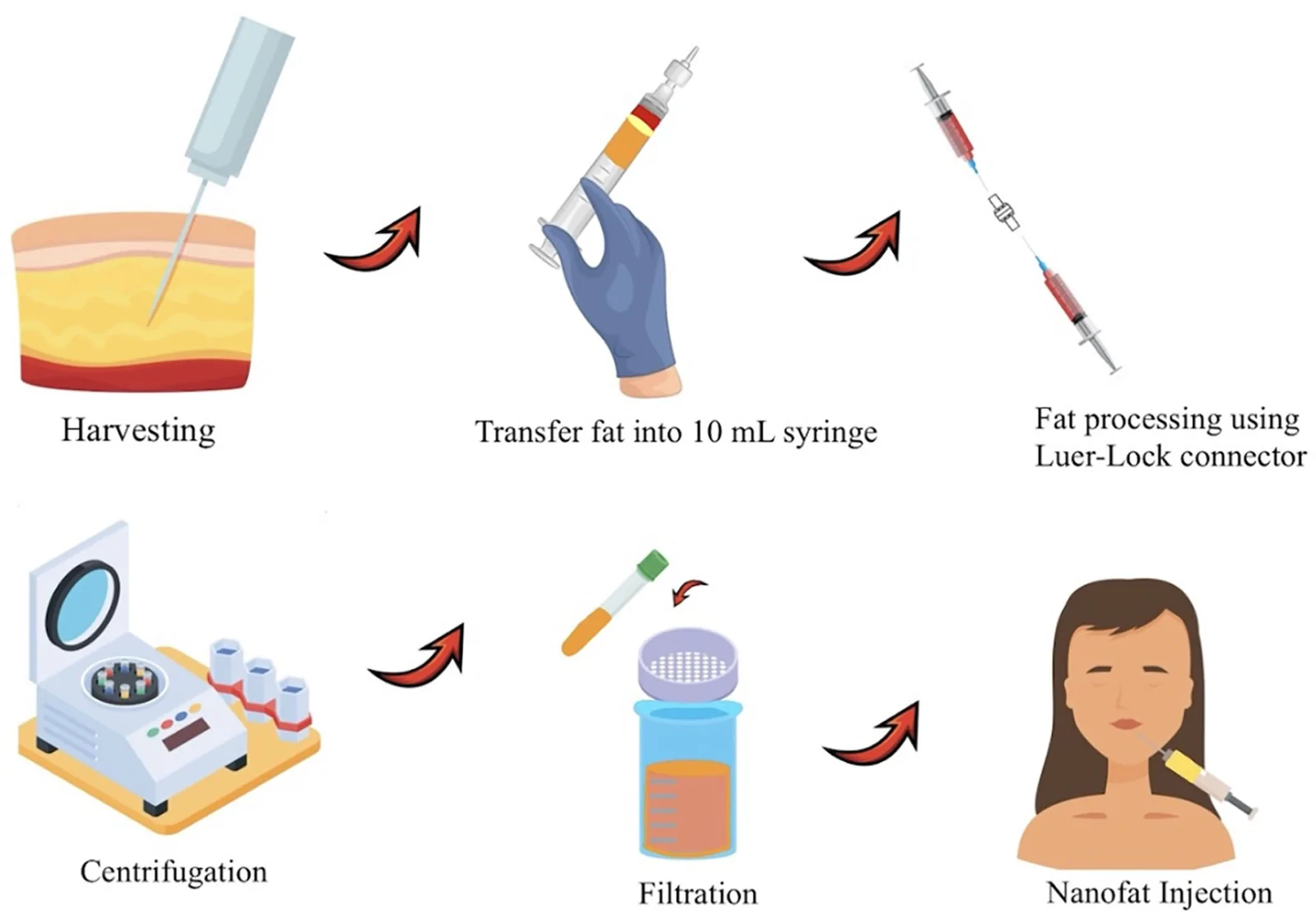
A major advancement came in 2013 when Tonnard et al introduced nanofat, a specialized form of autologous fat grafting designed to harness the regenerative potential of adipose-derived stem cells (ASCs) (Fig. 1 ). 5 Unlike traditional fat grafts, which primarily restore volume, nanofat undergoes mechanical emulsification and filtration, breaking down fat lobules while preserving the stromal vascular fraction (SVF)—a biologically active component rich in ASCs, growth factors, and extracellular matrix proteins. 6 This cellular-rich composition enables nanofat to act as a “stem cell depot,” facilitating tissue regeneration, neovascularization, and extracellular matrix remodeling in response to local tissue demands. 7 , 8
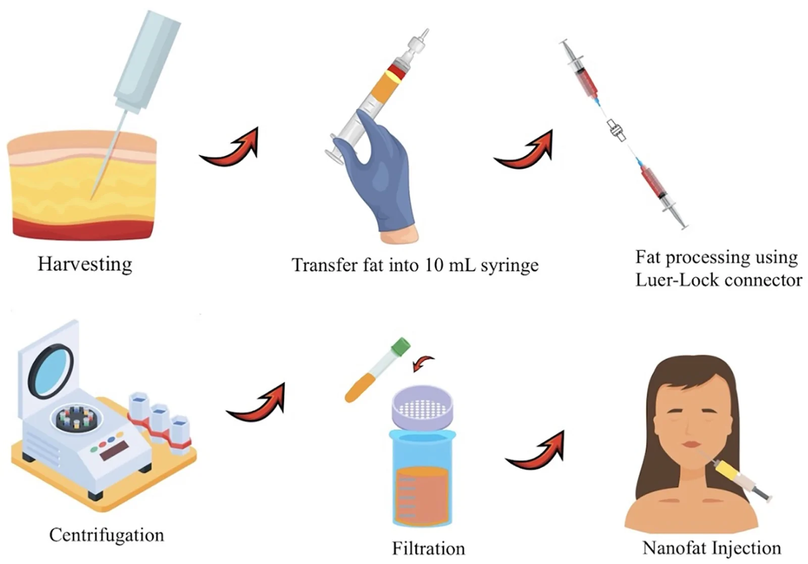
Fig. 1. Nanofat generation and methodology.Over the past decade, fat processing techniques have advanced to improve graft quality, cell viability, and regenerative outcomes. 9 The original nanofat technique introduced by Tonnard et al relied on mechanical emulsification to preserve SVF, a key component in regenerative signaling. 5 Since then, enzymatic digestion methods have emerged, enabling more precise isolation of ASCs and higher SVF yields. 10 Although these increased yields may offer potential advantages in efficacy, enzymatic approaches are subject to greater regulatory scrutiny due to concerns around manipulation and intended use. 11 Collectively, both mechanical and enzymatic methods have fueled the expansion of fat-based therapies that not only restore volume but also modulate the wound healing environment and support tissue regeneration. 12
Despite its regenerative potential, nanofat initially struggled to gain widespread clinical adoption. Early barriers stemmed from its lack of immediate, visible effects, and prevailing trends favoring high-volume fat transfers and synthetic fillers. 13 At the time, plastic surgery prioritized dramatic augmentations, with patients seeking exaggerated fullness in areas like the lips, cheeks, and buttocks. Nanofat, relying on gradual skin regeneration rather than instant volume, did not align with these expectations and was met with skepticism. Recently, however, interest in nanofat has surged, aligning with a shift toward more natural outcomes. The late 2010s and early 2020s marked a growing demand for subtle, long-lasting enhancements, influenced by social media and evolving patient preferences. 13 Additionally, the “era of great deflation” driven by trends like Ozempic and thinner beauty standards has further increased interest in less invasive, organic treatments. 14 Patients now favor options that avoid the rigid, artificial look of implants or fillers. Nanofat, with its availability, biocompatibility, cost-effectiveness, and low donor-site morbidity, make it a compelling option in this evolving landscape. 5
This systematic review provided a comprehensive assessment of nanofat’s clinical outcomes and complication rates across diverse medical applications. By synthesizing the current literature, we aimed to equip clinicians with evidence-based insights to facilitate safe and effective integration of nanofat into clinical practice.
A comprehensive literature search was conducted on December 21, 2024, using PubMed, Embase, Scopus, Cochrane, and Web of Science, following the PRISMA guidelines. 15 The search included the keywords “nanofat,” “nano-fat,” and “stromal-cell transfer.” Studies were included if they met the following criteria: full-text availability; original research; report of outcomes and complications; and application in facial rejuvenation, burns, scarring, alopecia, or radiation-induced tissue damage. Studies using SVF gel were also included, as SVF represents the key regenerative component within nanofat and is central to its therapeutic effects. 16 Exclusion criteria included studies that did not specifically focus on nanofat, as well as discussion articles, conference abstracts, ongoing studies, reviews, letters, and commentaries. Additionally, reference lists from included studies were manually screened for any relevant articles.
All identified articles were screened based on titles and abstracts. Full-text articles were then reviewed independently by 2 authors, C.G.O. and O.S. Any discrepancies in study selection were resolved through discussion between the authors. In cases where consensus could not be reached, a third reviewer was consulted (D.J.G.).
Data extraction was performed independently by C.G.O. and O.S. using a standardized form. Extracted data included study design, sample size, patient demographics, nanofat preparation techniques, clinical outcomes, and reported complications.
All statistical analyses were conducted using Excel and Python, leveraging Matplotlib for data visualization. Studies were categorized into 3 primary outcomes: patient satisfaction, Patient and Observer Scar Assessment Scale (POSAS), and Vancouver Scar Scale (VSS). For each outcome, the mean difference (MD) and corresponding 95% confidence interval (CI) were calculated for continuous variables to determine effect sizes.
Heterogeneity was assessed using Cochran’s Q test and the I 2 statistic. A random-effects model was applied when heterogeneity was statistically significant ( P < 0.05) or when I 2 exceeded 50%, indicating substantial variability across studies. A 2-tailed P value of less than 0.05 was considered statistically significant for all analyses.
The initial search of the databases yielded 1044 articles. Of these results, a total of 687 articles were removed due to duplication. After the remaining articles were screened by title and abstract, 321 were eliminated, and the full texts of 46 articles were assessed for eligibility. As a result, 28 articles were excluded from the review based on the established exclusion criteria. This left 18 studies for inclusion, encompassing data from 768 patients (Fig. 2 ). 17 – 35
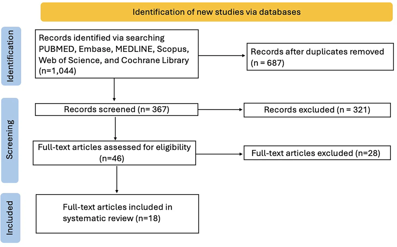
Fig. 2. Flow diagram illustrating the stepwise approach taken in the systematic review process, conducted in accordance with PRISMA guidelines.
A total of 9 studies (n = 512 patients) assessed patient satisfaction, all with statistically significant findings. Moderate heterogeneity was observed across these studies with respect to patient satisfaction ( I 2 = 52.45%, Q = 5.67). Under a random-effects model, heterogeneity slightly decreased ( I 2 = 50.35%, Q = 5.42). The pooled MD for patient satisfaction was 31.55%, with a highly significant P value ( P = 0.00005, P < 0.05), indicating that the satisfaction rate is statistically different from the null hypothesis of 50%. Across facial rejuvenation, burns, scarring, and alopecia, the mean patient satisfaction rate was 88.94% (95% CI: 80.70%–97.19%), demonstrating consistently high levels of patient-reported outcomes across diverse indications (Fig. 3 ).
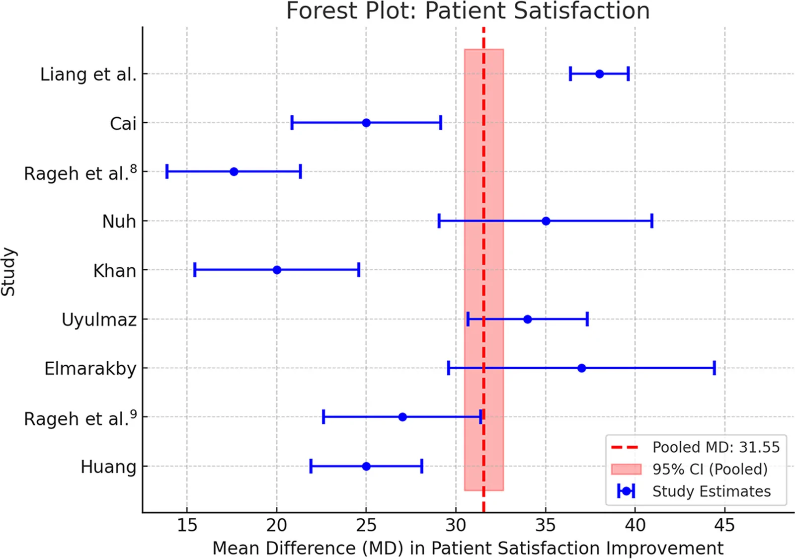
Fig. 3. Forest plot of standard mean differences and 95% CI of the mean percentage change in patient satisfaction.
A total of 3 studies utilized POSAS as a patient outcome, encompassing 106 patients (Figs. 4 , 5 ). POSAS demonstrated low-to-moderate heterogeneity ( I 2 = 27.89%, Q = 2.14), with no substantial change in heterogeneity when applying a random-effects model. The pooled MD for patient POSAS was 2.80 ( P = 0.0007, P < 0.05), and the observer POSAS was 2.55 ( P = 0.001, P < 0.05), indicating a statistically significant reduction in POSAS scores following nanofat treatment. The mean pretreatment patient POSAS score was 6.71, which decreased to 3.92 posttreatment, showing an average difference of 2.80 (95% CI: 0.82–4.77). The mean pretreatment observer POSAS was 6.12, which decreased to 3.56 posttreatment, showing an average difference of 2.55 (95% CI: 0.26–5.37) reflecting a substantial improvement in scar quality and patient-reported outcomes.
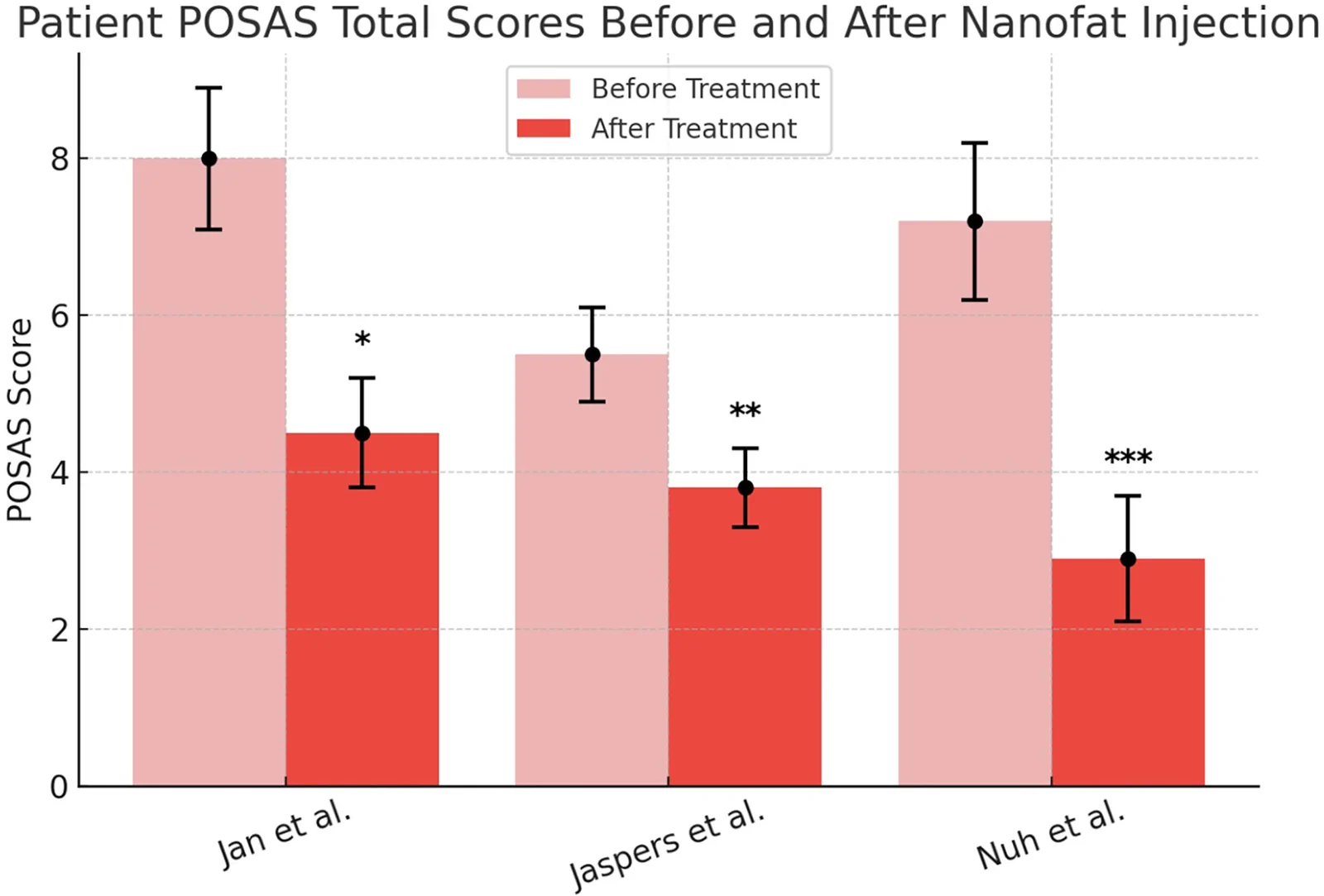
Fig. 4. Patient POSAS total scores before and after nanofat injection.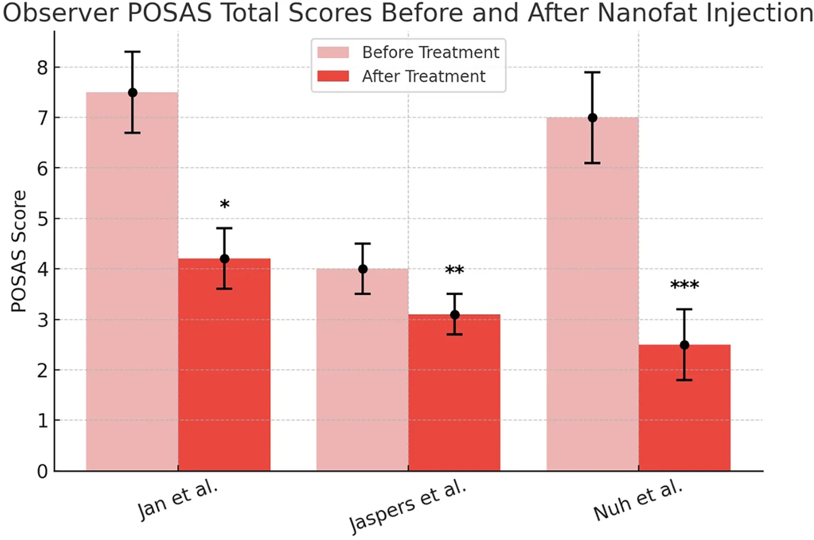
Fig. 5. Observer POSAS total scores before and after nanofat injection.
A total of 4 studies representing 72 patients implemented VSS as a patient outcome in their study. Statistically significant differences were found before and after nanofat treatment was administered for all studies included in this review (Fig. 6 ). VSS demonstrated low to moderate heterogeneity ( I 2 = 39.12%, Q = 3.28), which slightly decreased with the application of a random-effects model ( I 2 = 36.80%, Q = 3.12). The pooled MD for VSS was 3.20 ( P = 0.0003, P < 0.05), indicating a statistically significant reduction in VSS scores following nanofat treatment. The mean VSS score decreased from 8.50 pretreatment to 5.51 posttreatment, demonstrating an average improvement of 2.98 (95% CI: 0.91–5.05) reflecting a substantial improvement in scar characteristics and overall scar quality (Fig. 7 ).
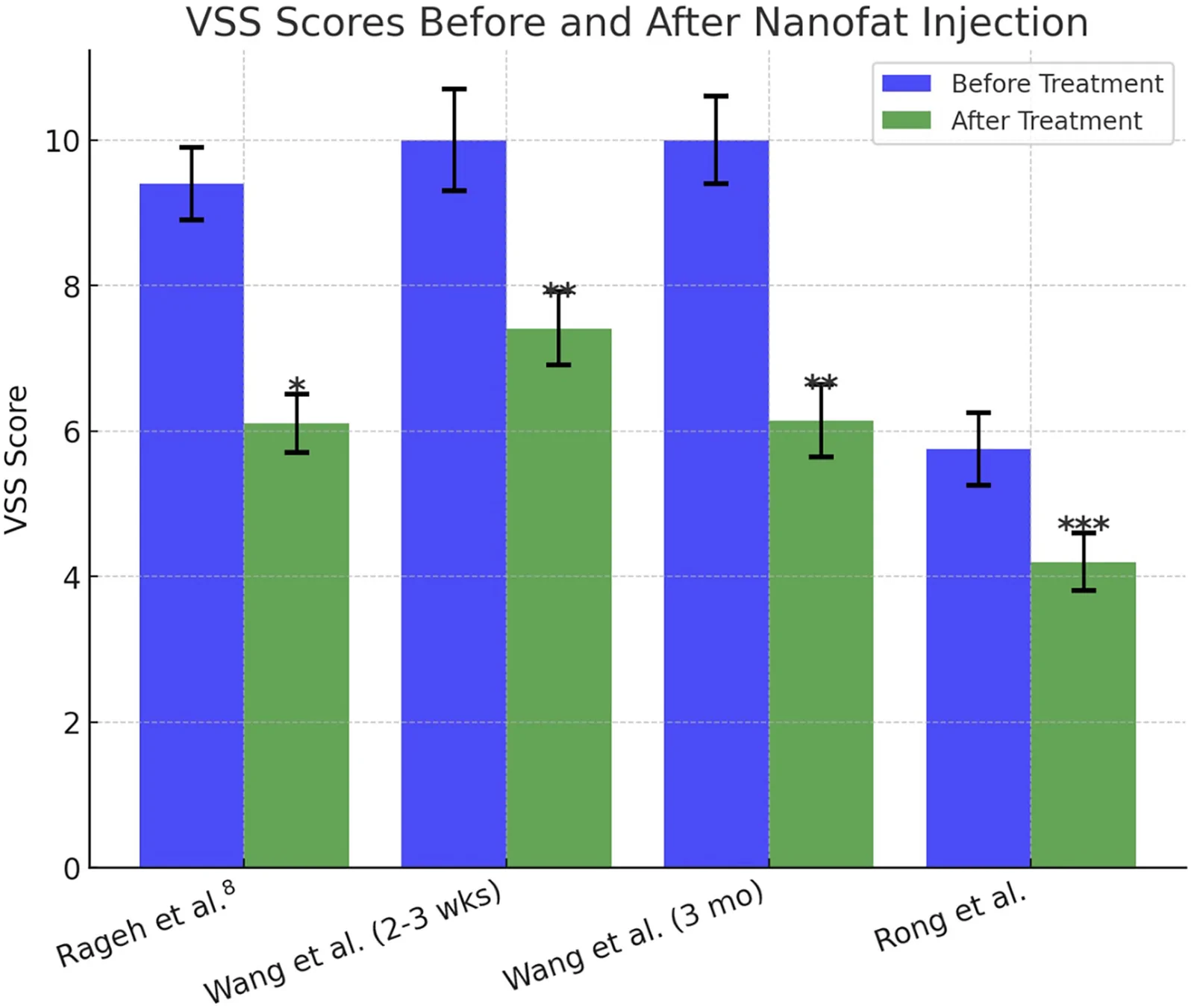
Fig. 6. VSS scores before and after nanofat treatment.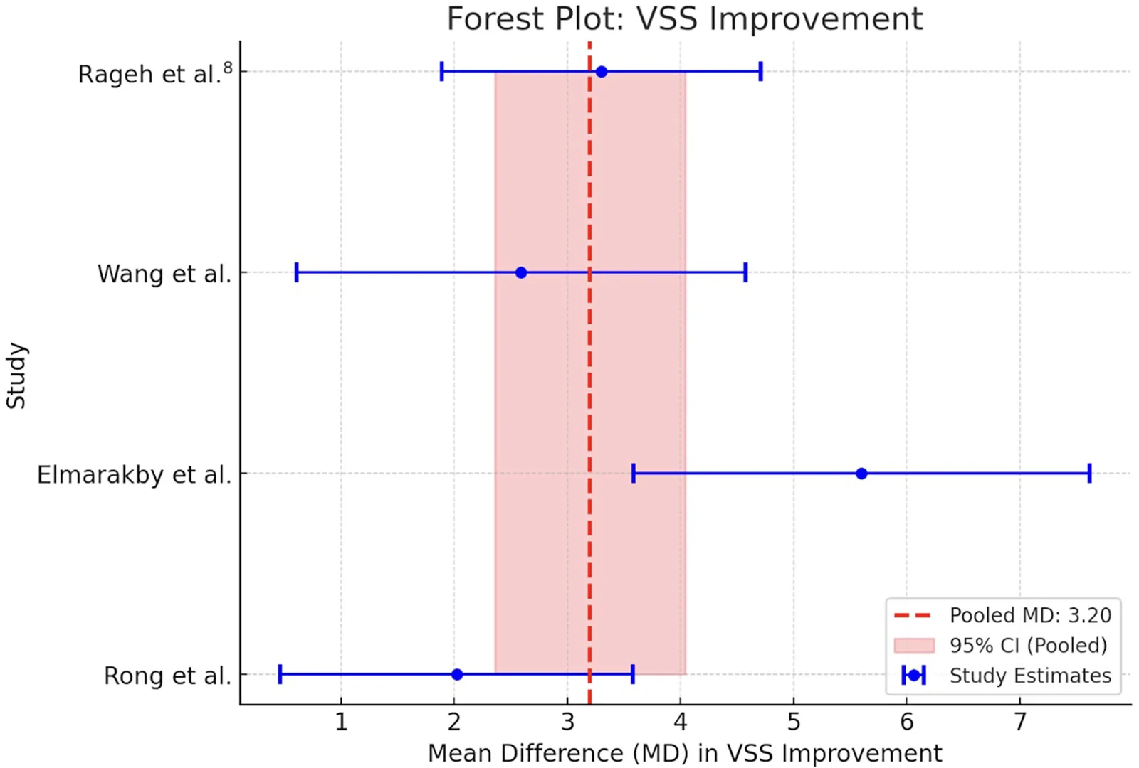
Fig. 7. Forest plot of standard mean differences and 95% CI of the mean percentage change in VSS improvement.
Facial rejuvenation aims to restore a youthful appearance by addressing signs of aging, such as sagging skin, wrinkles, and volume loss. This systematic review includes 4 studies involving 296 patients and 68 mice, all focusing on facial rejuvenation. Akbari et al 30 assessed wrinkle volume, depth, area, and percentage of area before and after nanofat grafting, finding significant improvement in all parameters. Wrinkles with greater volume, depth, and area loss showed the most pronounced improvement. Zhu et al 31 examined the impact of SVF and found that it led to higher fibroblast counts, increased transforming growth factor beta 1 expression, and thickened dermal collagen compared to tretinoin, fat, and phosphate-buffered saline. Additionally, Cai et al 18 conducted histological evaluations on neck wrinkles, showing increased collagen density in the treated area after SVF injection.
Although the face has an inherent ability to heal, postburn facial scarring can lead to significant psychosocial consequences, especially in patients with darker skin types who are prone to hyperpigmentation. 26 Given the visibility of facial scars, even minor imperfections can have a profound emotional impact. This review examined the use of nanofat as a potential treatment for postburn scars across four studies, involving a total of 135 patients. The studies measured outcomes using the VSS, POSAS, and patient satisfaction, with all showing favorable results. Rageh et al 19 further enhanced the reliability of their findings by incorporating the Antera 3D camera alongside the VSS. Across all studies, there was a statistically significant reduction in the total scores for both VSS and POSAS, indicating the effectiveness of nanofat in improving burn scar quality.
Eyebrows, eyelashes, and the scalp are integral to facial expression, self-image, religious beliefs, and psychosocial well-being. 23 Nanofat offers potential as a therapeutic option for alopecia, as it has demonstrated promising results in stimulating hair growth and improving scalp health. This review includes 2 studies on alopecia, encompassing a total of 27 patients. Outcomes such as POSAS; patient satisfaction; and hair density, thickness, and keratin scores were evaluated. Kim et al 32 assessed hair density in the SVF-treated area and found a significant increase at both 3- and 6-month follow-ups compared to untreated areas. Additionally, keratin scores on the scalp showed significant improvement in the SVF-treated area ( P = 0.032) at 6 months.
Radiation therapy, commonly used in cancer treatment, can lead to radiation-induced tissue damage, resulting in skin fibrosis, chronic inflammation, and soft tissue atrophy. 35 These complications significantly impact both function and aesthetics, making reconstruction challenging. Nanofat, rich in adipose-derived stem cells and growth factors, offers a promising regenerative therapy to counteract these effects. This review includes 2 studies, encompassing 20 patients and 24 rats, which assessed outcomes using Late Effects of Normal Tissues-Subjective, Objective, Management (LENT-SOM) and histological analysis of skin samples. Rigotti et al 33 used LENT-SOM and observed that of the 11 patients initially classified as LENT-SOM grade 4 (irreversible functional damage), 4 progressed to grade 0 (no symptoms), 5 to grade 1, and 1 to grade 2. Sowa et al 34 conducted histological assessments of irradiated skin samples and found significant dermal thickening, atrophy, and collagen deposition in the subcutaneous fatty layer compared to the no-radiation control group. In the group treated with SVF gel, less fibrosis was observed compared to the groups treated with fat or melanoma cell adhesion molecule (CD146).
Scarring is an inherent part of the wound healing process, but abnormal scarring can lead to aesthetic concerns and psychological distress. 36 Nanofat has shown potential for treating various types of scars, including burn scars, postsurgical scars, and acne scars. This review includes 6 studies on the use of nanofat in scarring, representing a total of 198 patients, with 2 studies (49 patients) specifically focusing on acne scars. The studies used a variety of outcome measures, including VSS, visual analog scale (VAS), patient satisfaction, Goodman and Baron scores, and FACE-Q. Two of the studies combined nanofat with other therapies: one combined nanofat with fractional CO 2 lasers, and the other with platelet-rich plasma. Rong et al 29 used an Antera 3D camera for objective evaluation of scar characteristics, validating the VSS and VAS scores reported in their study. Their findings showed that, when evaluating scar depressions, the SVF gel–treated side exhibited improved color, texture, melanin, and hemoglobin levels compared to the control side. ( See table, Supplemental Digital Content 1 , which displays the summary of nanofat studies by clinical application, https://links.lww.com/PRSGO/F263.)
Complications were minimal across the included studies, with 12 of 18 (67%) reporting no adverse effects. Among the 6 studies that did report complications, the most common issues were minor erythema, edema, bruising, and blistering, all of which resolved within 3 weeks. Jan et al 26 reported mild edema in 30 (62.5%) patients, which resolved within 2 weeks. Jaspers et al 27 noted minor superficial hematomas and 1 case of severe postoperative pain, which was alleviated quickly. Rageh et al 20 reported that all patients reported mild pain, erythema, and crust formation which faded away within 6.68 ± 0.95 days after the session. Huang et al 25 reported that 48 (93%) patients experienced temporary erythema for an average of 2–3 weeks (Table 1 ).
Table 1. Nanofat Complications and Management
| Complication | Percentage (No. Patients) | Management |
|---|---|---|
| Edema | 3.39 (26) | Cold compress, head elevation, and lymphatic drainage massage |
| Erythema | 5.73 (44) | Cold compress, topical steroids, and avoidance of direct sunlight |
| Bruising | 1.82 (14) | Cold compress, arnica gel, and gentle massage |
| Blistering | 0.26 (2) | Topical antibiotics, sterile dressing, and avoidance of friction |
This systematic review analyzed efficacy and safety across 5 distinct clinical applications: facial rejuvenation, scarring, alopecia, burns, and radiation-induced tissue damage. Across all outcomes included in this study—patient satisfaction, POSAS, VSS, VAS, transforming growth factor beta 1 expression, hair density and thickness, and histological analysis of skin samples—nanofat and SVF gel demonstrated statistically significant improvements.
Patient satisfaction remained consistently high with an overall satisfaction rate of 88.94% across 512 patients. Among internationally accepted scar quality measuring tools, nanofat resulted in a statistically significant improvement in scar quality, with a mean patient POSAS reduction of 2.8 and an observer POSAS reduction of 2.55 across 106 patients with burns and alopecia. Likewise, VSS scores improved by 2.98 points among 72 patients, underscoring nanofat’s effectiveness in scar remodeling and regeneration.
Beyond its regenerative potential, nanofat exhibits an excellent safety profile. Twelve studies reported no complications, and among the 6 studies that did report adverse events, erythema was most common. Notably, no studies documented major complications such as granulomas, fat necrosis, or significant inflammation. Additionally, despite nanofat’s high oil content, no cases of oil cyst formation were reported—a notable finding compared to microfat, which has been associated with oil-related side effects. 37 This suggests that nanofat may possess distinct structural properties that mitigate oil-related complications.
Overall, our findings support nanofat’s utility in treating burns, alopecia, radiation-induced soft tissue damage, and scarring. However, its efficacy as a monotherapy for facial rejuvenation remains less well established, and in clinical practice, it is frequently combined with adjunctive treatments. Uguten et al 38 speculated that this may be due to the need for additional stimulation, such as thermal or mechanical injury, to sufficiently activate regenerative pathways and achieve visible results.
A key finding in this review was the heterogeneity in nanofat preparation techniques, with significant variations in donor site selection, centrifugation speeds, and emulsification methods. The lower abdomen, medial and lateral thighs, gluteal region, and flanks were the most commonly used donor sites, with the abdomen being the most frequently harvested location (Table S1). However, some studies suggest that alternative donor sites may offer superior viability and patient tolerance. For example, Faikh-Gomez et al 39 reported that patients who underwent abdominal fat harvesting experienced greater postprocedural pain than those who had fat extracted from the thigh. Similarly, Alam et al 40 found that nanofat derived from the medial thigh exhibited higher cell viability and survival rates compared to abdominal fat. However, this review does not provide sufficient evidence to confirm these findings, highlighting the need for further comparative studies.
Centrifugation protocols also varied widely, ranging from 1000 rpm for 2 minutes to 2700 rpm for 15 minutes. However, 3000 rpm for 3 minutes was the most commonly used speed (33% of studies), aligning with previous findings from Coleman et al 1 and Demirel et al, 41 both of whom demonstrated that this protocol optimally preserves adipocyte and SVF integrity in both in vitro and in vivo studies.
In contrast to centrifugation protocols, emulsification methods were more consistent. Tonnard et al established mechanical emulsification as the foundational method for nanofat preparation, and it remains the predominant technique, used in 83% of the studies included in this review. 5 This involves the repeated passage of lipoaspirate between two syringes connected by a female-to-female Luer-Lock connector, typically performed 20–30 times. The resulting shear stress disrupts adipocytes while preserving the SVF. Studies have shown that this method upregulates multipotency markers, suggesting enhanced regenerative potential. 42 Its procedural simplicity, speed, and exemption from extensive regulatory oversight make mechanical emulsification highly practical for clinical use. 43 – 46
In contrast, enzymatic digestion, typically employing collagenase or related enzymes, isolates SVF through chemical breakdown of the extracellular matrix. 47 This yields a more purified and higher concentration of regenerative cells; however, it is more time-intensive and thus significantly more expensive and subject to stricter regulatory oversight. 48 In the United States and Europe, cellular therapies must meet criteria for minimal manipulation and homologous use to avoid classification as advanced therapy medicinal products, making enzymatically processed SVF more difficult to implement clinically. 7 , 47 , 49 – 51
Given the variability in nanofat preparation across the studies, we propose a standardized processing protocol informed by the collective evidence synthesized here. 17 – 35 , 52 Harvesting should begin with lipoaspiration from regions of abundant adipose tissue such as the abdomen, thighs, or flanks using sterile technique. The aspirated fat is then mechanically emulsified by transferring it between two 10-mL syringes connected via a Luer-Lock adapter, repeated 20–30 times until a uniform emulsion is achieved. The emulsified product is subsequently centrifuged at 3000 rpm for 3 minutes at room temperature, and the middle stromal-rich layer is isolated and passed through a 500-µm filter for final refinement.
This review has several limitations. The number of included studies was limited, and few were randomized controlled trials, reducing the strength of causal inferences. Inconsistencies in study design, sample size, follow-up duration, and outcome measures further limited comparability. Additionally, some applications, particularly radiation-induced tissue damage and alopecia, had small patient cohorts, limiting analytical power.
Future research should prioritize comparative studies of nanofat processing methods to identify optimal techniques across indications. Establishing standardized protocols will not only enhance clinical efficacy and safety but also facilitate broader regulatory acceptance and integration into regenerative practice.
This systematic review highlights the evolving role of nanofat in regenerative and aesthetic medicine, examining its methodology, clinical outcomes, and safety profile across diverse applications, including facial rejuvenation, burns, alopecia, scarring, and radiation-induced tissue damage. The evidence presented underscores nanofat’s efficacy and tolerability, reinforcing its relevance in an era where patients are increasingly seeking natural, minimally invasive solutions. Furthermore, its cost-effectiveness has the potential to democratize access to cosmetic and reconstructive treatments, broadening its impact beyond traditionally served populations. Ongoing advancements in regenerative medicine suggest that this innovation may not only complement but redefine the standard of care, potentially surpassing conventional treatments in the years to come.
The authors have no financial interest to declare in relation to the content of this article.
声明:中文精读 · 仅供学术参考。内容来自公开文献检索,不代表本人观点,不构成诊疗建议。 医疗美容需在正规医疗机构由执业医师实施。
基于相同主题推荐 · 继续深挖
PRISMA 系统综述(2024-12),纳入 18 项研究、768 例患者;纳米脂肪为机械乳化/过滤的自体脂肪,富集 SVF 与脂肪来源干细胞,侧重再生而非单纯容积充填。
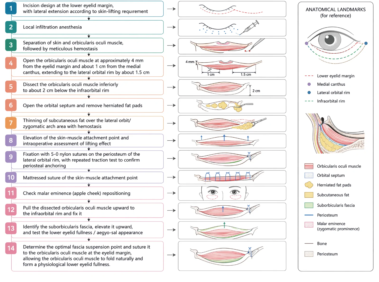
V3中面部提升经睫毛下切口,沿内眦线、外眦线、颧脂肪垫下界三个向量悬吊,可同时改善中面部下垂、下睑袋及睑颊轮廓不连续。
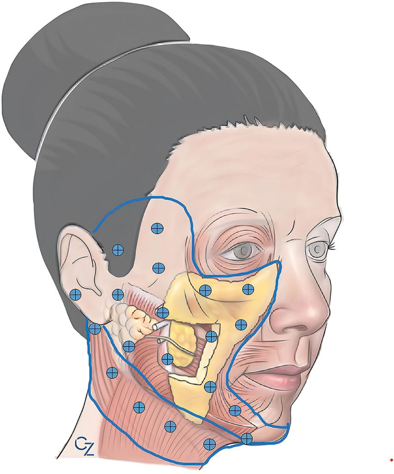
背景:多平面面部年轻化(深层除皱 + 容积/脂肪处理 + 皮肤重铺)创伤较大,术中出血与术后淤青影响恢复与满意度。氨甲环酸(TXA)具抗纤溶、减少出血作用,本文评估其在多平面面部年