蘑菇型自体增强乳房固定术多中心回顾(198 例)
2026年10月10日19 min read
Plastic and Reconstructive Surgery Global Open
结论与实践要点
本文为回顾性多中心病例系列,纳入 2018 年 1 月至 2023 年 12 月共 198 名接受蘑菇型自体增强乳房固定术(可联合或不联合假体及脂肪移植)的女性,平均 44.3 岁(范围 18–74)。主要结局为安全性与初步形态:总体并发症率约 13%,其中 1 例血肿需再次手术干预,17 例因增生性瘢痕或乳头不对称接受局部修整,未观察到乳头乳晕复合体坏死。该术式以中央深蒂的蘑菇形皮瓣旋转填充上极,旨在提升乳房并改善上极容积。
局限在于回顾性设计与多中心异质性,缺乏长期随访与标准化患者报告结局,并发症统计依赖各中心自发记录,可能存在低估;所谓“多中心”由同一作者系列构成,外部验证有限。该技术适用于轻度至中度下垂、希望以上极自体组织填充者;对重度下垂或上极组织量不足者效果受限,是否联合假体或脂肪移植需个体化评估,并应在知情同意中说明修整概率。
Background:Numerous procedures have been described to lift breast tissue and to improve aesthetic appearance of the upper pole through auto-augmentation mastopexy procedures, with a wide range of techniques. This study introduces a novel mastopexy procedure designed to optimize volume in the central and upper breast pole using a mushroom-shaped flap.
Methods:This article presents a modified approach to fill the breast’s upper pole using a median, deep-based pedicle flap. This is a retrospective, multicentered study, based on a series of cases conducted by the author. The perioperative management and the surgical technique are described. A systematic approach was used to identify acute and subacute complications during the postoperative follow-up period.
Results:From January 2018 to December 2023, 198 women were included in the study and underwent mushroom mastopexy with or without implant and/or lipofilling. The mean age was 44.3 years (range, 18–74 y). The general complication rate was 13%, and we observed 1 hematoma that required reintervention. In 17 patients, local surgical revision was necessary due to hypertrophic scarring and nipple asymmetry. No nipple–areola complex necrosis was observed.
Conclusions:Mushroom mastopexy lifts the breast while restoring maximum volume to the upper pole and can be combined with breast augmentation using implants or lipofilling. This technique is simple, reliable, and produces excellent aesthetic results with a low complication rate.
Question: What is this technique and what does it offer?
Findings: We present a modified approach to enhance the breast’s upper pole using a median, deep-based pedicle flap. This is a retrospective, multicentered study, based on a series of cases conducted by the author. The perioperative management and surgical technique are described. A systematic approach was used to identify acute and subacute complications during the postoperative follow-up period.
Meaning: The mushroom auto-augmentation mastopexy is a reproducible, safe, and sophisticated surgical option for patients who wish to improve both the volume and contour of their breasts.
Mastopexy is the second most frequently performed aesthetic breast surgery, after augmentation by implants, with a 7% increase in the number of procedures between 2022 and 2023. 1 Factors such as gravity, repeated weight changes, pregnancy, and aging frequently contribute to breast ptosis, characterized by tissue descent to the lower pole and diminished volume in the upper pole. During breast reshaping, ptotic tissue is lifted and tightened, while the skin envelope is reduced. When breast volume is small to moderate, the use of an implant may be an effective option to restore upper-pole fullness. 2 However, some patients may refuse implants, necessitating alternative techniques such as fat grafting. Various mastopexy/reduction approaches with different skin incisions and pedicle designs, such as the periareolar technique, the “inverted-T scar” (Wise pattern), vertical scar, or L-shaped scar technique, have been described over decades. 3 , 4 A special approach represents the auto-augmentation mastopexy. In this article, we present our approach using deep and superomedial flap techniques for mastopexy with auto-augmentation, which allows the entire breast volume to be preserved while changing its shape. Additionally, we evaluate the complication rates associated with this technique. Our approach was developed based on experience with 198 consecutive women seeking breast shape correction, either with or without the use of implants or lipofilling.
For this retrospective study conducted from January 2018 to December 2023, all patients who underwent mushroom mastopexy, with or without a simultaneous implant or lipofilling surgery, were included. Surgery was performed at one of the following 3 centers in Belgium: the Plastic and Maxillo-facial Surgery Unit of the CHU de Liège, the Plastic Surgery Unit of the CHC MontLégia, or the private clinic of Mont-Saint-Martin. Smoking cessation was required for a minimum of 4 weeks before the surgery. We included both primary and secondary surgery, mushroom mastopexy alone, composite mushroom mastopexy (associated with implant or fat grafting), and mushroom mastopexy for symmetrization with contralateral reconstruction. Exclusion criteria were patients with ptosis due to tuberous breast deformity or Poland syndrome, patients lost to follow-up, patients with an American Society of Anesthesiology score greater than II, patients with any uncontrolled chronic illness and/or medical history of diabetic mellitus or metabolic syndrome, patients with body mass index (BMI) higher than 30 kg/m 2 , patients requiring breast reduction surgery with removal of more than 300 g, and patients with previous mastopexy or breast reduction. Written and verbal informed consent was obtained from all the patients. Baseline characteristics such as age, BMI, smoking, and previous bariatric surgery were recorded. Patients who underwent mastopexy were subdivided into 6 groups: mastopexy alone, mastopexy with fat grafting, mastopexy with implant and fat grafting, mastopexy with implant left in situ, mastopexy with implant removal, and mastopexy with implant removal and fat grafting. A diligent process of identifying acute and subacute complications was undertaken throughout the postoperative follow-up period. This encompassed cases requiring reoperation due to major complications or aesthetic concerns, including hematoma, delayed wound healing, infection, seroma, and hypertrophic scarring. For statistical evaluation, the Fisher test was used to examine potential differences between groups. The statistical significance was set at a P value of less than 0.05.
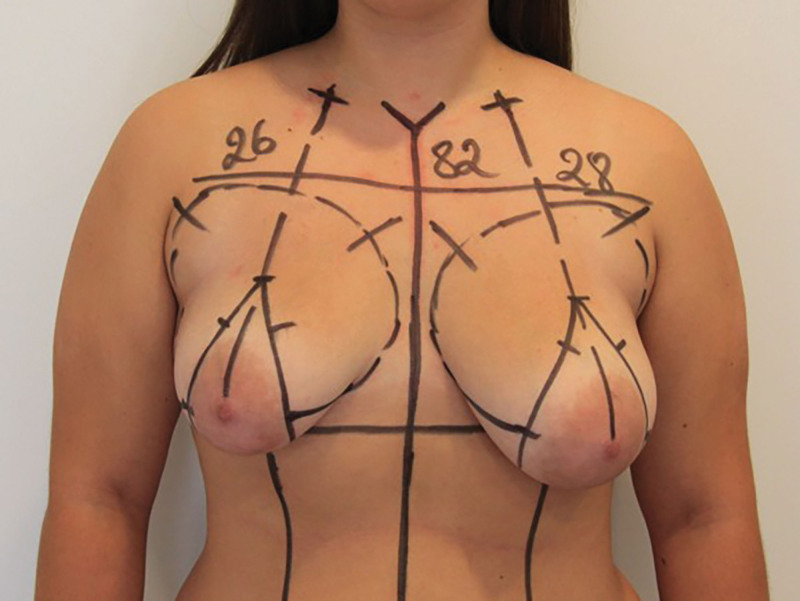
Skin markings were made preoperatively while the patient was in a standing position. First, the median line and a midclavicular vertical line were marked on the meridian of the breast, passing through the middle of the clavicle at the center of the areola on each side (ie, a hemiclavicular nipple–areola complex [NAC] line). The midpoint of the circumareolar marking was drawn at the point of best aesthetic projection over this midclavicular line (17–20 cm). This point depends on several parameters, including the patient’s size, torso width and height, and desired volume. It is decided at the surgeon’s convenience. A line parallel to the midline is drawn at 9–11 cm bilaterally below the inframammary fold (IMF). Below the areolar point, the vertical lines of the pattern are estimated by slightly pulling and rotating the breast inward and upward for the medial line, and outward and downward for the lateral vertical line. These 2 points are located at 9 cm, and the marking is completed by connecting the IMF line to the horizontal endpoint of the pattern. The footprint of each breast is also defined by a drawing (Fig. 1 ).
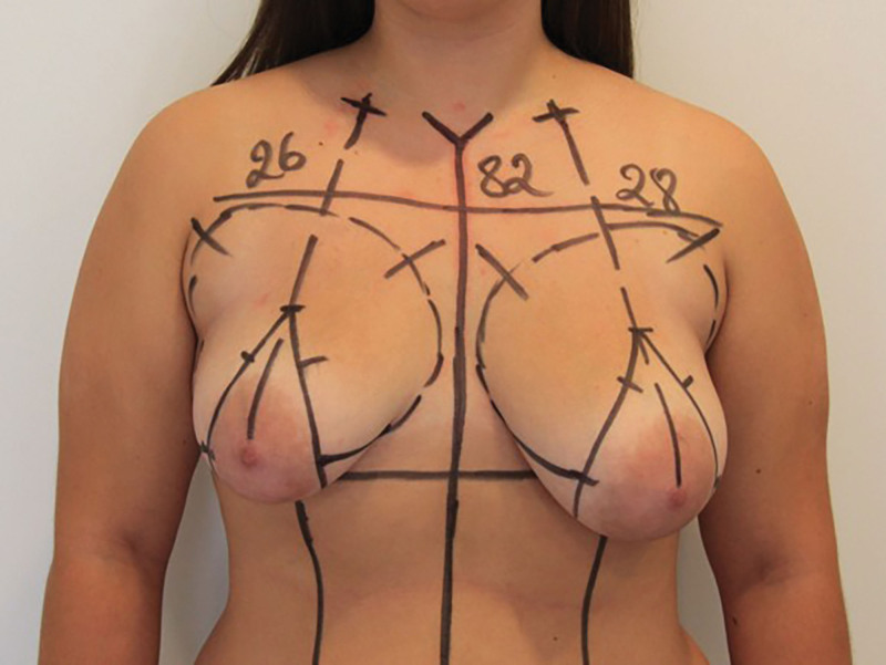
Fig. 1. A photograph of preoperative markings with an inverted-T pattern. The distance between the nipple and the mid-clavicle is noted on each side, as well as the patient’s size.倒 T 形术前标记,标注乳头至锁骨中点距离。
The procedure was performed with the patient lying in the dorsal decubitus position, with the back slightly raised. One gram of second-generation cephalosporin was administered intraoperatively. We used a local infiltration solution to inject the incisional line and de-epithelialized zone with saline solution, adrenaline (1:200,000), and 1% Xylocaine. After achieving the hemostatic effect of the infiltration, the area was subjected to de-epithelialization. A transverse parenchymal incision was made through the breast tissue along the lower incision. A full parenchymal incision was made on the medial and lateral vertical line of de-epithelialized lower breast tissue down to the IMF. This dissection then delimits the mushroom-shaped flap (Fig. 2 ). The flap was raised, and dissection with cautery was continued in a cephalad direction above the pectoralis fascia up to the second intercostal space to create a pocket for the auto-augmentation flap to settle into easily. Analgesic pectoral nerve block I and pectoral nerve block II were performed bilaterally. 5 To achieve symmetry, the auto-augmentation flap could be trimmed accordingly. The auto-augmentation flaps on both sides were inset at the same height relative to the ribs, with a rotational movement (Fig. 3 ). Then, the medial and lateral pillars were reapproximated over the flap using Polysorb 0. The neo-areola was circumscribed with a 42-mm areola marker. After confirming hemostasis, the circumareolar incision was closed with a continuous suture of Stratafix 3-0. We close the vertical lift in a double layer without any drains. We apply skin adhesive above the sutures (Prineo). A diagram explaining the technique is available in Figure 4 . When the tissue was removed, it was always sent for pathological examination. All patients were discharged on the same day. Patients received a second-generation oral cephalosporin antibiotic, 500 mg, twice daily for 5 days. A bra must be worn for 6 weeks after the procedure; it is carefully fitted to avoid excessive compression. Clinical results are shown in Figures 5 – 7 . Videos demonstrating the author’s operative technique are available. ( See Video 1 [online] , which shows incision lines.) ( See Video 2 [online] , which shows the harvesting of the superomedial and deep pedicle.) ( See Video 3 [online] , which shows the dissection of the pocket for the auto-augmentation flap, the rotation of the flap, and the deep sutures.) ( See Video 4 [online] , which shows the neo-areola marking and the superficial sutures with the final result.)
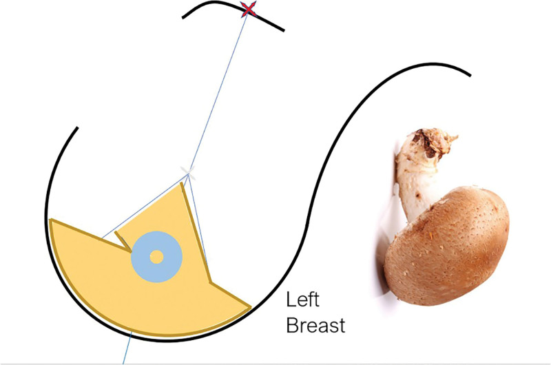
Fig. 2. Schematic illustration of the mushroom flap in the left breast.左乳蘑菇型皮瓣示意图。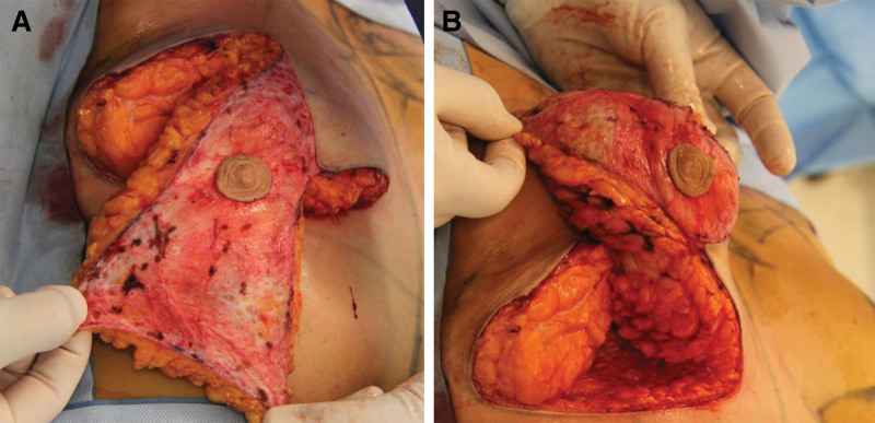
Fig. 3. Intraoperative view of the mushroom flap and its rotation in a right breast. A, Position after dissection. B, Position in the pocket, showing the rotation.右乳蘑菇皮瓣解剖与旋转术中观(A 解剖后,B 入腔旋转)。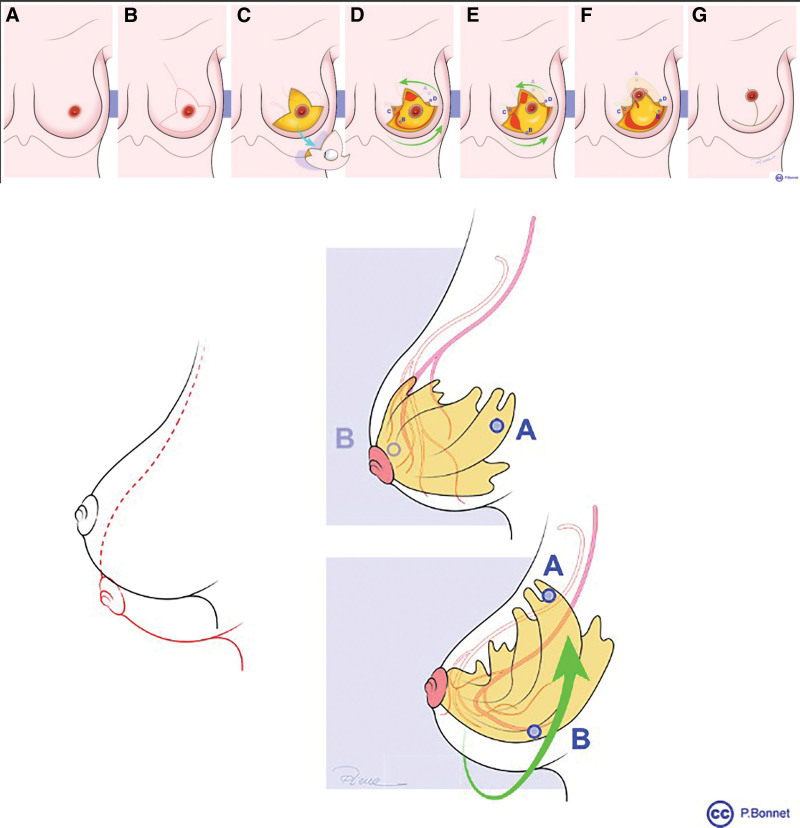
Fig. 4. Schematic illustration of the surgical technique of the mushroom auto-augmentation flap mastopexy. A, Left breast without marking. B, Preoperative markings with the future point of the areolar complex. C, De-epithelialization of the marked area. D, Incision of the flap and creation of the upper pole pocket. E, Rotation of the flap. F, Flap in place in the upper pole. G, Closure with sutures. Note: The schematic drawings do not display exact distances and metrics.蘑菇型自体增强固定术分步示意图(标记至缝合)。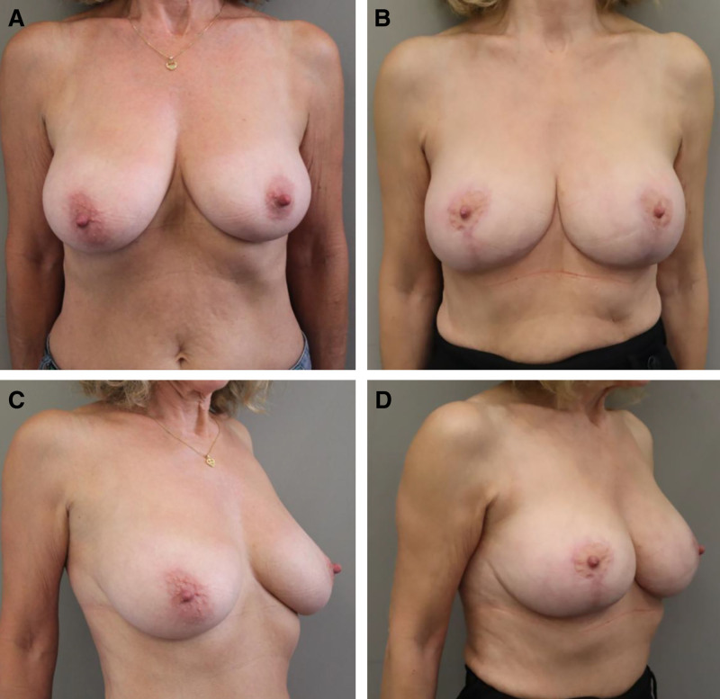
Fig. 5. Preoperative and 10-month postoperative photographs of a woman who underwent bilateral inverted-T mastopexy using the mushroom auto-augmentation flap (frontal view before [A] and after [B], and three-quarter view before [C] and after [D]).双侧倒 T 固定术术前与术后 10 个月(正面及四分之三位)。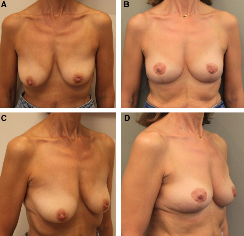
Fig. 6. Preoperative and 12-month postoperative photographs of a woman who underwent bilateral inverted-T mastopexy using the mushroom auto-augmentation flap (frontal view before [A] and after [B], and three-quarter view before [C] and after [D]).双侧倒 T 固定术术前与术后 12 个月。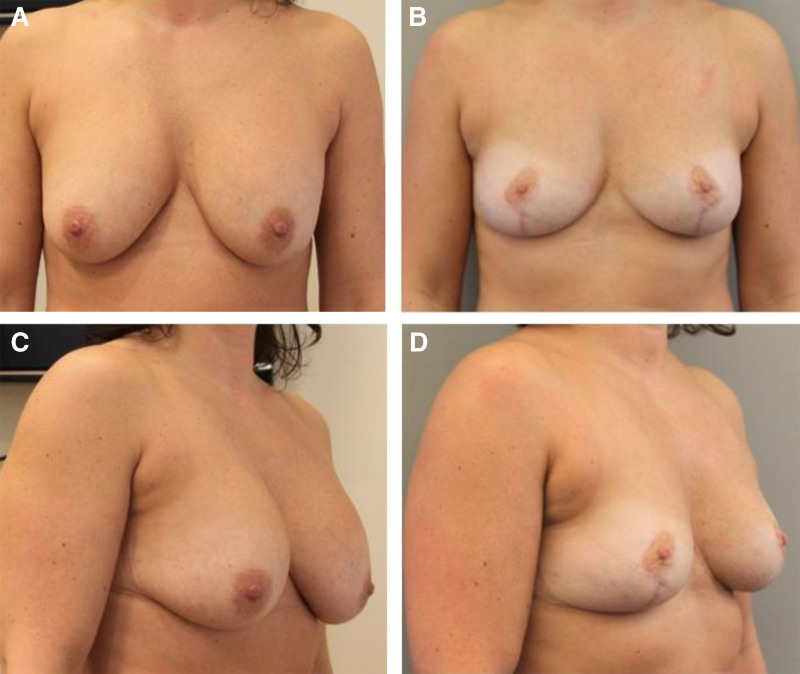
Fig. 7. Preoperative and 12-month postoperative photographs of a patient who underwent bilateral inverted-T mastopexy with explantation of ruptured 280-cc prepectoral implants, combined with mushroom flap auto-augmentation and fat grafting of 130 cc to the right breast and 100 cc to the left breast (frontal view before [A] and after [B], and three-quarter view before [C] and after [D]).取出破裂假体并联合蘑菇皮瓣与脂肪移植,术前与术后 12 个月。补充视频 / 附件(原文未随文提供原图,可于出版方页面查看)gox-14-e7774-s001.mp4Video 1 which shows the incision lines.补充视频 / 附件(原文未随文提供原图,可于出版方页面查看)gox-14-e7774-s002.mp4Video 2 shows the harvesting of the supero-medial and deep pedicle.补充视频 / 附件(原文未随文提供原图,可于出版方页面查看)gox-14-e7774-s003.mp4Video 3 shows the dissection of the pocket for the auto-augmentation flap, the rotation of the flap and the deep sutures.补充视频 / 附件(原文未随文提供原图,可于出版方页面查看)gox-14-e7774-s004.mp4Video 4 shows the neo-areola marking and the superficial sutures with the final result.
A total of 206 patients underwent surgery from January 2018 to December 2023; however, 8 patients were excluded: 1 with ptosis due to tuberous breast deformity, 1 with Poland syndrome, and 6 with less than 1 month of follow-up. In total, 198 patients were included in the study, including 33 unilateral mastopexies and 165 bilateral mastopexies. Patient ages ranged from 18 to 74 years (the mean age was 44.3 y), and weights ranged from 45 to 124 kg (mean weight was 64.5 kg). The mean BMI was 23.9 (range 17.23–40.48 kg/m 2 ). Twenty-eight patients were active smokers but were told to quit smoking 1 month before the surgery. Twenty patients were postbariatric patients with massive weight loss (>20 kg). The patients were subdivided into 6 groups: 135 underwent mushroom mastopexy alone, 25 underwent mastopexy with fat grafting, 2 underwent mastopexy with implant and fat grafting, 1 underwent mastopexy with implant left in situ, 22 underwent mastopexy with implant removal, and 13 underwent mastopexy with implant removal and fat grafting. The patient characteristics are summarized in Supplemental Digital Content 1. ( See table, Supplemental Digital Content 1 , which shows patient characteristics, https://links.lww.com/PRSGO/E900.) The mean clavicle-to-NAC distance preoperatively was 25.68 cm. Follow-up (from the date of operation to the last consultation in the name of the referring surgeon) ranged from 1 to 66 month (average 12.45 mo). Due to the moderate resection of soft tissue (<300 g/breast) and the relatively low occurrence of seroma or hematoma, we do not use drains for this procedure. There was 1 major complication with a hematoma that needed an urgent reoperation (0.5%). Six (3%) patients required antibiotics for wound infection, with no need for surgical drainage. Eighteen (9%) patients experienced delayed wound healing, requiring dressings (either Iso-Betadine, Iruxol, or Flaminal). The most common area of dehiscence was at the upper and lower junction areas of the vertical scar. All cases resolved without further morbidity. There was 1 hypertrophic scar that required corticosteroid injection into the scar. There was no NAC necrosis reported in this study (Table 1 ). Seventeen (8.58%) patients underwent an aesthetic reintervention for scar correction. The scar corrections were all under local anesthetic. No signs of breast cancer were observed in the final pathology when tissue was removed. There was no statistically significant difference between the different surgical groups and the complication rate ( P < 0.005). ( See table, Supplemental Digital Content 2 , which shows the test of the association between the type of operation and the presence of each of the complications, https://links.lww.com/PRSGO/E901.) Ptosis correction was satisfactory in every case, both in the patients’ opinions and in the author’s assessment. The secondary ptosis observed at 6 months postoperatively in most patients (n = 148, 74%) who undergo traditional mastopexy has not been observed in this series.
Table 1. Complications Following Auto-augmentation Mushroom Mastopexy (n = 198 Patients)
| Complications | No. Patients, n (%) |
|---|---|
| Hematoma | 1 (0.5) |
| Hypertrophic scarring | 1 (0.5) |
| Local infection | 6 (3) |
| NAC ischemia | 0 |
| Wound dehiscence | 18 (9) |
Many surgeons have focused on enhancing the upper pole of the breast to restore a more youthful appearance using autologous tissue transfer, as reported in recent studies. 6 – 9 The autologous inferior-based flap technique was first described by Ribeiro in 1975. 10 This method involves relocating excess breast tissue from 1 area to a region with insufficient volume, with the transplanted tissue serving as an autoprosthesis and facilitating proper blood flow to the lower part of the breast. Hammond and O’Connor 11 introduced a circumvertical mastopexy technique with auto-augmentation of the upper pole, using a lower island flap transposition. In this procedure, both the periareolar and vertical skin segments are de-epithelialized, and a vertical incision is made through the skin to access the breast parenchyma. The flap is then transposed superiorly to augment the upper breast pole. 11 Raafat et al 12 and Calobrace and Gabriel 13 have demonstrated improved outcomes by incorporating fat grafting into the procedure. Also, many surgeons are of the opinion that mastopexy with simultaneous augmentation can be performed safely and reliably. 14
Anchoring remains a key procedure in auto-augmentation mammoplasty, as the flap must remain stable against gravity. We believe that maintaining the pedicle with the 2 pillars joined together provides a strong base for the pedicle when it is raised and prevents the pedicle from descending and the loss of volume in the upper pole. The principle is the same as in breast reduction surgery.
Ultimately, the blood supply remains very robust. The centrally based pedicle, with its preserved breast septum, receives its blood supply directly from the fourth, fifth, and sixth intercostal perforating vessels of the internal mammary arteries, together with vascular contributions from the lateral thoracic artery and thoracoacromial artery by means of the pectoralis major muscle. 15 Concerning the blood supply of the NAC, we performed only a superomedial and profond-based pedicle, resulting in a very satisfying projection. This technique preserves the entire volume of the breast and relocates it mainly to the upper pole. The plane of detachment and the scar caused by the relocation of the lower breast segment to the upper pole allow for a stable result over time.
This technique is also effective when removing implants after several years. Combining it with lipofilling allows a certain volume to be maintained without implants.
As a limitation of this study, we have no long-term outcomes (>2 y) to observe potential deformation of the new shape, as well as questionnaire-based assessment regarding upper-pole fullness. We are nevertheless adding 1 of our 5-year postoperative follow-up photographs, although it was not included in this retrospective series, as it was taken before this series. ( See figure, Supplemental Digital Content 3 , which shows preoperative 12-mo and 5 y postoperative photographs of a patient who underwent bilateral inverted-T mastopexy, front and three-quarter photography, https://links.lww.com/PRSGO/E902.) In the future, it would be interesting to compare the stability of the shape with other techniques, both through photographs and other technical possibilities such as breast surface 3D imaging systems. A general drawback of all kinds of inverted-T designs remains the abundant presence of scars. 16 However, we believe this kind of scar pattern is the most promising in breast shaping and modeling; therefore, we used this technique for all grades of ptosis and skin elasticity.
Auto-augmentation mastopexy represents a sophisticated surgical option for patients aiming to enhance both breast volume and contour. This study illustrates the application of the mushroom flap technique, which can be effectively and safely combined with volume-enhancing procedures such as implant augmentation or autologous fat transfer.
The authors have no financial interest to declare in relation to the content of this article.
The authors thank Prof. Bonnet for his contribution to the preparation of the manuscript and the creation of the drawings.
All procedures performed in studies involving human participants were in accordance with the ethical standards of the institutional research committee and with the 1964 Declaration of Helsinki and its later amendments or comparable ethical standards.
声明:中文精读 · 仅供学术参考。内容来自公开文献检索,不代表本人观点,不构成诊疗建议。 医疗美容需在正规医疗机构由执业医师实施。
基于相同主题推荐 · 继续深挖
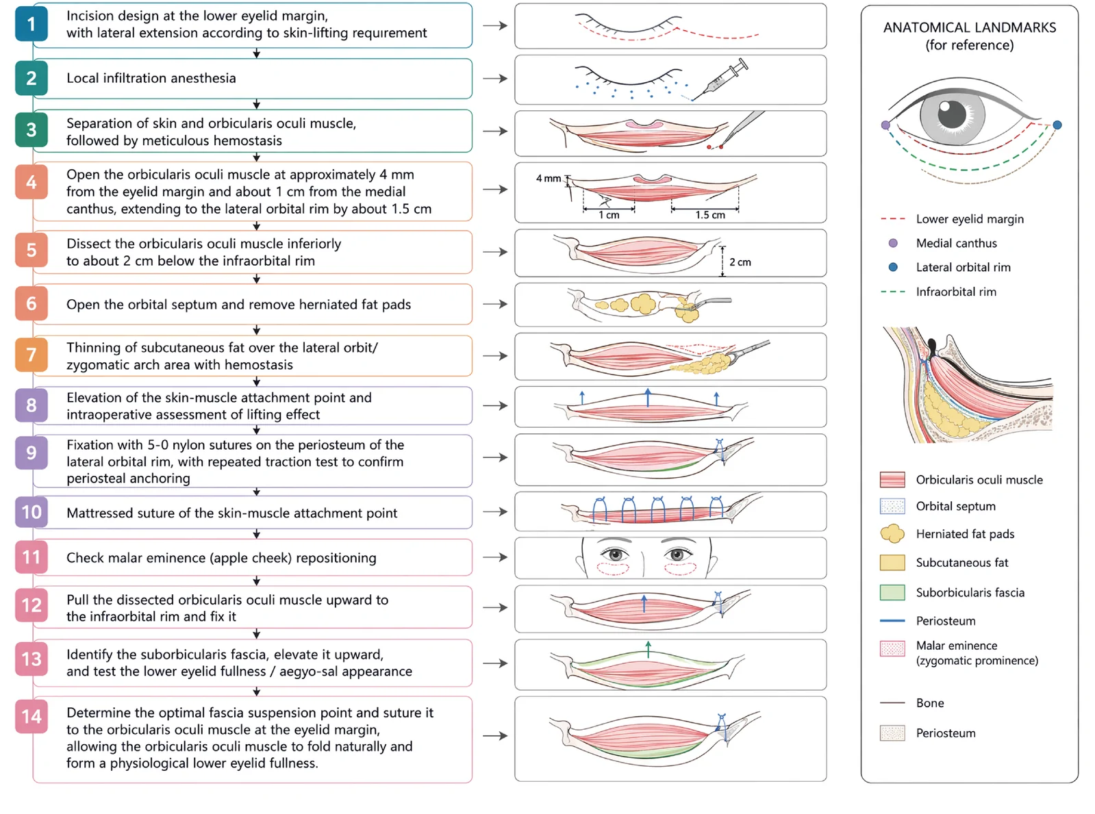
V3中面部提升经睫毛下切口,沿内眦线、外眦线、颧脂肪垫下界三个向量悬吊,可同时改善中面部下垂、下睑袋及睑颊轮廓不连续。
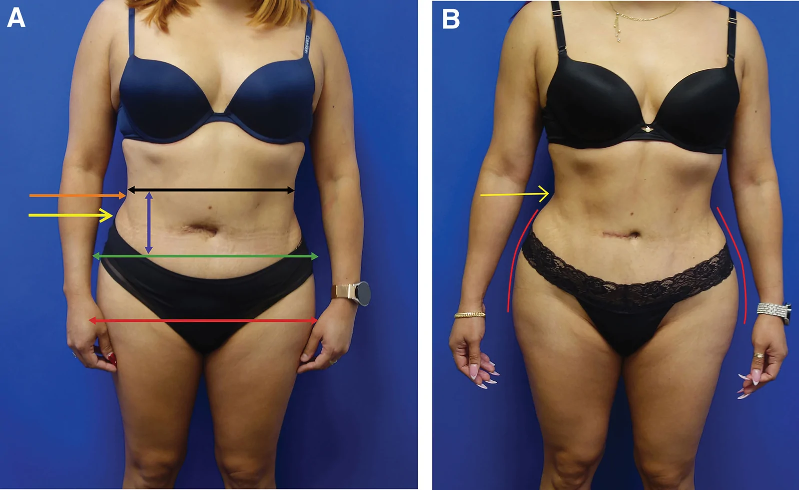
回顾性大样本病例系列(2016年11月至2025年1月),共1999例连续手术,中位年龄36岁(IQR 31至42),中位BMI 29(IQR 26至32),最短随访6个月。
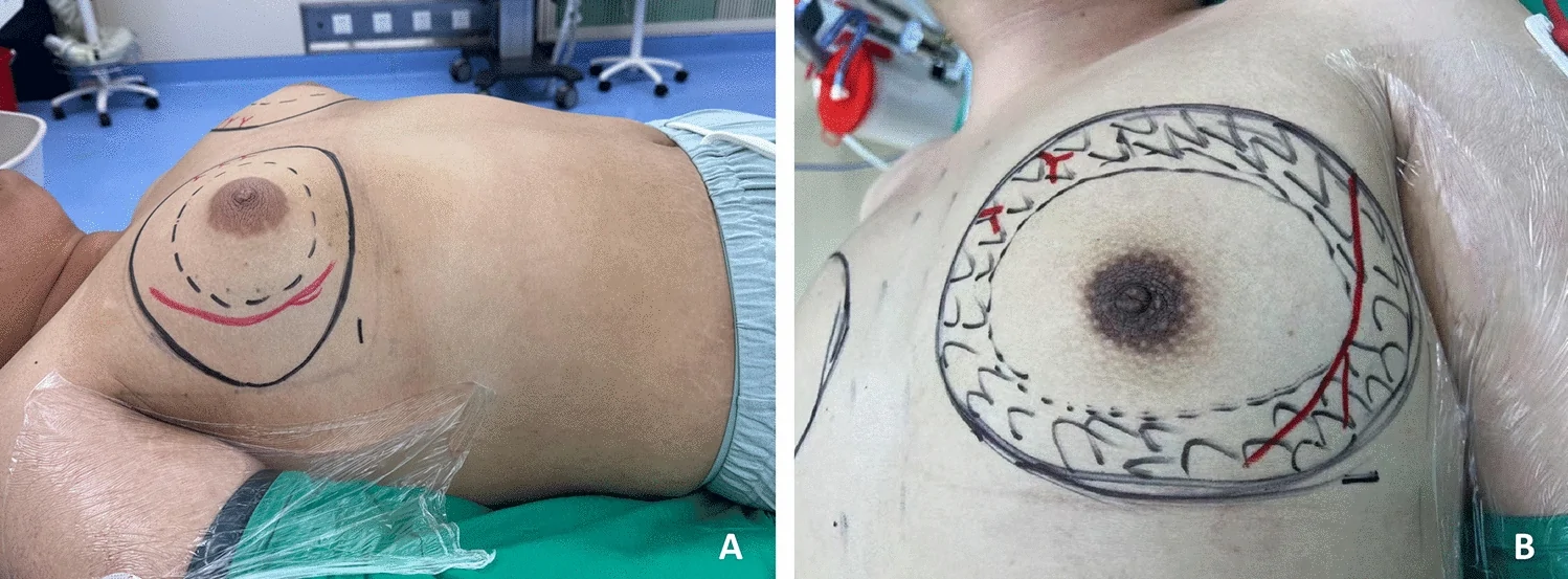
单中心、单臂前瞻性病例系列(证据等级 III),纳入 2024 年河北医科大学第一医院 786 例男性乳腺发育内镜腺体切除术患者(773 例双侧、13 例单侧),平均随访 96.5