无创微柱脂肪注射在 1362 例中的技术演进:聚焦维持时间与轮廓塑形
2026年10月10日16 min read
Plastic and Reconstructive Surgery Global Open
Plastic and Reconstructive Surgery Global Open
结论与实践要点
本文为单作者技术经验总结(非对照研究),报告 1362 例采用“无创微柱”脂肪注射进行面部轮廓塑形的临床实践。技术要点为:以 17–20 G 钝侧孔套管,在每一新的血管化层次单次通过,每针沉积 0.025–0.05 mL 脂肪,形成细条状均匀填充以减少团块与吸收不均;注射前于受区预注射血管收缩剂并勾画亚单位。文中以颏部、上睑沟等病例照片(随访 8 个月至 4 年)展示容积维持情况。
局限显著:属回顾性技术论述,缺乏随机对照与标准化容积保留率测量,所谓“持久”主要依据个案影像与术者主观判断,脂肪吸收存在个体差异且难以预估;样本量大但为单中心单术者经验,推广需谨慎。该技术对上睑沟、睑颊交界等精细区域有参考价值,临床应重视术前亚单位规划与适度保守注射,避免过度矫正,并向求美者说明吸收与可能需要补充注射的边界。
Background:The field of regenerative surgery has been revolutionized by minimally invasive fat injection methods. Over the past decades, techniques have evolved significantly. However, concerns regarding long-term volume retention and creating the desired result remain a challenge for many surgeons.
Methods:Our experience with 1362 patients has led to the development of a refined microfat injection technique that achieves durable facial contouring with a single treatment. Our method eliminates the need for overinjection, reducing the risks of contour irregularities and unpredictable fat resorption. The technique is knife-free, minimizing scarring and improving patient satisfaction.
Results:Meticulous preoperative planning is paramount. Targeted areas are drawn on the face and delineated in subunits. These recipient sites undergo preinjection with a vasoconstrictive agent, mirroring the planned fat volume. This allows intraoperative assessment and precise volume refinement. Fat is harvested, processed, and injected in microcolumns using blunt side-port cannulas ranging from 17 to 20 G cannulas, with each pass depositing 0.025 to 0.05 mL in a new vascularized plane. This systematic microcolumnar deposition of small threads of fat ensures uniform augmentation, optimal vascularization, and long-term volume preservation.
Conclusions:This technique is particularly advantageous to delicate facial areas requiring high precision, including the upper eyelid sulcus, lower eyelid/lid–cheek junction, and creating refined chin definition with augmentation. By emphasizing a precise fat placement technique, we achieve natural, long-lasting results. This article presented the rationale, evolution, and technical details of our microfat injection approach, highlighting its special applications with safe, enhanced, and predictable outcomes in facial contouring.
Question: Can a precise, knifeless microfat injection technique provide durable and natural facial contouring with a single treatment?
Findings: A systematic microcolumn injection method eliminates the need for overinjection, enhances vascularization, and minimizes fat resorption. Preinjection with a vasoconstrictive agent optimizes volume assessment, whereas blunt cannula placement in vascularized planes ensures uniform augmentation.
Meaning: This refined fat grafting approach offers long-lasting, predictable results with minimal invasiveness, making it particularly effective for delicate areas like the upper eyelid sulcus, lid–cheek junction, and chin augmentation.
Fat injection techniques provide a minimally invasive means of volume enhancement and contour refinement, revolutionizing the field of regenerative surgery. 1 It may be used to treat asymmetries or volume loss that may have resulted from aging, trauma, or congenital anomalies. 1 It is also regenerative and can improve the appearance of skin by softening the appearance of scars and burns while also stimulating antiaging effects in the skin dermis. 1 Fat grafting techniques may vary but always consist of several key steps: harvest, processing, and engraftment. 2
Coleman, Khouri, and many others have contributed significantly to the field of regenerative medicine surgery. 3 – 9 Coleman should be credited for bringing fat injections to the forefront through the Coleman technique, which standardized the processes of fat harvesting, refinement, and placement of fat grafts. 10 Fat grafts now yield a higher number of viable adipocytes and maintain better cellular function, resulting in more reliable and consistent clinical outcomes. 11
The evolution of our method has been informed by the treatment of 1362 patients who underwent fat injections to the face. Our primary goal has been to achieve optimal facial contour and volume retention over years with a single treatment. There is no need for overinjection with our method, which can lead to fat globules that are more prone to central necrosis due to insufficient vascularization. 12 A defining aspect of our approach has also been the elimination of scalpels, which are sometimes used to create an entry site for both harvesting and injection cannulas, thus reducing the risk of visible scarring. The purpose of this study was to describe our knifeless microcolumn fat grafting technique based on the senior author’s experience with more than 1300 patients.
The role of standardized photography in plastic surgery is well established and is used in examination and follow-up of our patients. 13 , 14 In the context of fat injection, high-quality preoperative and postoperative images provide critical documentation, guide surgical planning, and allow for long-term follow-up to assess volume retention. Additionally, photography offers insight into skin quality changes following fat injection, an area of growing interest, given that superficial injections have demonstrated potential improvements in dermal texture. 5 Anecdotally, we have also noted improvement of skin quality in our patients.
Multiple precautions are taken to optimize safety and minimize complications. A vasoconstrictive agent is preinjected into the targeted areas planned for fat injection at least 10 minutes before fat injection. In the periorbital region, 0.25% Xylocaine with 1:400,000 epinephrine is used; for all other facial areas, 0.125% Xylocaine with 1:400,000 epinephrine is used. The volume of vasoconstrictive agent administered matches the anticipated fat graft volume to facilitate accurate intraoperative assessment. This step serves to reduce the risk of fat embolization, minimize intraoperative bleeding, and mitigate postoperative ecchymosis, thereby promoting a faster recovery.
Infections have not been observed in our series, a finding we attribute to meticulous adherence to sterile technique, pretreatment with intravenous antibiotics, and the inherently robust vascular supply of the face.
Meticulous preoperative planning is paramount. The areas targeted for augmentation are delineated into aesthetic subunits, allowing precise control over the final shape. A facial diagram is used to document the planned injection volumes for each subunit, which serves as a surgical guide in the operating room and a reference for postoperative assessment. ( See Video [online] , which demonstrates the knifeless microcolumn fat grafting technique in a patient with a secondary cleft lip deformity and facial contour deformity secondary to a left partial maxillectomy.)
Recipient sites undergo preinjection with the vasoconstrictive agents described earlier, mirroring the planned fat volume. This step allows for an intraoperative assessment of volume distribution, providing an opportunity to refine injection amounts as needed. This step in our method allows surgeons to determine how much fat to inject in each area, which is a commonly asked question.
The facial aesthetic subunit diagraming is another key feature of our method. The diagram is updated intraoperatively to record precise injection volumes, a crucial component of the surgical record that informs long-term follow-up and refines the surgeon’s ability to estimate ideal injection volumes.
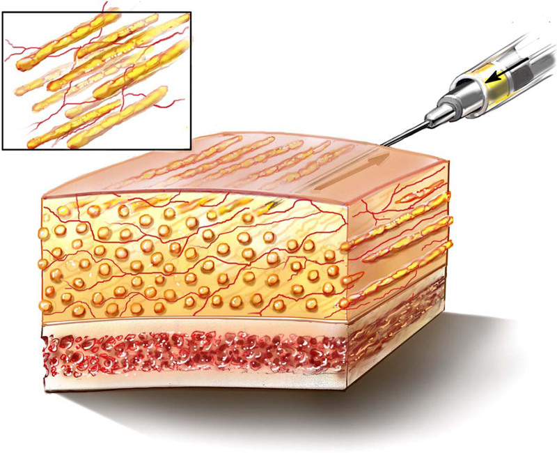
Precise injection technique is essential for achieving durable, natural-appearing results. Overinjection is deliberately avoided, as we do not anticipate volume loss over time with our method. Fat is injected using blunt side-port cannulas in microquantities ranging from 0.025 to 0.05 mL per pass with a 1 mL Luer lock syringe. The 20 G cannulas are used for the periorbital area, whereas 19G, 18 G, and 17 G are used in other facial areas. The microcolumnar deposition ensures adequate vascularization of each fat thread, reducing the risk of resorption and contour irregularities. 12 , 15 Each pass is carefully placed in a distinct tract, ensuring resistance is felt with each cannula passage to confirm the placement of fat in a new vascularized plane (Fig. 1 ). With our method, systematic and deliberate injections are performed in a consistent manner, either from left to right or right to left, ensuring uniform augmentation and predictable aesthetic outcomes. Random injections into the recipient areas are strictly avoided.
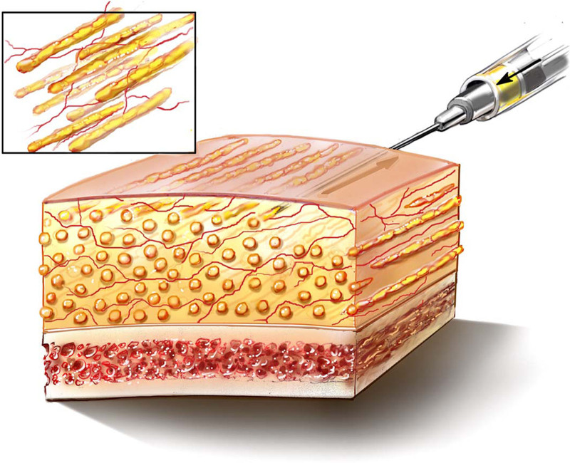
Fig. 1. Fat injection technique. The cannula is passed once in each area to ensure that the thread of fat graft is well vascularized and does not form into a collection of fat.脂肪注射技术:每区单次通过以保证脂肪条血管化。As the fat is injected, the facial diagram is updated to record the exact amount of fat injected into each specific area. This is a key portion of the operative record as it is relied upon as the patient is followed up postoperatively. This also helps to train the surgeon’s eye to determine the amount of fat to inject in subsequent patients. Fat grafting is extremely technique-dependent, and each surgeon should determine the success of their individual technique by following their long-term results.
补充视频 / 附件(原文未随文提供原图,可于出版方页面查看)gox-13-e7118-s001.mp4Video 1. This video demonstrates the knifeless micro-column fat grafting technique in a patient with a secondary cleft lip deformity and facial contour deformity secondary to a left partial maxillectomy.
Fat donor site selection significantly impacts injection feasibility and long-term graft retention. 16 The medial thighs are preferred, given the superior longevity we have had of the harvested fat and its compatibility with the small cannulas used in this method. If additional volume is required, the flanks serve as an alternative source, whereas the abdomen is avoided due to variability in fat cell consistency and a higher tendency to clog fine cannulas. 17 Donor sites are preinfiltrated with 0.125% of Xylocaine 1:800,000 epinephrine to vasoconstrict before harvesting.
Our “smooth flow” (Micrins, Renaissance Surgical, Inc. Lake Forest, IL) harvesting cannulas with a proprietary internal and external finish are 2 mm in diameter and have multiple 18 G openings. We have designed them to harvest viable fat cells that can be easily injected through our 20 G cannulas for use in the upper and lower eyelid and periorbital areas, decreasing the risks of irregularities. We have not observed lumps or bumps in our patients using this method and this instrumentation.
Fat is never centrifuged but rather is carefully removed from the 10-mL syringe through the large end and rolled on Telfa nonadherent gauze to remove all the oils and tumescent injection that is found in the aspirate. Thus, the fat that is subsequently loaded into the 1-mL syringes is concentrated, which is another reason to not over inject.
The microcolumn regenerative microfat injection technique has broad applications, particularly in areas that demand high precision, such as the deep sulcus of the upper eyelid, the lower eyelid and lid–cheek junction, the contiguous cheek region, and refined chin contouring. Each of these areas presents unique anatomical challenges that require an exacting and meticulous technique.
Fat injections for chin augmentation provide enhanced definition, increased projection, and vertical height, while also refining contour irregularities (Fig. 2 ). In our experience, optimal results are achieved with volumes typically not exceeding 10 mL. A vasoconstrictive agent is preinjected to predict volume and shape before fat grafting. Using a 17-G blunt cannula, microcolumn threads of fat are placed at multiple depths, from the periosteal layer to the subcutaneous plane. Long-term photographic follow-up has demonstrated stable volume retention and continued skin quality improvement.
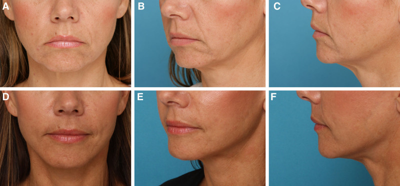
Fig. 2. Chin enhancement. Preoperative (A–C) and 3-year postoperative (D–F) images of a 48-year-old woman who underwent one treatment of microcolumn fat injection (7 mL in the chin/perimental region) in combination with a rhytidectomy. Postoperative images demonstrate fat volume retention, increased projection and vertical height, and smoother contours.颏部增强:48 岁女性单次微柱注射(7 mL)联合除皱术,术后 3 年。
Aging or congenital volume deficiency in the upper eyelid sulcus often results in a hollowed appearance (Fig. 3 ). In cases where brow lift surgery is planned, uncovering the upper sulcus can further accentuate this hollowness. Fat grafting in this region effectively restores volume while maintaining a natural, youthful contour.
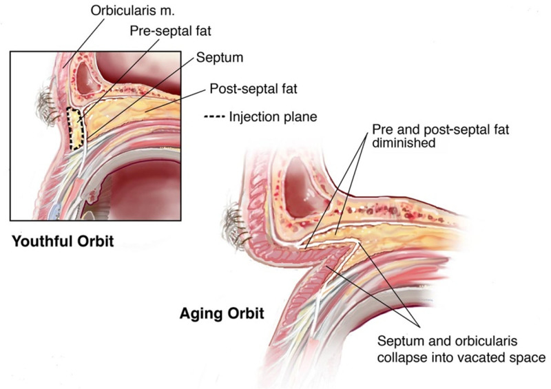
Fig. 3. In the aging orbit, diminished pre- and postseptal fat contributes to the hollow upper eyelid sulcus.老化眼眶:眶隔前后脂肪减少致上睑沟凹陷。Intraoperative assessment following vasoconstrictive preinjection allows for final volume adjustments, ensuring optimal contour restoration with a single treatment (Fig. 4 ). We use a 20-G blunt cannula for preseptal injections through medial and lateral brow access points. The injection is performed in the preseptal space, a placement confirmed in previous retrospective studies. 18 , 19 Our own cadaveric studies have informed our surgical technique, 20 and our studies in the orbit have verified that the fat is placed in this preseptal space using our method. Figure 5 demonstrates these results at 1 and 4 years postoperatively in a 44-year-old patient following 2-mL fat injection into the upper eyelid sulcus bilaterally performed in conjunction with an endoscopic brow lift and forehead rejuvenation.
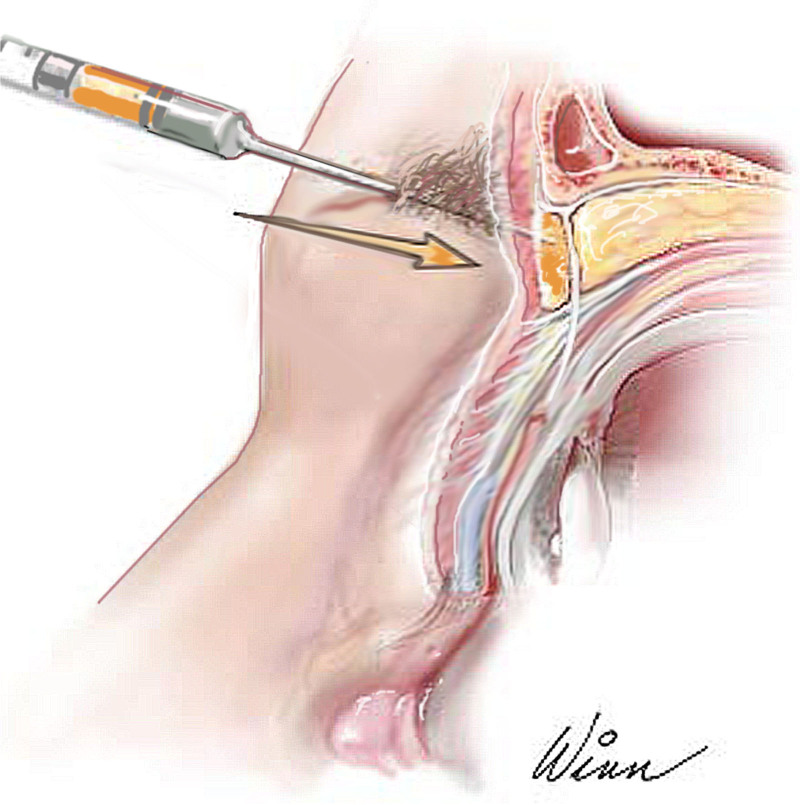
Fig. 4. Preseptal microfat injection using the knifeless method.经无创法行睑前间隙微脂肪注射。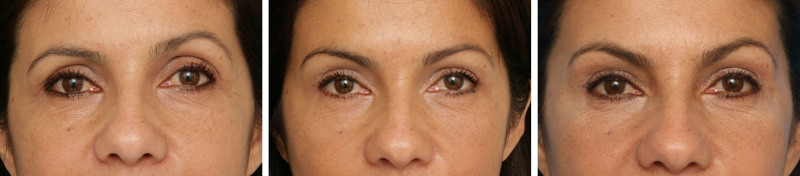
Fig. 5. A 44-year-old patient who underwent an endoscopic brow lift with a bilateral 2-mL microcolumn fat injection to the upper eyelid sulcus. Postoperative images at 1 and 4 years demonstrate sustained fat retention following a single treatment.44 岁患者内镜眉提升联合双侧 2 mL 上睑沟微柱注射,术后 1 与 4 年。
Precise correction of lower eyelid hollowness, tear trough deformities, and cheek volume deficiencies is critical in facial rejuvenation. Although our previous technique for eyelid and midface rejuvenation through extended blepharoplasty effectively addressed fat compartment abnormalities and orbitomalar lid–cheek junction deformities, it was limited by the challenge of determining the optimal volume of fat resection. 21 Marking the treatment area preoperatively guides fat placement to harmonize facial proportions. A vasoconstrictive preinjection equal to the planned fat volume is administered at least 10 minutes before grafting, allowing for anatomic assessment and reduction of procedural risks. The fat is deposited in microcolumns/threads at the periosteal level via a 20-G blunt cannula. Strict adherence to the microcolumn technique ensures even distribution and prevents overinjection, thereby creating a natural contour addressing asymmetries (Fig. 6 ).
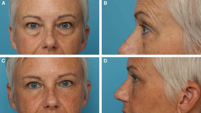
Fig. 6. Periorbital rejuvenation. Preoperative (A and B) and 8 months postoperative (A and D) images of a 56-year-old woman with lower eyelid microcolumn fat injection in addition to an upper and lower blepharoplasty and forehead rejuvenation.56 岁女性下睑微柱注射联合上下睑整形与额部年轻化,术前与术后 8 个月。
Our technique incorporates distinct innovations in cannula size, fat processing, injection volume per pass, and a knifeless approach for cannula insertion. Depth of fat injection varies significantly with other established techniques. Our method places injections in the orbital region at the periosteal level and not subcutaneously. In the nasolabial area, we do go subcutaneously to avoid the angular vasculature that is in the intermediate level. In the posterior mandible, we inject deep to and into the masseter muscle along the angle, whereas in the anterior mandible, we inject at the periosteal level. Although our experience with this technique has been successful, consideration of other potential alternative fat harvesting techniques is warranted. For example, some authors have isolated and added the stromal vascular fraction (SVF), a heterogenous cell population derived from adipose tissue, to their fat grafts. 22 The addition of SVF-enriched fat grafts has been shown to significantly improve survival rates of fat grafts, potentially due to the angiogenic and adipogenic properties of SVF. 23 However, isolation of SVF via enzymatic or mechanical methods may be expensive and time-consuming. 24 Furthermore, there are currently no standardized protocols for SVF isolation and application, posing challenges for consistent clinical outcomes.
The advent of regenerative fat grafting has significantly expanded the capabilities of plastic surgery, offering long-term volume restoration with minimal invasiveness. Our knifeless microcolumn technique has proven highly effective in achieving optimal contour and durability, with outcomes that endure over time. Critical elements of this approach include preinjection of a vasoconstrictive agent to minimize risks, meticulous surgical planning with standardized photography, and precise microthread injection techniques. Our experience with over 1300 patients has consistently demonstrated reliable, aesthetically superior outcomes, reinforcing the value of this method in the evolving landscape of fat grafting.
The authors have no financial interest to declare in relation to the content of this article.
Patients provided written consent for the use of their images.
The authors express their deepest gratitude to the surgeons at Bach Mai Hospital in Hanoi, Vietnam, as well as Nuoy Reconstructive International for their collaboration and dedication to this project.
声明:中文精读 · 仅供学术参考。内容来自公开文献检索,不代表本人观点,不构成诊疗建议。 医疗美容需在正规医疗机构由执业医师实施。
基于相同主题推荐 · 继续深挖
微柱脂肪注射以钝针分层微量沉积,单根脂肪条充分血管化

这项前瞻性、开放标签、随机、多中心(德国11个中心)上市后研究纳入121名受试者,按1:1随机接受 Belotero Volume(CPM-V,n=58)或含利多卡因的 CPM-V
回顾性研究,纳入49例下颌后缩2.5–6 mm的初次隆鼻患者