1470nm激光辅助吸脂治疗男性乳房发育的审美结局与并发症分析
2026年10月10日24 min read
Plastic and Reconstructive Surgery Global Open
Plastic and Reconstructive Surgery Global Open
结论与实践要点
本研究为一项前瞻性、单盲随机对照试验,纳入30例按Simon标准分型的男性乳房发育患者,随机分为常规吸脂组(n=15)与1470nm激光辅助吸脂(LAL)组(n=15),以BREAST-Q量表、视觉模拟评分(VAS)及医师美学评估为主要结局。结果显示,LAL组BREAST-Q评分与美学改善程度均高于常规吸脂组(P<0.005、P<0.001),术后疼痛VAS更低(P<0.001);常规吸脂组血肿与皮肤回缩发生率更高(P=0.014、P<0.001)。超声随访提示乳腺下脂肪厚度在术后6个月内持续下降。研究提示LAL在该人群中具备可重复性,并可能减少部分术后并发症。
需注意本研究为单中心、小样本(每组15例)的前瞻性RCT,缺乏多中心验证,且LAL组手术操作时长更长(P=0.022),对术者经验有一定要求。文中提及的胶原收缩与组织收紧属术后在体观察推论,并非离体或体外机制验证。对于体积较大、触诊质硬的真性腺体增生,文中仍建议以皮下腺体切除为主要处理方式。临床决策应结合Simon分型、皮肤松弛程度及患者预期综合判断,相关长期稳定性仍需更大样本随访。
Background:Traditional surgical treatment for gynecomastia has multiple complications. Recently, energy-based therapies have gained popularity over conventional surgical treatments, including liposuction using radiofrequency energy, power-assisted liposuction, ultrasound-assisted liposuction, and laser-assisted lipolysis (LAL). This work aims to assess LAL efficacy and glandular tissue treatment using a 1470-nm laser for gynecomastia to correct breast volume, flaccidity, and skin tightening without its removal.
Methods:This prospective, single-blinded randomized controlled trial was conducted on 30 patients enrolled in the National Institute of Laser Enhanced Sciences outpatient clinic who had gynecomastia classified according to the Simon standard. The participants were randomly allocated to 2 groups: group 1 (n = 15) received conventional liposuction (suction-assisted liposuction), and group 2 received LAL (1470-nm diode laser) treatment. Patients were assessed using the BREAST-Q scale.
Results:All patients achieved satisfactory results. The BREAST-Q scores were significantly higher in group 2 compared with group I ( P < 0.005). Visual analog scale scores significantly decreased in group 2 ( P < 0.001) compared with group 1. Group 2 had significantly improved aesthetic outcomes compared with group 1 ( P < 0.001). In group 1, skin retraction and hematoma incidence were significantly greater than in group 2 ( P = 0.014, <0.001).
Conclusions:The LAL described is safe and reproducible. It showed a higher BREAST-Q value, a significantly better aesthetic outcome, and fewer postoperative complications.
Question: Is laser-assisted liposuction and therapy of glandular tissue with a 1470-nm laser effective in the treatment of gynecomastia?
Findings: Laser-assisted liposuction showed shorter operative time, less postoperative pain, higher BREAST-Q value, significantly better aesthetic outcomes, and fewer postoperative complications.
Meaning: Laser-assisted liposuction should be used more often in the treatment of gynecomastia.
Gynecomastia is a glandular, adipose, or combined condition. The condition is reported to affect up to 65% of men, ranging from moderate excess to over-feminization of the thorax. 1 Gynecomastia can be caused by hyperthyroidism, cirrhosis, hypogonadism, obesity, renal failure, HIV, and the use of specific therapeutic agents, such as anabolic steroids, cimetidine, chlorpromazine, diazepam, or illicit substances. 2 , 3 An increase in the estrogen-to-androgen balance is suggested to play an essential role. 4
The traditional surgical treatment for gynecomastia entails the abrupt excision of the mammary gland through a semicircular incision on the areola edge. 5 However, the cosmetic outcome of this method of surgery is frequently suboptimal as a result of complications such as scarring, “doughnut” deformities, nipple necrosis, nipple inversion, or loss of sensation in the nipple. 6
Recently, energy-based treatments have gained popularity over conventional surgical treatments. These treatments include power-assisted liposuction, ultrasound (US)-assisted liposuction, liposuction using radiofrequency energy, and laser-assisted lipolysis (LAL), providing the following advantages: outstanding patient tolerance, rapid recovery, and the supplementary advantage of cutaneous compression. 7
The recently introduced LAL method integrates thermal energies with liposuction. By using this new technique, it is possible to access the following objectives for gynecomastia correction surgery: stimulation of skin tightening, reduction of blood loss, and eradication of adipose and glandular tissue. Due to its liquefaction effect on adipocytes, LAL enhances subsequent aspiration and diminishes operator fatigue. In addition, it has a coagulative impact, which reduces trauma and hemorrhage. This reduces the risk of hemodynamic complications and enables the removal of a larger volume of fat, thereby improving the recovery time. 8 , 9
This investigation evaluates the efficacy of LAL and glandular tissue therapy with a 1470-nm laser in treating gynecomastia. The goal was to improve skin tightening, flaccidity, and breast volume without excision.
This prospective, single-blinded randomized controlled trial included 30 patients attending the outpatient clinic at the National Institute of Laser Enhanced Sciences, Cairo University, with bilateral gynecomastia. All patients provided written consent, were informed, and received a detailed explanation of the procedure. The study was conducted with the approval of the ethics committee at the National Institute of Laser Enhanced Sciences, Cairo University, Cairo, Egypt (NILES-EC-CU-23/1/4).
The gynecomastia patients included in the study were aged 18–45 years with grade IIa or IIb gynecomastia according to the Simon classification. 10 All of them were in generally good health, and they had no history of significant medical problems.The exclusion criteria were patients with gynecomastia grade III according to the Simon classification, with a body mass index higher than 35, and patients with breast masses. Routine laboratory investigations were also performed, as well as hormonal profile, to rule out any pathological causes of gynecomastia. The clinical details of all patients were obtained, including age, body mass index, chief complaints, and medical history.
The participants were randomly allocated to 2 groups (15 in each) on a 1:1 basis using a computer-generated randomization sequence. In group 1, patients were administered conventional liposuction or suction-assisted liposuction. In group 2, patients were administered LAL (using a 1470-nm diode laser). Sequentially numbered and sealed envelopes were used to conceal the allocation of patients. After the induction of anesthesia, the envelopes were transported to the operating room. A nurse not involved in patient care unsealed the envelope to assign the patient to the appropriate group. The evaluator was blinded to the group allocation when performing US, visual analog scale (VAS), or BREAST-Q assessments.
The patient was positioned in the supine position with arms abducted to 90 degrees. The breast tissue on both sides, along with the periphery, was infiltrated with Klein basic tumescent solution. 11 A 3 mm stab incision was made by scalpel in the mid axillary line midway between the areola and axilla and used as port for insertion of infiltration as well as suction cannula. Thirty minutes after tumescent infiltration, liposuction was initiated using a suction device with a negative pressure of 1 bar. Depending on the thickness of the adipose tissue layer, liposuction of the deeper layers was performed with a 4-mm Mercedes tip cannula, followed by superficial or fine-contouring liposuction using a 3-mm cannula. The entire breast, as well as its periphery, was treated using this technique. After liposuction, the residual fibroglandular tissue was extracted from the subareolar area via a circumareolar incision extending from the 3 o’clock to 9 o’clock position. A substantial thickness of subareolar tissue was left in place to maintain the blood supply as well as the contour of the nipple-areolar complex. After securing hemostasis, wounds were closed by using a 0/4 Proline suture with negative pressure drains in situ. Patients were advised to put on a front-closure compression vest as early after surgery as feasible for a minimum period of 3 months.
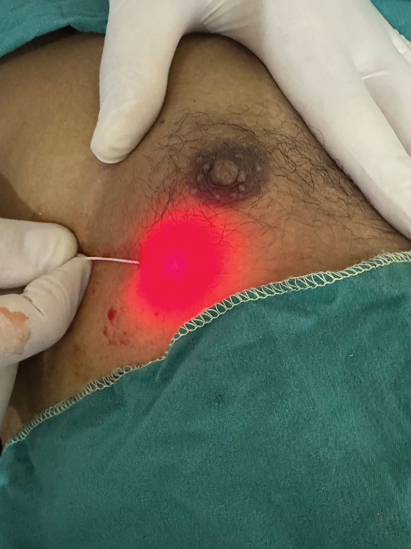
Treatment of the patients in this group included heating both the glandular and fat components of gynecomastia, followed by conventional liposuction. The lasering procedure was started 30 minutes after the administration of the same tumescent solution as used in the other treatment group. A 3-mm stab incision was made by an 11-blade scalpel in the mid-axillary line midway between the areola and axilla to introduce a special cannula 9 cm long and 1 mm in diameter with only a front opening to tunnel the treated area; then, we passed a 600-μm diameter fiber optic connected at its distal end to the 1470-nm diode laser (VELAS by GIGA Laser, Wuhan, China) through the same cannula (Fig. 1 ). Next, tunneling maneuvers were completed with the laser firing, allowing easy passage of the laser cannula in the breasts, whether soft or considerably firm.
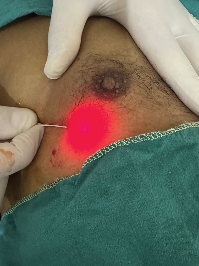
Fig. 1. Laser fiber optic of diode 1470 sub glandular for lipolysis.1470nm二极管激光光纤置于腺体下层面进行溶脂的操作示意。The operator denoted the site of the distal end of the fiber optic tip, which was placed 3 mm out of the cannula, with the red aiming beam visible within the breast tissue (Fig. 2 ). The lasering was done along these lines, with the operator using their nondominant had to control both the cannula speed of movement and depth. The deep plane was irradiated first, followed by the subdermal plane for effective dermal contraction and to ensure safety. The cannula was moved faster at this level so that the fiber tip was in contact with the subdermis for a short time to eliminate dermal burn. During the procedure, the operator could feel and hear the popcorn effect.
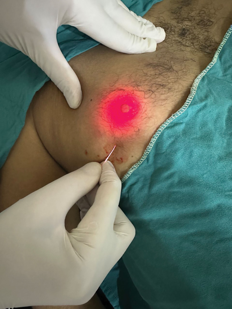
Fig. 2. Laser fiber optic of diode 1470 intraglandular for gland ablation.激光光纤进入腺体内对腺体进行消融的操作示意。The last area to be irradiated was glandular tissue and subglandular fat, which requires US monitoring. The operator moved the fiberoptic tip to cover almost the entire area of the glandular tissue. Immediately after the procedure, the US showed a narrow hypoechoic rim (0.3–0.5 cm) that extended around the glandular tissue, which was considered the end point of the lasering. Once the laser irradiation was finished, liposuction was performed in the exact same manner as in group 1 (Fig. 3 ).
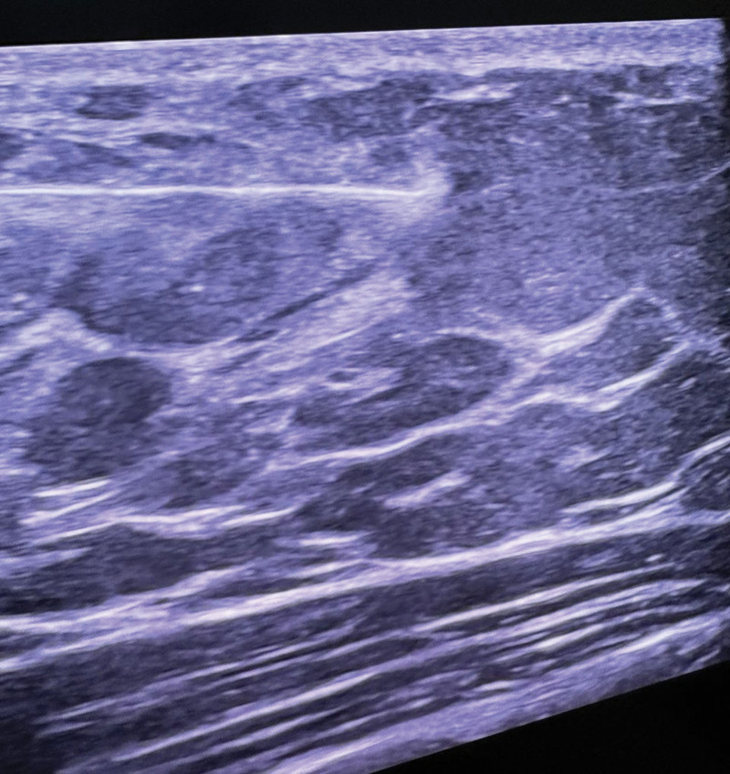
Fig. 3. Laser fiber optic diode 1470 by ultrasound for lipolysis.在超声引导下以1470nm激光光纤行溶脂。
The parameters used were as follows: 1470-nm wavelength, 8–10 W power with continuous emission, 600-μm optic fiber diameter. The total time depended on the gynecomastia volume, with the total energy delivered per patient bilaterally ranging from 800 to 1500 J. The clinical endpoint was softness felt by palpation, which indicated the proper liquefaction of the adipocytes.
An infrared thermometer was used to constantly monitor skin temperature externally. The temperature never exceeded 42°C. During the procedure, a cooling apparatus maintained a temperature of −10°C, ensuring the continuous flow of cold air at 600 L/min.
The efficacy of the treatment was evaluated at fixed regular assessments and follow-ups at 1, 3, and 6 months postoperatively. Clinical and operative data, including operative procedure duration, hospital stay length, and complication incidence, were evaluated to interpret the clinical results.
The pain was evaluated using a VAS, with the following categories: 0 to 24 (poor), 50–74 (good), 25–49 (average), and 75–100 (very good). 12 Additionally, the aesthetic outcomes were assessed by an independent, blinded plastic surgeon who evaluated the digital photographs. The following criteria were used to assess the breasts: overall shape, overall shape to the chest wall, nipple-areola position and shape, skin tension, residual skin and/or glandular excess, overresection, psychosocial alleviation, and scar quality. Each item was assigned a score of 0 (not accepted), 1 (accepted), or 2 (sufficient). The degree of improvement was used to grade the results as exceptional (>75%), good (51%–75%), middling (26%–50%), and poor (<25%).
The patient’s satisfaction was evaluated at each visit via a questionnaire, assessing the patient’s motivations for the surgery and satisfaction with the appearance of their breasts. Patient satisfaction was evaluated using a 5-point Likert scale (1 = extremely dissatisfied; 5 = extremely satisfied). 13 These criteria were derived from the BREAST-Q scale. 14 Hematomas, seromas, nipple-areola sensation deficit, and nipple-areola loss were classified as absent (0), mild (1), or moderate to severe (2) complications. Figure 4 shows gynecomastia before LAL and liposuction, and Figure 5 shows the results after LAL and liposuction.
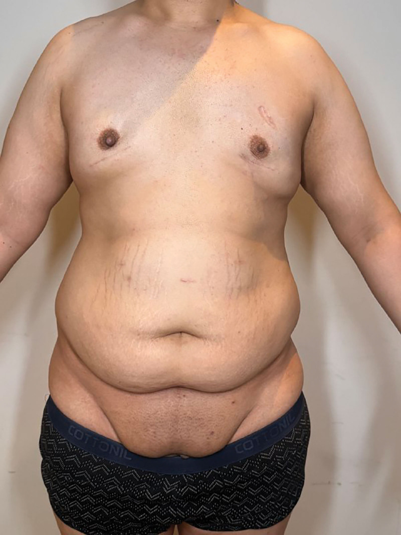
Fig. 4. Case 1: before LAL and liposuction for gynecomastia.病例1:激光辅助吸脂治疗男性乳房发育术前外观。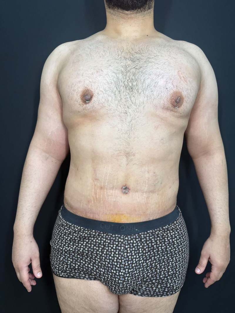
Fig. 5. Case 1: after LAL and liposuction for gynecomastia.病例1:治疗后外观,可见胸围与轮廓改善。
Ultrasonography using SonoSite MicroMaxx US equipment (SonoSite Inc.) with a high-frequency multilinear probe was performed for all patients in supine position: preoperatively, intraoperatively during surgery, postoperatively, and during follow-up. Figure 6 shows 2 dimensions of the gland before and after LAL where the decreased dimensions were observed. The before and after sonograms of the glandular tissue thickness were measured by a blinded radiologist.
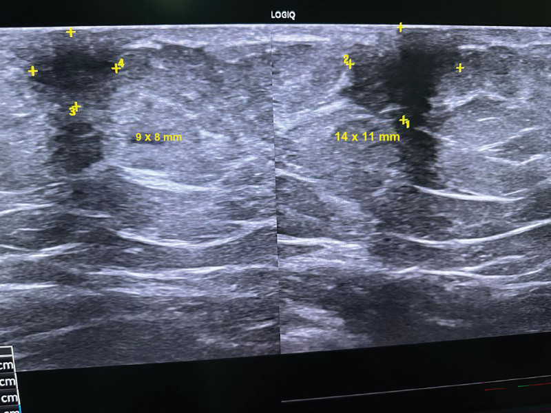
Fig. 6. US dimensions of the glandular element before and after LAL: right side before (14 × 11 mm) and left side after (9 × 8 mm).超声测量腺体成分:右侧术前14×11mm,左侧术后9×8mm。
The sample size calculation was performed using G*Power 3.1.9.2 (Universität Kiel, Germany). The sample size was calculated according to the subjective evaluation of all patients at the 6-month assessment point for determining an aesthetic standpoint, where 18 patients (64.3%) scored the result “very good” and 6, “good” (21.4%), according to a previous study. 12 Two cases were added to overcome dropout, based on the following considerations: an α error of 0.05, a study power of 80%, and an allocation ratio of 1:1. Therefore, 30 patients were allocated (with 15 patients in each group).
The statistical analysis was conducted using SPSS v28 (IBM, Armonk, NY). The Shapiro–Wilks test and histograms were used to evaluate the normality of the data distribution. Quantitative parametric data were analyzed using an unpaired Student t test and presented as the mean and SD. The Mann-Whitney test was implemented to evaluate quantitative nonparametric data, which were presented as median and interquartile range. Qualitative variables were analyzed using the χ 2 test or Fisher exact test, and they were presented as frequency and percentage (%) as applicable. A 2-tailed P value less than 0.05 was considered statistically significant.
The study assessed the eligibility of 59 patients; 22 patients were unable to meet the criteria, and 7 patients declined to participate. Furthermore, the remaining 30 patients were randomly assigned to one of the 2 groups, each consisting of 15 patients. Statistical analysis was conducted on all patients from the allocated group (Fig. 7 ).
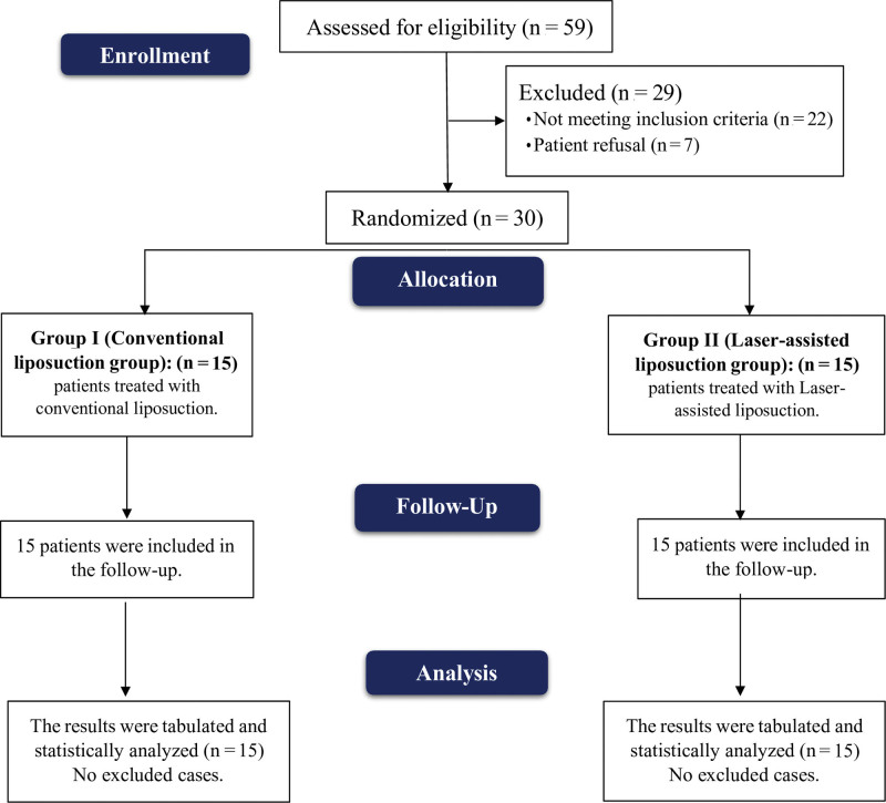
Fig. 7. CONSORT flowchart of the studied groups.本研究两组受试者的CONSORT随机分配流程图。The study groups differed insignificantly regarding patients’ characteristics and laboratory investigations (Table 1 ).
Table 1. Patient Characteristics and Laboratory Investigations of the Studied Groups (n = 30)
| Group 1 (n = 15) | Group 2 (n = 15) | P | |
|---|---|---|---|
| Age, y | 41.13 ± 12.74 | 37.8 ± 15.2 | 0.520 |
| Weight, kg | 73.6 ± 6.22 | 72.33 ± 6.3 | 0.582 |
| Height, m | 1.6 ± 0.07 | 1.61 ± 0.05 | 0.656 |
| BMI, kg/m 2 | 28.74 ± 2.96 | 27.96 ± 2.1 | 0.412 |
| Hb, g/dL | 12.2 ± 1.47 | 12.1 ± 1.03 | 0.881 |
| WBCs, ×10 9 /L | 6.89 ± 2.37 | 7.69 ± 1.87 | 0.313 |
| Platelets, ×10 9 /L | 289.6 ± 60.8 | 310.7 ± 62.1 | 0.335 |
No significant difference between the groups was observed regarding the morphology and volume of gynecomastia, adverse effects, and the duration of the disease, as per Simon classification (Table 2 ).
Table 2. Clinical Data of the Studied Groups (n = 30)
| Group 1 (n = 15) | Group 2 (n = 15) | P | |
|---|---|---|---|
| Simon classification | |||
| IIa | 7 (46.7) | 9 (60) | 0.464 |
| IIb | 8 (53.3) | 6 (40.3) | |
| Duration of disease, mo | 6.27 ± 1.44 | 6.47 ± 1.73 | 0.733 |
The operational duration of group 2 was significantly longer compared with that of group 1 ( P = 0.022). Nevertheless, there was no statistically significant difference in the length of hospital stay between the 2 groups (Table 3 ).
Table 3. Operative Data of the Studied Groups (n = 30)
| Group 1 (n = 15) | Group 2 (n = 15) | P | |
|---|---|---|---|
| Operative time, min | 97.7 ± 11.1 | 108.6 ± 13.5 | 0.022 * |
| Hospital stays, h | 30.8 ± 3.59 | 28.27 ± 3.2 | 0.054 |
| Volume suctioned | 311 ± 65 | 296 ± 9.87 | 0.048 |
Group 2 exhibited a significantly lower VAS score than group 1 ( P < 0.001). Group 2 demonstrated a considerably greater BREAST-Q score ( P < 0.005) than group 1 (Table 4 ).
Table 4. VAS and BREAST-Q Scale of the Studied Groups (n = 30)
| Group 1 (n = 15) | Group 2 (n = 15) | P | |
|---|---|---|---|
| VAS | 7 (5.5–8) | 2 (1.5–3) | <0.001 * |
| BREAST-Q | 3 (2–3.5) | 5 (4–5) | 0.005 * |
Group 2 had significantly improved aesthetic outcomes compared with group 1 ( P < 0.001). In terms of patient satisfaction, the groups that were examined did not reveal any statistically significant differences (Table 5 ).
Table 5. Aesthetic Outcome and Patient Satisfaction of the Studied Groups
| Group 1 (n = 15) | Group 2 (n = 15) | P | |
|---|---|---|---|
| Aesthetic outcome | |||
| Excellent | 7 (46.7) | 13 (86.7) | <0.001 * |
| Good | 5 (33.3) | 2 (13.3) | |
| Fair | 3 (20.0) | 0 (0) | |
| Patient satisfaction | |||
| Very dissatisfied | 1 (6.7) | 0 (0) | 0.078 |
| Dissatisfied | 2 (13.3) | 0 (0) | |
| Neutral | 5 (33.3) | 1 (6.7) | |
| Satisfied | 3 (20) | 4 (26.7) | |
| Very satisfied | 4 (26.7) | 10 (66.7) |
Regarding complications, the incidence of hematoma and skin retraction was significantly higher in group 1 than in group 2 ( P = 0.014, <0.001). However, no significant difference was noticed between the 2 groups in terms of the incidence of the other complications (Table 6 ). No patients from either group required secondary revisions.
Table 6. Incidence of Complications in the Studied Groups
| Group 1 (n = 15) | Group 2 (n = 15) | P | |
|---|---|---|---|
| Hematoma | 5 (33.3) | 0 (0) | 0.014 * |
| Skin retraction | 8 (53.3) | 0 (0) | <0.001 * |
| Seroma | 4 (26.7) | 1 (6.7) | 0.141 |
| Site burn | 3 (20) | 0 (0) | 0.067 |
| Nipple-areola sensation deficit | 2 (13.3) | 0 (0) | 0.143 |
| Nipple-areola loss | 1 (6.7) | 0 (0) | 0.309 |
| Edema | 4 (26.7) | 2 (13.3) | 0.316 |
The average thickness of the gynecomastia reduced significantly with statistical significance ( P < 0.05) following the laser-assisted liposuction at week 4. The average thickness of the left subcutaneous fat in centimeters at baseline; 15 to 30 days; and 3, 4, and 6 months was 14.11, 10.46, 9.83, 7.31, and 7.10, respectively. The right side’s average values were 14.50, 10.22, 9.80, 7.31, and 7.10, respectively. The ultrasonographic fat thickness of both sides of the breast decreased consistently over the entire period following LAL, even though specific values were not statistically significant. The glandular tissue showed numerous hyperechoic zones at the first follow-up after a 1-week US examination showed that the thickness of the subareolar glandular tissue was slightly more than the original baseline thickness in two-thirds of patients, the other one-third of whom saw a decrease in size. In all patients, the glandular tissue was tender and less mobile.
During the subsequent 4-week follow-up, the glandular tissue exhibited moderate tenderness, with a reduction in size and continued mobility restrictions. The US revealed a heterogeneous echo pattern with a narrower hypoechoic rim. In all cases, the tumor size decreased during the 6-month follow-up. The US showed a nearly homogeneous echo pattern that, at specific locations, fused with the echoes of regular tissue.
To the best of our knowledge, no other research study has targeted the glandular part of gynecomastia with laser-assisted liposuction alone. We found that the subjective assessment of results at 6 months by the patients was very good in 23 patients; good in 7, and fair in 2, and no results were assessed to be bad. In the last evaluation after 6 months, the reduction of the chest perimeter had an average of 12.5 cm, together with the decrease in the diameter of the areolas. The objective evaluation of the photographs by the surgeons was very good in 26 cases, good in 5 cases, and regular in 1 case.
Trelles et al 12 described a technique that was found to be both safe and reproducible. In all patients, there were no incidents during the postoperative period. Six (21.4%) patients rated the result as “good,” 3 as “fair” (10.7%), and 1 as “poor” (3.6%) after 6 months. Eighteen patients (64.3%) rated the result as “very good.” The photographs were appraised by physicians as “very good” in 22 (78.6%) patients, “good” in 5 (17.9%), and “fair” in 1 patient (3.6%).
In terms of complications, group 1 exhibited a significantly higher incidence of hematoma and skin retraction ( P = 0.014, <0.001) compared with group 2. However, there was no significant difference between the 2 groups in terms of seroma, site burn, nipple-areola sensation deficit, nipple-areola loss, and edema.
Aboelatta et al 15 reported that in group 1, no hematomas were observed, and 2 patients experienced seromas. One patient experienced a transient tingling sensation in the nipple and areola of 1 breast. All patients experienced moderate postoperative ecchymosis. Mild to moderate postoperative edema resolved within 8–10 weeks. Two patients in group 2 developed hematomas. In addition, 3 patients were diagnosed with seroma. All patients experienced moderate to severe postoperative ecchymosis, which resolved within 15–20 days. Mild postoperative edema resolved within 6–8 weeks. In both groups, no areola necrosis, infection, hypertrophic fibrosis, or areola dish-container deformity was observed. Throughout the duration of the investigation, no patients underwent secondary operations or experienced recurrences.
The 1470-nm laser irradiation in continuous node achieves a rapid thermal effect, coagulating the collagen fibers. When this effect is produced at the level of the fibrous septa of the fatty tissue and at the reticular dermis level, it leads to the tightening of the underlying tissue. 16 , 17 In its thermal spread, the 1470 nm wavelength originates a dispersion when it interacts with the fatty tissue, which achieves the closing of blood and lymphatic vessels by coagulation. Thanks to this effect, the risk of hematomas and seromas is practically null, which justifies not applying drainages after the surgery. 18 , 19 Based on previous findings, drains are not necessary if a compressive bandage is used to prevent accumulation of exudate and the possibility of formation of seromas. 20
In our experience, LAL is effective and successfully reduces fat while coagulating blood vessels. This specific action enhances the recovery from sagging by stimulating collagen production, ensuring that this effect does not result in loose skin. It should be judged within two months after surgery. Although the tissue recovery process continues for several weeks after laser treatment, the formation of new collagen fibers can also take several months. The tightening of collagen fibers, with great probability, is responsible for the remodeling and correction of skin sagging. Other authors point out that more than 25% of the cases in which adequate volume reduction was achieved required surgical excision for sagging skin. 21 In our work, we can affirm that the treatment of gynecomastia assisted by laser reduces the need for glandular tissue excision.
According to the observations of this study, LAL as a combined surgical technique can be performed whether the goal is to eliminate fat and fibrotic tissue underlying the mammary gland or to resolve skin sagging or excess skin, which usually needs to be excised and requires redesigning the pectoral appearance. When gynecomastia is voluminous and the breast is hard on palpation, subcutaneous mastectomy is indicated as the treatment of choice, as it reduces the diameter of the areola, tightens the skin, and improves the appearance of the flaccid pectoral area. 22 At the lower pole, it is recommended that the LAL maneuvers exceed the inframammary line by 2 cm or 3 cm, in order to avoid projecting a shadow onto the chest by the fold of the skin. 23
The study was conducted at a single-center with a small sample size. Consequently, it is advisable to study a larger sample size and a larger cohort.
We conclude that the LAL described is safe and reproducible. It showed shorter operative time, less postoperative pain, higher BREAST-Q value, significantly better aesthetic outcomes, and fewer postoperative complications.
The authors have no financial interest to declare in relation to the content of this article.
声明:中文精读 · 仅供学术参考。内容来自公开文献检索,不代表本人观点,不构成诊疗建议。 医疗美容需在正规医疗机构由执业医师实施。
基于相同主题推荐 · 继续深挖
前瞻性单盲随机对照试验,30例按Simon分型分为两组各15例。
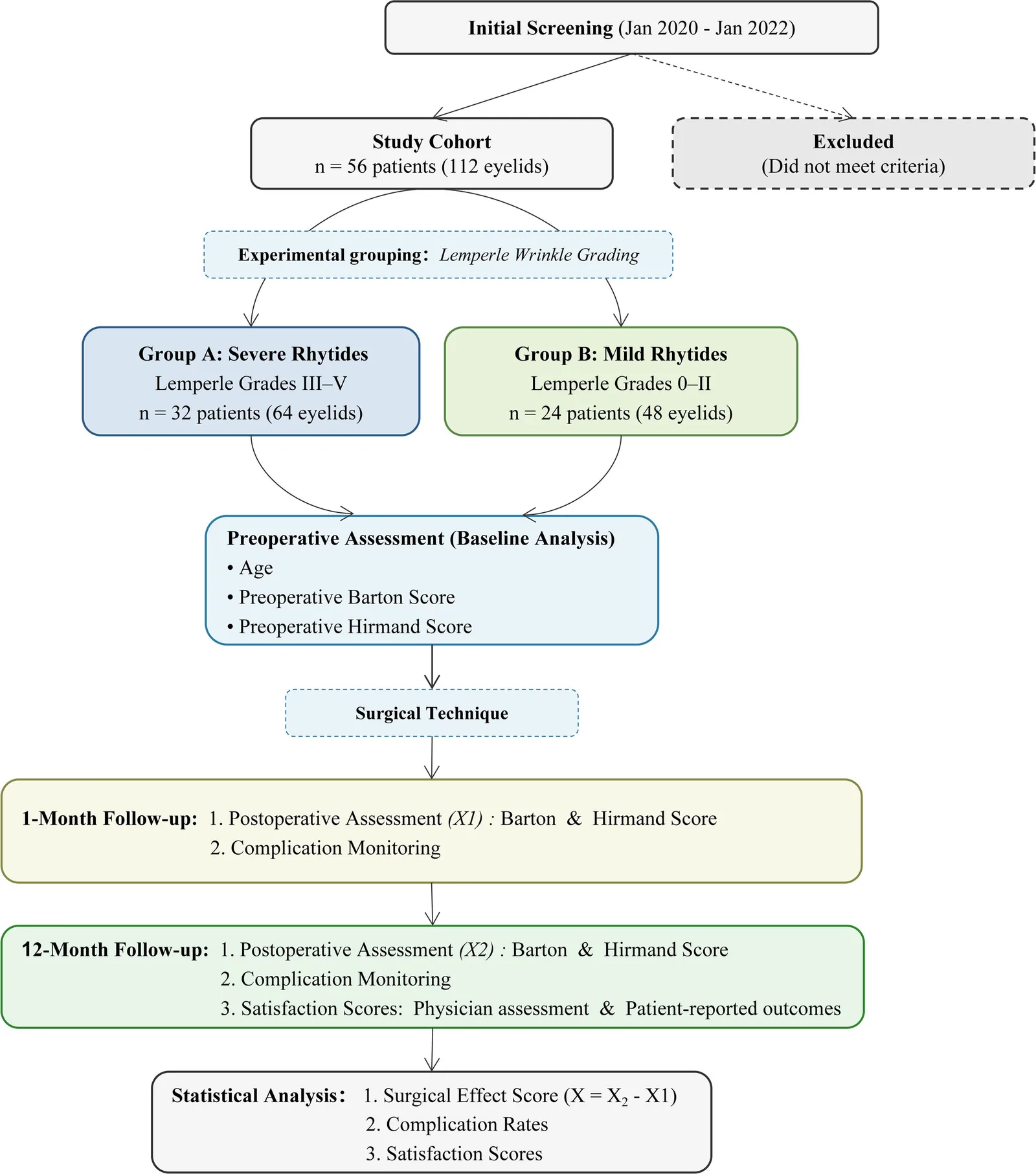
一项回顾性队列研究(56例/112眼)按术前 Lemperle 皱纹分级,将经皮肤下睑成形术患者分为重度动态皱纹组(A组,III–V级,n=32)与轻度组(B组,0–II级,n=2
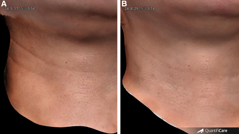
研究类型:回顾性单臂观察性研究(单作者、单中心),共纳入 20 例水平颈纹患者(女 14 例、男 6 例),平均年龄 35.4 岁(27–59 岁),Fitzpatrick II–