巨大减重后患者上臂塑形(brachioplasty)的术式与瘢痕位置:15项研究的文献综述
2026年10月12日29 min read
Plastic and Reconstructive Surgery Global Open
Plastic and Reconstructive Surgery Global Open
结论与实践要点
本文为叙述性文献综述(非Meta分析),按PICO策略检索PubMed、Embase与Web of Science,纳入1974年以来15项减重手术后臂部塑形的临床队列研究(多数来自意大利,美国3项,法国与埃及各1项),因异质性大、样本量小而未行定量合并,并以ROBINS-I工具评估偏倚风险。综述归纳出两类去除多余皮肤脂肪的方式——整块锐性切除与吸脂辅助臂部塑形(LAB),以及两类最终瘢痕位置——内侧(n=732,沿肱二头肌沟)与后内侧(n=141,内侧上髁至后腋襞连线)。
主要发现:LAB提示皮瓣游离更容易、总体并发症更低;一项研究整块切除20例、LAB 11例,LAB并发症显著更低且满意度更高。所引Meta分析显示内侧切除总并发症率43.7%(其他切口9.7%),异常瘢痕15.6%对2.98%,切口裂开8.7%对3.1%,血清肿8.41%对1.4%,美容性再次手术9.9%对3.8%。吸脂与更低血清肿(3%对13.53%)、淋巴水肿(1.6%对6.5%)、神经损伤(1.5%对5.5%)及异常瘢痕(7.5%对20.08%)相关,但可能因水肿增加无张力关闭难度。并发症率报道可高达56%,瘢痕外观不满意为常见主诉;有研究前10例内侧瘢痕患者自觉持续可见,改为后内侧放置后接受度提高。
作者提示:LAB联合后内侧瘢痕(日常活动中可见度较低、重力张力分布更均匀)加分步cut-as-you-go切除(临时钉合、控制张力)的组合值得关注;腋部冗余切除宜沿后腋襞展开、保留腋窝顶。局限性:纳入研究以观察性为主、证据等级低、偏倚风险高,并发症与瘢痕质量报告口径不一,尚缺前瞻性对照研究验证。
Background:A wide variety of brachioplasty approaches and modifications have been described to make the procedure safer and more appealing to patients and surgeons alike. There is, however, no consensus on the most appropriate surgical approach. The current report aimed at identifying the technique that yields optimal aesthetic outcomes and scar quality associated with highest patient satisfaction and lowest scar visibility and complication rates, in the growing population of patients with massive weight loss.
Methods:A PICO PubMed, Embase, and Web of Science literature search was conducted to identify clinical cohort studies published since 1974 about post-bariatric patients undergoing brachioplasty.
Results:Fifteen studies satisfied the inclusion criteria and were retrieved for review. Two main methods for excess skin and fat removal, en bloc sharp excision or liposuction-assisted brachioplasty, and 2 final scar placements, medial and posteromedial, with some variations, together with various axillary scars, have been reported. Higher overall complication rate was associated with excisional brachioplasty. Two main scar placements have also been reported: primarily medial, with various extensions to excise additional tissues in the axilla or the elbow region, and posteromedial.
Conclusions:Each described technique has advantages and disadvantages. Although quantitative analysis was not possible to conduct, it seems logical that combining advantages of liposuction-assisted brachioplasty with reduced posteromedial scar visibility, in addition to a stepwise “cut-as-you-go” approach, would be most favored, allowing safe procedure execution with optimal aesthetic outcomes and lowest complication rates.
Question: What is the safest and most effective brachioplasty technique for patients with massive weight loss in terms of tissue excision and scar placement?
Findings: Through a systematic review of studies on brachioplasty after massive weight loss, the authors found that liposuction-assisted brachioplasty combined with posteromedial scar placement significantly reduces complication rates and improves patient satisfaction compared with sharp excision and medial scars. A stepwise “cut-as-you-go” approach enhances safety and contour harmony.
Meaning: For optimal results after massive weight loss brachioplasty, combining liposuction-assisted excision with posteromedial scar placement and stepwise resection is recommended.
Brachial lipodystrophy with ptosis becomes increasingly evident with age, particularly after massive weight loss. 1 Renewed interest in upper arm contouring has paralleled recent advances in bariatric surgery. 2 – 6 In the 1930s, Thorek 7 was the first to describe reduction of pendulous arm deformity by medial arm fusiform skin and fat excision. Aesthetic brachioplasty was described in 1954 by Correa-Iturraspe and Fernandez. 8 Their technique yielded suboptimal results and was associated with frequent wound dehiscence, hematoma, seroma, and irregular arm contour. 1 , 9 – 11 Brachioplasty following massive weight loss was described by McCraw in 1974. 12
Baroudi, 9 Strauch et al, 13 and others have proposed various arm deformity classifications to guide in the selection of optimal treatment of excess fat and skin laxity. 10 , 11 Coefficient of Hoyer (the ratio between proximal and distal upper arm circumference) or the ratio of height of hanging skin to its thickness can assess skin laxity. 10 Bicipital groove to lowermost border of pendulous skin distance determines the grade of ptosis. 1 These guidelines are helpful; nevertheless, significant operative improvization tempered with clinical judgment may be imperative to properly correct both arm “bat-wing” and armpit contour. 3 , 10
A new classification and treatment strategy adapted from other authors 1 , 10 , 14 was recently published by Miotto et al. 15 Early stages with mild lipodystrophy and ptosis can be corrected by liposuction alone. Occasionally in selected patients, improved arm contour may be achieved by correcting volume deficiency in the bicipital groove with fat grafting. 16 For more advanced deformities, particularly following massive weight loss, dermolipectomy extending from the elbow to chest is necessary. 13 , 17 , 18 Most advanced cases with lateral thoracic laxity require extended brachioplasty to treat arms in combination with axilla, back, and chest. 4 , 19 , 20
Although described techniques are aimed at making the procedure safer and more appealing to plastic surgeons and patients, 2 , 10 , 11 , 18 , 21 there is no consensus on the most appropriate approach and specific indications for the use of adjuncts. Studies providing practitioners with clear evidence-based guidelines are also lacking. 5 , 22 The current review of various brachioplasty techniques that have evolved over time is aimed to identify the safest and most appropriate approach that yields the most optimal aesthetic outcome with the highest patient satisfaction, and lowest complication rates for the growing population of patients with massive weight loss.
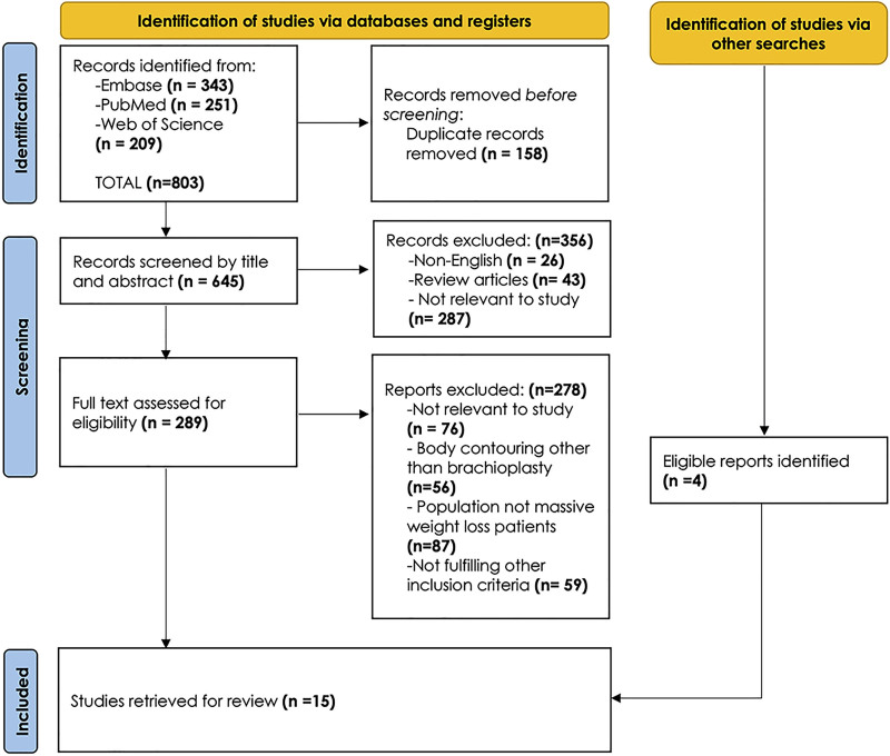
A PICO PubMed, Embase, and Web of Science literature search was conducted to identify clinical cohort studies of post-bariatric patients undergoing brachioplasty. Inclusion and exclusion criteria are detailed in Table 1 . The search PRISMA algorithm is shown in Figure 1 .
Table 1. Inclusion and Exclusion Criteria, and Literature Search Methodology
| Criteria | Inclusion | Exclusion |
|---|---|---|
| Study design | Peer-reviewed studies of arm contouring following massive weight lossSurgical technique including skin excision pattern and final scar placement | Reviews, case reports, comments, and letters to the editorsCohort studies of none patients with massive weight loss (patients with obesity) |
| Population | Cohort studies involving 10 or more patients | Studies involving less than 10 patients |
| Outcome | Objective and subjective patient-reported outcomeReport of complications | Studies with insufficient reported surgical details, patients; reported outcome, or complications |
| Methodological quality | Prospective or retrospective cohort studies with number of patients involved well defined | Studies in which the number of patients involved is not reported |
| Publication date | All studies published since 1974 | |
| Language | Studies published in English | Non-English publications |
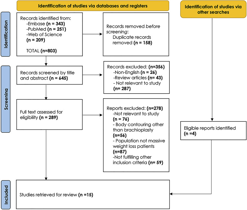
Fig. 1. PRISMA flow chart of retrieved studies. This work is licensed under CC BY 4.0. To view a copy of this license, visit https://creativecommons,org/licenses/by/4.0/. With permission from Page et al., BMJ 2021;372:n71. doi: 10.1136/bmj.n71.PRISMA文献筛选流程图(改编自Page et al., BMJ 2021;372:n71,CC BY 4.0)。Two independent investigators reviewed identified studies for relevance. Initial screening involved titles and abstracts. In case abstracts lacked sufficient detail, the methods section was reviewed. Disagreements in study selection were resolved by consensus or by consulting a third independent reviewer. Full texts of eligible studies were retrieved, and relevant data extracted using a standardized form. Meta-analysis was not performed due to large study heterogeneity in addition to the small number of patients involved, particularly those undergoing posteromedial brachioplasty.
In total, 15 studies were identified for review. Most were from Italy, 3 were from the United States, and there was one each from France and Egypt. Though ethnicity and skin type of patients involved were not provided; judging from clinical photographs, patients were mostly White.
Two methods for excess skin and fat removal are used: sharp en bloc excision and liposuction-assisted brachioplasty (LAB). Standard wet or superwet LAB was performed in 8 studies, 2 , 23 – 29 but, when there was incomplete deflation of skin, only excision was achieved. Simultaneous liposuction to the non-excised area was performed in 5 studies 18 , 20 , 22 , 24 , 30 and liposuction 3–5 months before brachioplasty in 1. 31 Skin avulsion or excision was performed in 5 studies. 23 – 25 , 28 , 29 Though reporting of complications lacked consistency, higher overall complication rate was associated with excisional brachioplasty. 20 , 22 , 27 LAB permitted easier flap dissection and reduced complications. 18 , 24 – 26 , 29 In one study, monobloc resection was performed in 20 patients, and LAB in 11. Incidence of complications was significantly lower with LAB associated with higher patient satisfaction. 23
Two scar placements are reported: medial (n = 732) at the bicipital groove along the medial intermuscular septum with various extensions to excise additional tissues in the axilla or the elbow region 2 , 18 , 20 , 22 , 23 , 25 – 28 , 32 – 34 and posteromedial (n = 141), defined by a straight line from medial epicondyle to posterior axillary fold. 24 , 26 , 30
High patient satisfaction was described in 8 studies: 5 with medial, 23 , 25 , 26 , 29 , 30 and 2 with posteromedial scars. 24 , 35 Patient-reported outcome measures were mostly about arm contour; scar quality was not a primary outcome. In 1 study, the scar was placed medially in the first 10 patients; it was constantly visible to them, friends, and family. For the next 40 patients, posteromedial scar placement was more acceptable and resulted in subjectively reported good scar quality. 31 Scar quality was objectively measured by the Vancouver Scar Scale and explicitly reported in only 1 study with posteromedial scars. 24
The summary of reviewed studies with level of evidence provided and risk of bias assessment are detailed in Table 2 and Figure 2 . ( See table, Supplemental Digital Content 1, which displays the details of identified studies, https://links.lww.com/PRSGO/E334.)
Table 2. Summary of Studies Retrieved for Review Describing Modality for Excess Skin and Fat Excision and Placement of Final Scar Together With Level of Evidence Provided
| Authors | Excess Tissue Excision | Scar | Outcome and Complications | Level of Evidence |
|---|---|---|---|---|
| Margara et al 25 | Completely defatted by liposuction; Skin only excision | MedialGroup A: standard; Group B: antecubital extension | Group A 3 temporary dysesthesias, resolved within 3 months | III |
| Nisi et al 28 | Excisional brachioplastyTriangular flaps de-epithelializedand fixed under upper flapLimited lipoaspiration | Medial | 2 unsatisfactory scars. No other complications | IV |
| Bianchi, et al 29 | Skin avulsion after extensive liposuction | Medial more posterior Lazy S, J-scar | No complicationsPatient and surgeon satisfaction high to very high | IV |
| Marchica et al 27 | Excisional brachioplastyTraditional arm lipoaspiration | Posteromedialaxillary Z-plasty if scar extends to chest wall | 50% overall complication rate; 25% poor scarring; 12.5% dog ears; 26.8% delayed healing; 5.4% seromas | IV |
| Elgazzar et al 35 | Excisional autoaugmentation brachioplasty | Posteromedialaxillary Z-plasty if scar extends to chest wall | Patients highly satisfied2 asymmetries; 2 hypertrophic scars; 1 keloid | IV |
| Simone et al 24 | Liposuction then skin excision including thin layer of subcutaneous tissue | PosteromedialAxillary Z-plasty | 1 (4.1%) wound dehiscenceNo other complicationsGood and excellent Vancouver Scar Scale | IV |
| Di Pietro et al 23 | 20 Excisional brachioplasty11 LAB | MedialFish tail | Incidence of complications significantly lower in LAB group (9% vs 60%, P < 0.01) | IV |
| Bocchiotti et al 26 | Partial liposuction to ease flap excision | Medial | 2 hematomas, 4 partial wound dehiscence, 1 seroma, 3 hypertrophic scars | IV |
| Gentileschi et al 18 | Tunnelization without suction of medial longitudinal ellipse; liposuction of posterior arm | Medial T or L in axilla | Small dehiscence for 1 patient and hypertrophic scarring for 2 patients | IV |
| Bossert et al 22 | Excisional brachioplastyOccasional liposuction to posterior arm | Medial | 46% overall complication rate | IV |
| Zomerlei et al 2 | Excisional brachioplasty | Medial fish tail, L, Z-plasty | Total complication rate (53.1%); most common: hypertrophic scarring (24.0 %), 8 requiring revision | IV |
| Migliori et al 32 | Excisional brachioplasty | Medial | 2 major seromas, 1 ulnar nerve compression | IV |
| Gusenoff et al 20 | Excisional brachioplastyOccasional liposuction to posterior arm | MedialExtension distally when indicated across elbow | 40.4% complication rate majority are seromas. 3 revisions for poor scar or recurrent skin laxity | IV |
| Cannistra et al 31 | Liposuction 3–5 mo before brachioplastyExcisional brachioplasty | Medial: first 10 patientsPosteromedial: Between medial axillary line and olecranon for next 40 patients; Z in axilla | Better scar quality for second group; location more acceptable to patients | IV |
| Hurwitz and Holland 30 | Excisional brachioplastyLipoplasty as needed in areas not being resected | Medial L with closed angle at dome of axilla | All patients pleased with results; 3 delayed healing, 4 seromas, 1 hypertrophic scar | IV |
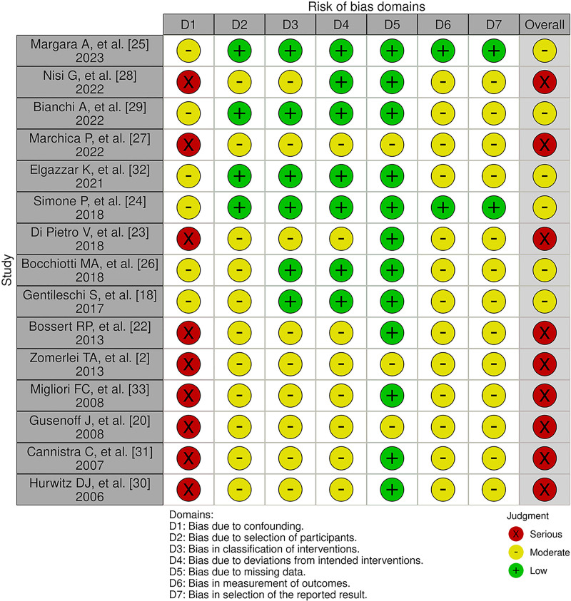
Fig. 2. Risk of bias assessment of retrieved studies for review with ROBINS-1 assessment tool of nonrandomized studies of interventions.纳入研究的偏倚风险评估(ROBINS-I工具)。
Brachioplasty for patients with massive weight loss almost invariably entails resection of excess redundant tissue. Factors determining outcomes are degree of skin and fat excision, modality of excision, and location of final scar while carefully striking a balance between resecting enough tissue for optimal contour and overresecting to avoid wound closure under tension. 36
Despite numerous refinements, complications are frequent and can be as high as 56%. Major complications are particularly high in post-bariatric patients. 2 , 16 , 33 , 37 Dissatisfaction with scar appearance is also a most common complaint. 38 , 39 Recently, complications have been reported to be much lower 5 , 37 ; nevertheless, a considerable rate with concerns regarding wound dehiscence, infection, seroma, edema, lymphocele, lymphedema, nerve injury, under-resection, banding, and residual contour deformity still persist. 18 , 19 , 28 , 33 , 38
Skin excess has been described to be both circumferential and longitudinal; excisions with distal to proximal and circumferential transverse vectors of pull have been recommended. 34 In fact, longitudinal skin excess has been overemphasized; it is mostly minimal and rarely a problem. 40 , 41 Techniques with only a transverse axillary component may be adequate for only moderate proximal deformity with appropriate skin tone. They are not suitable for patients with massive weight loss, in whom skin excess is mostly circumferential. 4 , 42 – 44 Apparent longitudinal excess is sagging of deflated skin envelope that can be corrected by circumferential reduction 42 (Fig. 3 ).
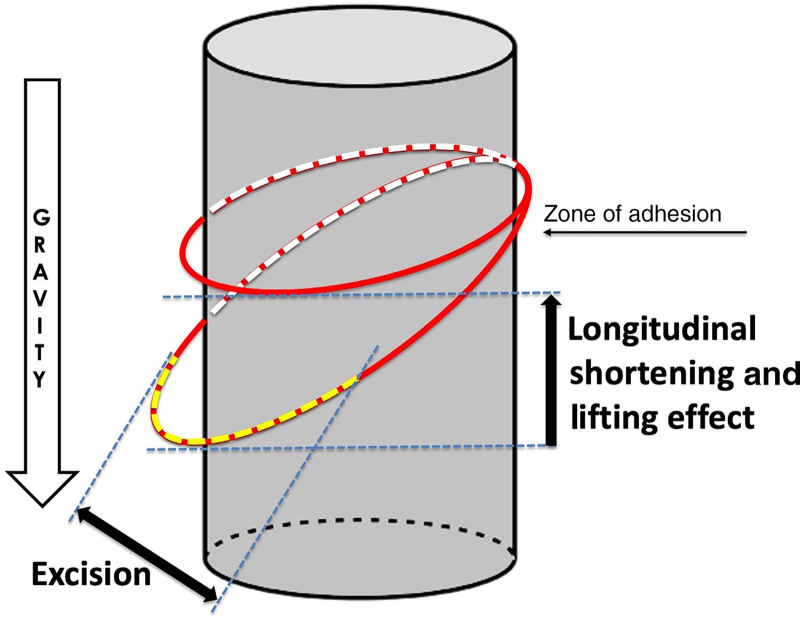
Fig. 3. Resection of redundancy for circumference reduction achieves simultaneous vertical lift.环周缩减式切除可在缩小臂围的同时获得垂直提升。Brachioplasty scars in most patients with massive weight loss are better when longitudinal; however, their exact position, key to patient satisfaction, remains debatable. 11 , 18 – 20 , 22 , 27 , 36 , 45 , 46 There is also no consensus on whether scars should be straight or sinusoidal, or what should be excised and from where. 45 Anthropometric studies demonstrated that the best way to determine skin excess is to project a truncated conical frustum from the axilla and deltoid to the elbow and wrist. 45 Any skin that lies outside this cone can be considered for removal. ( See figure, Supplemental Digital Content 2, which displays a [A] truncated cone [conical frustum] extending from the armpit and deltoid to the wrist. It defines the “ideal” anthropometric harmonious proportions and profile of the upper extremity. It shows where correction is needed. The medial excisional pattern [m] lies within this truncated cone, whereas the area that lies outside [pm] indicates the skin that needs to be excised. B, Ideal lowermost arm border. C, Final scar placement following posteromedial excision. D, Minimal scar visibility from behind, https://links.lww.com/PRSGO/E335.)
With the assumption that brachioplasty scar location does not contribute to scar quality, 13 , 47 and that scarring that never entirely fades away is inevitable, the medial straight scar popularized by Baroudi 9 is mostly performed and traditionally considered to be most acceptable, though it is seldom of high quality. 5 , 32 This scar is camouflaged in the bicipital groove shadow and is presumably well hidden, virtually imperceptible most of the time during composed posture, and acceptable during expressive conversations and daily activities. 1 , 10 , 18 , 25
Nevertheless, wound closure along the medial intermuscular septum can lead to eye-catching scar line depression. Instead, scar placement between the bicipital groove and posterior edge of the arm is suggested. 48 Regardless of variations in medial placement, such scars are constantly visible to patients. 31 In contradistinction, posteriorly based scars within the brachial sulcus, a shallow oblique depression located at the center of the humerus lateral border, though not visible to the patient, are almost always visible to everybody else looking from behind. 2 , 21 , 46 , 49
In an attempt to reduce tension and improve scar quality, Lockwood 50 advised suspension of the arm fascial system to clavipectoral fascia. Goddio 51 described de-epithelialization and tucking of the posterior skin flap beneath the anterior flap. Modified by Nisi et al 28 as the “jaws” brachioplasty, the technique reduces tension through transfixed points. El Khatib 1 , 52 suggested pulling down the upper excision margin and fixing it to deep fascia with permanent sutures. On the other hand, Regnault 42 described the necessity to modify longitudinal excisions depending on amount of skin excess. To prevent widened or hypertrophic scars and reduce linear contracture, Strauch et al 13 and De Souza Pinto et al 53 suggested a sinusoidal pattern. Fantozzi 45 proposed the triple-vector approach creating 2 asymmetric curves with medial and posterior flaps. S-shaped 34 and kris-knife 54 excision patterns have also been described to reduce scar tension. Distal curvilinear extension of medial arm incision to the anticubital surface is reported as well, to correct excessive sagging at the elbow. 25 Despite all tension distribution attempts, sinusoidal scars are the most aesthetically displeasing, with high complications and scar revision rates. 2 , 55 Moreover, deeply placed anchoring sutures may not be advisable, as vital structures particularly in the axilla may be injured. 13 Finally, silk-derived surgical scaffold (SERI; Allergan, Inc., Irvine, CA) was used to support the medial arm superficial fascial system as an adjunct to conventional brachioplasty. 52 , 56 Claims that this device results in faster maturation and better scar quality await confirmation.
Scar placement at the posteromedial aspect of the arm constitutes an important improvement in longitudinal brachioplasty. It has been reported to minimize frontal scar visibility when the arms are elevated. 13 , 38 , 39 , 52 , 53 , 57 , 58 Symbas and Losken 59 have reported their experience with the 2 currently most widely used approaches: the L-shaped medial resection of Hurwitz and Holland 30 and the double-ellipse posteromedial technique of Aly et al. 36 Even though their report is not comparative, their preference went to posteromedial scars that are not visible to patients even when looking in a mirror. After bicipital groove scar placement for their first 10 patients, Cannistra et al 31 elected to place scars well posteriorly along a line between the medial axillary line and olecranon for their next 40 patients and considered this to be more acceptable.
Although patients claim preoperatively not to be concerned about scar appearance, objection to scar length and location becomes readily expressed after surgery. 5 , 11 , 21 , 60 Unpredictable scar quality is the most common adverse outcome and remains a challenge for both surgeons and patients. Lack of skin elasticity in patients with massive weight loss is probably a major predisposing factor for pathologic scarring. 13 Many other factors including wound closure under tension, wound dehiscence, infection, and Fitzpatrick skin type contribute to poor scarring as well. Combination of gravitational forces, stress, thin skin, and a superficial fascial skin system can also lead to scar widening and hypertrophic scarring. 56
Scar shape and placement need not be “a matter of taste and philosophy” 36 ; it is about understanding tension forces in dynamics and how a more even spread of forces along the arm can reduce complications and long-term scarring problems. 11 , 45 Closer analysis of tissue displacement dynamics reveals that for wound closure in both medial and posterior linear scar placement, skin and subcutaneous tissues must be displaced and pulled. In standing position with the arms abducted for daily gesticulation, the pull is superior against gravity, resulting in high stress forces. Intensity of gravitational stress and its distribution are greatly reduced in the posteromedial scar location (Fig. 4 ). 24 , 31 , 38
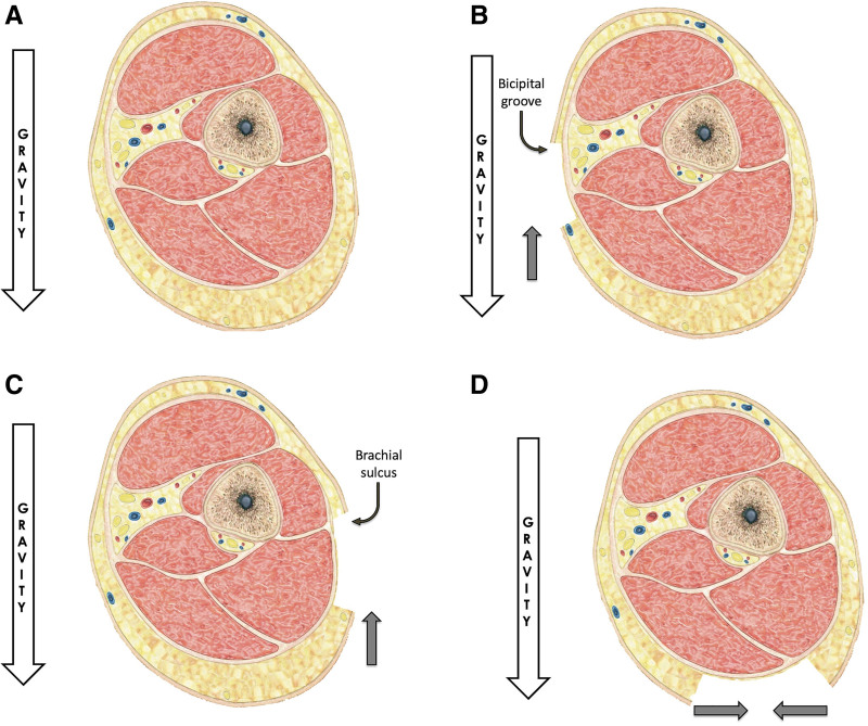
Fig. 4. Diagrammatic representation of gravity's effect on the arm in the abducted position with the various excisional approaches. A, Effect of gravity resulting in ptosis and “bat wing” deformity. B, Medial or (C) posterior excision patterns do not eliminate gravitational tension forces. D, Effect of gravity totally eliminated with direct excision of posteromedial tissues lying outside the conical frustum.重力影响示意图:A重力致臂下垂与蝙蝠翼畸形;B内侧、C后侧线性切除仍存重力张力;D直接切除锥形截头体外的后内侧组织可消除重力影响。Scar location may make its presence more or less acceptable. Posteromedial scar placement is more effective in scar concealment when the arms are elevated or are resting at the side. 5 , 13 , 24 , 56 Unfortunately, no reliable PROMs are available to confirm this observation. Though very little data are available regarding healing time comparing medial to posteromedial scar placement, medial scars are associated in general with higher complication rates, including higher incidence of wound dehiscence, 24%–40% risk of hypertrophic or wide visible scarring, and higher revision rates. They are also frequently associated with residual contour deformities and skin ptosis. 1 , 2 , 10 , 11 , 18 , 29 , 36 , 55 , 56 A recent meta-analysis of the impact of incision placement on complications demonstrated that medial excisional patterns are predominantly characterized by higher complication rates relative to all other incisions (43.7% versus 9.7%). They result in higher aberrant scarring (15.6% versus 2.98%), wound dehiscence (8.7% versus 3.1%), seroma formation (8.41% versus 1.4%), ptosis (9.44% versus 5.84%), overall reoperation for aesthetic purposes (9.9% versus 3.8%), and nerve-related complications including temporary or permanent dysesthesia, paresthesia, or confirmed nerve sectioning or compression (2.89% versus 1.39%). 5
With sharp en bloc excision along the bicipital groove, damage of medially located major lymphatics and sensory subcutaneous nerves can occur, even with careful dissection. Medial brachial and antebrachial cutaneous sensory nerves are at great risk. Ulnar and median nerves may also be vulnerable in cases where deep brachial fascia is violated. 15 , 18 , 20 , 22 , 45 , 59 , 61 – 63 With sharp excision along the brachial sulcus, 4 , 13 , 45 , 55 , 60 , 64 in addition to flattening of posterior natural rounding of the arm, 54 the subcutaneous posterior branch of the medial cutaneous antebrachial nerve may be injured, resulting in painful neuroma and elbow paresthesia. 15 , 18 , 23
Combined liposuction/excision brachioplasty offers a safer option. 15 , 19 Recent evidence suggests that LAB, whether liposuction is at the excision site, adjacent to it, or circumferential, does not lead to higher overall complication rates regardless of scar placement. 2 , 5 , 18 , 21 , 22 , 26 , 27 , 38 , 41 , 65 On the contrary, liposuction is associated with lower incidence of seroma formation (3% versus 13.53%), lymphedema (1.6% versus 6.5%), and nerve injury (1.5% versus 5.5%). It even results in lesser aberrant scarring (7.5% versus 20.08%). 4 However, concomitant liposuction may cause some difficulty in wound closure without tension due to exaggerated fluid infiltration and edema formation, leading to underresection. It also increases slightly the risk of wound dehiscence, skin necrosis, and hematoma formation. 4 , 5 , 21 , 40 Suction-assisted lipectomy a few months before brachioplasty has been suggested whenever debulking is required. 31
Skin-only excision or avulsion combined with aggressive liposuction of the area to be excised (Fig. 5 ) has been described to significantly decrease complications and improve patient satisfaction. It preserves underlying structures and decreases bleeding compared with sharp dissection. It also allows easy wound closure. 4 , 18 , 21 – 24 , 41 , 45 , 47 , 49 , 57 , 64 , 65 Worth noting is that contrary to recommendation of tapering and feathering liposculpture of adjacent areas, 24 edges of the liposuctioned zone should remain very distinct, creating a dished-out deformity as described by Knotts et al 57 to result in smooth contour.
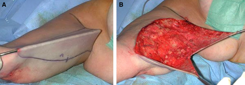
Fig. 5. LAB. A, Complete defatting of the posteromedial area. B, Skin-only excision.LAB示意:A后内侧区域彻底去脂;B仅切除皮肤(保留皮下结构、减少出血)。Regardless of excisional technique and final scar placement, wound closure is not met infrequently with difficulty. If delayed until after complete excision, edema invariably settles complicating wound suturing, particularly if the extent of excision has been overestimated. Easy wound closure in this situation necessarily entails undercorrection. 20 Moreover, uneven tension along the suture line results in distressing arm banding. Preoperatively existing banding can also be exacerbated. 33 Stepwise skin excision and the “cut-as–you-go” approach with temporary staples allow correction and readjustment of skin excision and closure with controlled tension. It greatly reduces the risk of banding that is very difficult to correct subsequently. 36
Correcting axilla deformity is an essential component of brachioplasty. From simple geometric analysis, it cannot be achieved by just medial elliptical excision along the bicipital groove; minimal excision is possible near the vertex angle proximally in Strauch zones III and IV where most skin excess occurs. For wider excision, a large vertex angle would be necessary; this creates stress during suturing with unfavorable “dog-ear” conical deformations. Additional excision with resultant T, L, W, and Z scars in the armpit are unavoidable for restoration of upper arm and axillary contour. 11 , 15 , 30 , 46 , 66 , 67 However, scars across the axilla are not to be taken lightly. Despite claimed advantages, they do not yield optimal upper arm and axillary contour in many patients. 11 , 15 , 30 , 66 , 68 With Z-plasty recommended to deepen the axillary dome, 22 underresection is the norm. Moreover, coning deformity develops at each closed angle of the Z that does not fit well in a dynamic concave axillary dome. 30 Fish tail incision extended over anterior and posterior axillary folds may be more appropriate 11 ; however, scars across the anterior fold are highly objectionable to many patients. 42 On close examination, excess tissue is actually in the posterior axillary fold in continuity with excess in the posteromedial arm. Excision is thus better centered along this fold, sparing the axillary dome and extending to the lateral chest wall whenever necessary 65 , 69 (Fig. 6 ). With this approach, a normal axillary hair pattern, much appreciated by male patients, is preserved. 36
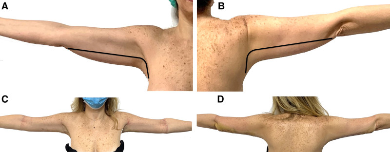
Fig. 6. Excision pattern in the posterior axillary fold. A and B, Redundant tissue in the posterior axillary fold in continuity with excess in the posteromedial arm. Excision pattern centered along this fold sparing axillary dome and extending to lateral chest wall. C and D, Totally nonvisible scar with arms abducted both in anterior and posterior views.后腋襞切除模式:切除沿后腋襞展开、保留腋窝顶并可按需延伸至胸壁;C/D示上臂外展时瘢痕在前后观均不可见。
This review is not without limitations. The few studies reviewed are largely observational, providing a low level of evidence and carrying high risk of bias. Moreover, in addition to a high degree of heterogeneity and incomplete data reporting, there is large disparity across studies pertaining to what constitutes true complication or undesirable outcome, as well as in reporting scar quality and patient satisfaction. Reporting of complications lacked consistency, limiting data synthesis and interpretation. Use of standardized scales of surgical complications should be encouraged for future studies.
The main focus of various brachioplasty technical modifications has been to improve arm, axilla, and upper trunk contour, optimize scar placement, and minimize complications. Nevertheless, none of the described techniques is without drawbacks, complications, and need for possible revisional touch-ups. Though scar position has to be discussed critically with patients, it remains somewhat unclear which combination of excision technique and scar position offers the best outcome. It seems that posteromedial scar placement, in addition to reduced visibility to the patients as well as others during daily gesticulation, reduces tissue stress, theoretically leading to better scar quality. Furthermore, rather than sharp excision, liposuction of the area to be excised followed by skin-only excision offers added safety and greatly reduces complication rates. Finally, along with surgical and artistic judgment, “cut–as-you-go” stepwise skin excision results in optimal outcomes with smooth and harmonious contour. There is, however, a need for prospective comparative studies to validate this approach.
The authors have no financial interest to declare in relation to the content of this article.
声明:中文精读 · 仅供学术参考。内容来自公开文献检索,不代表本人观点,不构成诊疗建议。 医疗美容需在正规医疗机构由执业医师实施。
基于相同主题推荐 · 继续深挖

这项前瞻性、开放标签、随机、多中心(德国11个中心)上市后研究纳入121名受试者,按1:1随机接受 Belotero Volume(CPM-V,n=58)或含利多卡因的 CPM-V
鼻部塑形疗效与安全性:12个月前瞻性研究/PMC13100342_fig1.jpg)
这项单中心前瞻性研究纳入24名中国女性受试者,接受CaHA凝胶鼻部注射(骨膜上/骨膜浅层,单侧单次总量≤2.0 mL,平均0.93±0.22 mL),全部完成12个月随访,无脱落。
研究设计:单中心、单术者回顾性图表复习,2017 年 1 月至 2018 年 1 月连续 191 例除皱手术中 110 例随访满 100 天者纳入分析,平均随访 404.5 天