Triple-C SMAS 折叠除皱术:110 例回顾性研究的安全性与满意度数据
2026年10月13日29 min read
Plastic and Reconstructive Surgery Global Open
Plastic and Reconstructive Surgery Global Open
结论与实践要点
本文发表于 Plastic and Reconstructive Surgery Global Open(DOI:10.1097/GOX.0000000000002575),报告资深作者 2017 年 1 月 9 日至 2018 年 1 月 8 日间施行的 triple-C SMAS 折叠除皱术。研究为单中心回顾性图表复习:12 个月内连续施行 191 例除皱,排除随访不足 100 天者后 110 例纳入分析。受术者以女性为主(107 例女性、3 例男性),女性平均年龄 62.1 岁、男性 65.0 岁;初次除皱占 81.8%(90 例),二次占 14.5%(16 例),三次占 3.6%(4 例);平均随访 404.5 天。所有手术均在门诊手术室于局麻加镇静下完成。
技术层面,作者将 SMAS 处理设计为三层放射状 C 形折叠:折叠区位于耳前约 3 cm,自耳轮根部延伸至耳垂水平;第一层以 5–6 根埋藏的间断 2-0 Mersilene(不可吸收缝线)完成,是全层的承重结构,常规折叠 2–3 cm 组织;第二、三层改用连续锁边水平褥式 2-0 Vicryl(可吸收线),每层再折叠 0.5–1 cm,其目的一是埋藏第一层的不可吸收线结以降低可触及与挤出风险,二是对 SMAS 及深部软组织做平整与轮廓塑形。向量总体沿颧大肌约 45 度走行,但依据术前松弛部位个体化调整,面颈部各区域(颈、下颌缘、口角、鼻唇沟、中面)可分别施力。颈部常规经颏下小切口开放吸脂塑形,存在颈阔肌带与前侧松弛者加做颈阔肌切断与中线折叠(3-0 Vicryl)。皮肤重新铺放后几乎不加张力,女性置 10-French Jackson-Pratt 引流、男性置 15-French;术后次日拔引流,第 3 天可淋浴,第 7 天拆线,多数患者 1–2 周恢复社交活动。
结局数据显示总体并发症率较低:大血肿 2 例(1.82%)、小血肿 3 例(2.73%)、血清肿 5 例(4.54%)、轻度感染 1 例(0.91%,口服抗生素治愈)、增生性瘢痕 12 例(10.91%,曲安奈德联合 5-氟尿嘧啶注射治疗)、伤口裂开 2.73%、持续水肿 1.82%、轮廓不规则 0.91%、瘢痕修整 3.63%。面神经方面仅 1 例暂时性失用(0.91%,颧支,发生于一例三次除皱患者,保守治疗 6 个月内恢复),全文无长期面神经损伤记录,Great Auricular(耳大)神经损伤记录为 0.00%。缝线相关事件中 3 例(2.73%)为 Vicryl 线沿切口线挤出,而用于 SMAS 折叠的缝线未见挤出或可触及。总体满意率 96.4%,2 例(1.82%)接受再次除皱。手术时长方面,单纯除皱 35 例平均 162.3 分钟,联合手术 65 例(共 143 项辅助手术)平均 195.8 分钟。
对临床落地的启示在于:该术式把「承重」与「平整」分离——不可吸收缝线提供长期悬吊力但深埋于第二、三层可吸收线之下,理论上兼顾持久与低并发症;作者引述其既往双 C 折叠 1532 例资料:前 614 例两层均用 Mersilene 时挤出率为女性 1.9%、男性 3.0%,后 918 例两层均改 Vicryl 后挤出率降至 0%,但作者认为 Vicryl 长期可靠性不足,故折中为「深层不可吸收线 + 浅层可吸收线覆盖」。适应证上,这类折叠式提升避免了 SMAS 下剥离,理论上降低面神经损伤与皮瓣坏死风险(作者引用的系统综述汇总 183 项研究,提示 SMAS 折叠相较深平面技术在面神经损伤、皮肤坏死、血肿、血清肿方面风险更低;深平面技术暂时性面神经损伤率文献估计可达 3.6%),适合追求自然外观、恢复期短的门诊患者;但对重度下面部松弛需大量组织迁移者,折叠量受韧带限制的短板同样存在。
证据边界需明确三点:其一,本文为回顾性、单中心、单术者病例系列,无对照组,并发症与满意度均依赖图表回顾和主观评估(满意度仅在术后 8 周单次评定);其二,文中支持双层连续锁边缝合强度的关键论据来自猪皮离体标本的张力测试,属体外动物实验证据,仅能提示缝合构型在受控牵拉下的失效差异,不能外推为在体长期固定效果或临床优越性;其三,随访 404.5 天尚不足以评价提升的远期维持时间,满意率 96.4% 亦不能等同于客观容貌改善幅度。读者宜将其视为单术者大样本真实世界经验报告,而非技术间比较的结论性证据。
Various methods are used to reposition the superficial musculoaponeurotic system (SMAS) during facelift procedures. This study presents a novel, radially oriented, layered SMAS plication: the triple-C SMAS plication. This technique utilizes customizable vectors in the pattern of a “C” to plicate the SMAS in 3 layers to lift and tighten the deep structures of the face.
Methods:A retrospective review was performed of patients undergoing the triple-C SMAS plication over a 1-year period. Patients with a length of follow-up less than 100 days were excluded from the study. Demographic data, operative data, complication rates, and satisfaction rates were assessed.
Results:One hundred ninety-one consecutive patients underwent a triple-C SMAS plication over a 12-month period. One hundred ten patients met inclusion criteria. Average follow-up was 404.5 days. Complications assessed included temporary facial nerve neuropraxia (0.91%), major hematoma (1.82%), minor hematoma (2.73%), seroma (4.54%), great auricular nerve injury (0%), postauricular skin slough >2 cm (1.82%), and infection (0.91%). Two revision procedures were performed (1.82%). Patient satisfaction rate was 96.4%.
Conclusions:Traditional SMAS plication techniques involve single-layer, straight-line plications to lift the lower face and neck, limiting their versatility. The triple-C SMAS plication represents a novel technique to safely and effectively elevate the deep structures of the face in a radial pattern to restore a more youthful contour to the malar area, jawline, and neck. This represents a unique strategy for face lifting by which excellent results can be consistently obtained.
Facelift surgery represents the most powerful procedure in the plastic surgeon’s armamentarium to address the stigmata of aging of the face and neck. Since the technique was first described at the turn of the 20th century, facelift surgery has undergone a series of critical refinements as our knowledge of facial anatomy and aging processes has advanced. Initial descriptions of the technique involved elliptical skin excisions to tighten the skin and soft tissues of the face. 1 – 3 However, these methods were significantly limited in their scope and efficacy. Later descriptions would employ various degrees of skin undermining and excision. 4 , 5 Although these methods could tighten the skin envelope more effectively, they did not address the superficial musculoaponeurotic system (SMAS) and the deeper fat compartments of the face, thus lacking in longevity and reliability.
The last 50 years saw tremendous advances in our knowledge of facial anatomy, and with them, the foundations of contemporary facelift surgery. Skoog 6 described the first deep-plane technique in 1974. Recognizing the strength and durability of the deeper structures of the face, Skoog 6 elevated and advanced the skin, superficial fascia, and platysma as a single, composite unit. In 1976, expanding Gray’s concept of the fascia superficialis , 7 Mitz and Peyronie 8 defined the anatomy of the SMAS as a superficial fascial system that invested the facial mimetic musculature and was continuous with the platysma, temporoparietal fascia, and galea.
Further advances in our understanding of facial anatomy bolstered the foundations of modern facelift techniques. In 1989, Furnas 9 established the concept of the retaining ligaments of the face—consistent, fibrous condensations that tether the skin to deeper facial structures. In 2006, Rohrich and Pessa 10 demonstrated that facial fat is organized into distinct, anatomic subdivisions. According to these authors, the retaining ligaments of the face can be identified at the borders of these compartments.
Our current understanding of the processes of facial aging has been critical in shaping current practices. We now know that aging is far more complex than a purely gravitational process. Ultimately, aging leads to descent, atrophy, and radial expansion of superficial and deep facial structures. 11 The fundamental goals of facelift surgery are therefore to harmoniously elevate the structures of the face to highlight the natural contours that define the malar area, jawline, and neck.
The ideal facelift procedure achieves these objectives safely and efficiently in a simple and reproducible fashion, while minimizing downtime. With these goals in mind, we present our technique for natural facial rejuvenation—the triple-C SMAS plication. This unique strategy employs a layered, radially oriented SMAS plication utilizing permanent and absorbable sutures to reliably lift and tighten the superficial and deep structures of the face.
With the patient in the upright position, incisions, the extent of skin flap dissection, and platysmal bands are marked. All procedures are performed in our outpatient operating facility with local anesthesia and sedation (10 mg Diazepam orally, 1–2 mg Midazolam intravenously). Dilute local anesthetic solution is prepared with 15 cubic centimeters (CCs) 2% lidocaine with 1:200,000 epinephrine, 15 CCs 0.25% bupivacaine with 1:200,000 epinephrine, 10 CCs 8.3% sodium bicarbonate, 0.1 CC 1:1,000 epinephrine, and 50 CCs normal saline. This solution is infiltrated throughout the area of the planned dissection using a 27-gauge needle. Approximately 70 CCs are administered over each side of the face and neck.
An incision is made just posterior to the submental crease. Dissection proceeds sharply under direct visualization to the level of the thyroid cartilage. Open liposuction is then performed with a 2.7 or 3.7 mm cannula to contour the neck. Care is taken to remain 1 to 2 finger breadths inferior to the body of the mandible to avoid excessively thinning the skin flap before vertical translocation ( See Video 1 [online] , which demonstrates neck liposuction).
In patients with platysmal banding and anterior laxity, the platysma is horizontally transected a distance of 2–3 cm on either side, inferior to the hyoid. For patients with subplatysmal fat deposits, the platysma is minimally undermined and subplatysmal defatting is performed before midline platysma plication with 3-0 Vicryl sutures (Ethicon Inc., Bridgewater, NJ). For patients with minimal anterior laxity and platysmal banding, the neck is primarily addressed laterally; however, open liposuction and contouring of the neck is performed in the majority of patients ( See Video 2 [online] , which demonstrates the platysmaplasty procedure).
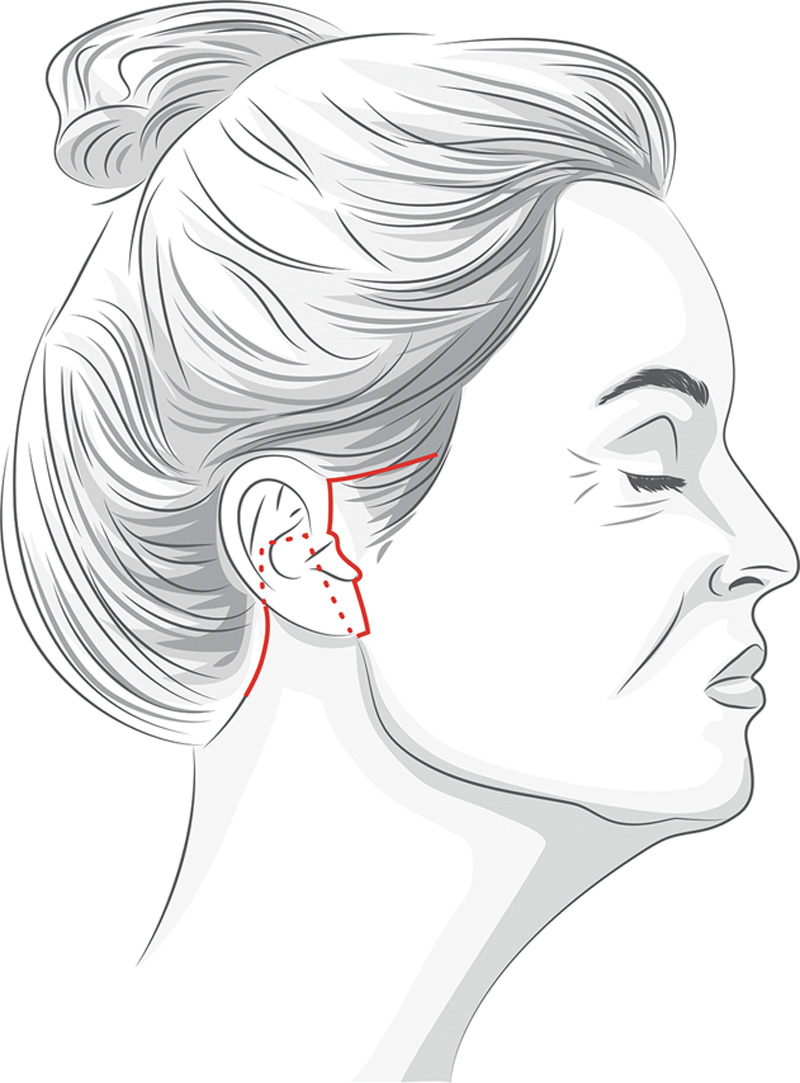
Dissection over the face begins with a temporal then posttragal incision that travels around the lobule and then posteriorly along the auriculomastoid groove with a back cut at the level of the root of the helix and then inferiorly along the retroauricular hairline (Fig. 1 ). In men, a pretragal incision is utilized. Incisions along the hairline are placed 1–2 mm posterior to the fine, vellus hairs. Incisions are made with a No. 15 blade, beveled across the hair shafts. Dissection begins retroauricularly with a No. 15 blade. Anteriorly, dissection over the parotid begins with a No. 15 then No. 10 blade under direct visualization to carefully define the skin-SMAS plane. Anterior to the parotid, dissection is performed with facelift scissors to the level of the lateral canthus (Fig. 2 ).
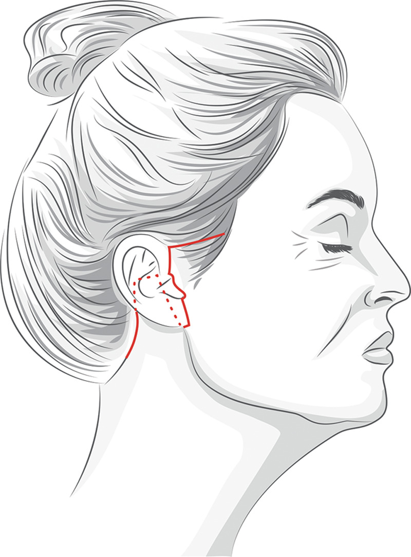
Fig. 1. Temporal and retrotragal incision design.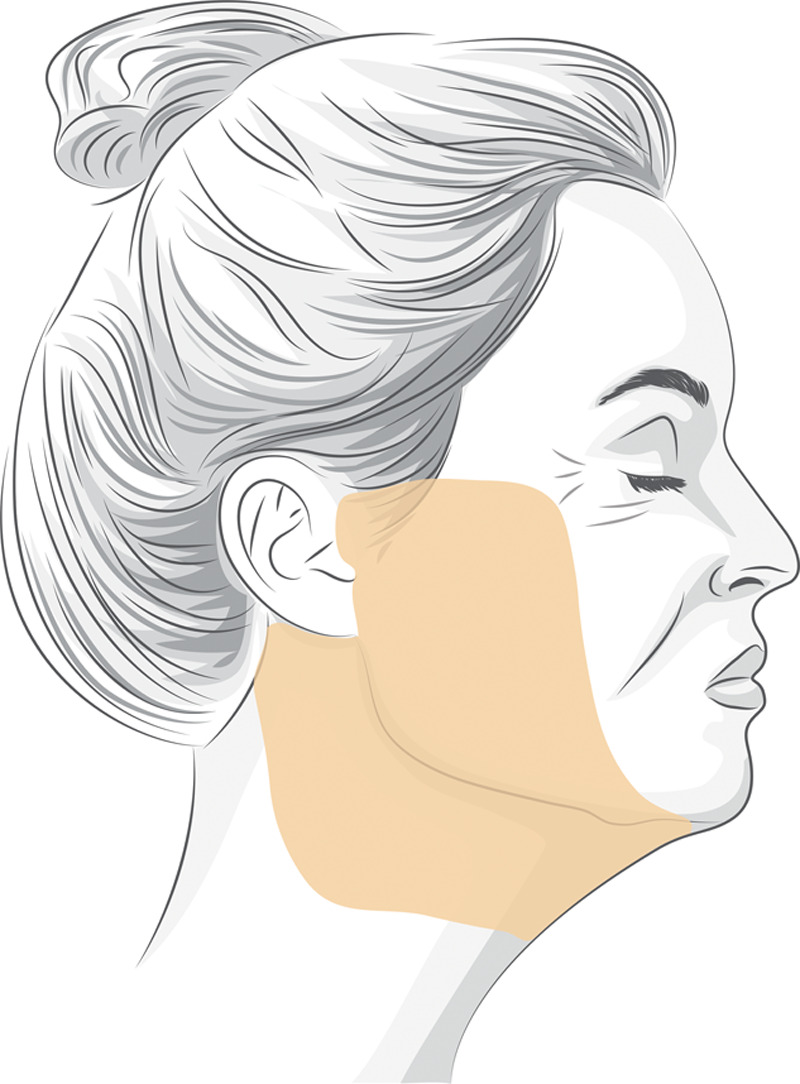
Fig. 2. Area of proposed skin flap undermining.With skin flaps elevated, hemostasis is meticulously obtained. The triple-C SMAS plication is then designed. A radial, “C” shaped SMAS plication is designed approximately 3 cm anterior to the ear, extending from the root of the helix to the level of the lobule posteriorly (Fig. 3 ). This area will correspond to the subsequent SMAS plication. The degree of SMAS translocation and plication will depend on the laxity of the face and neck.
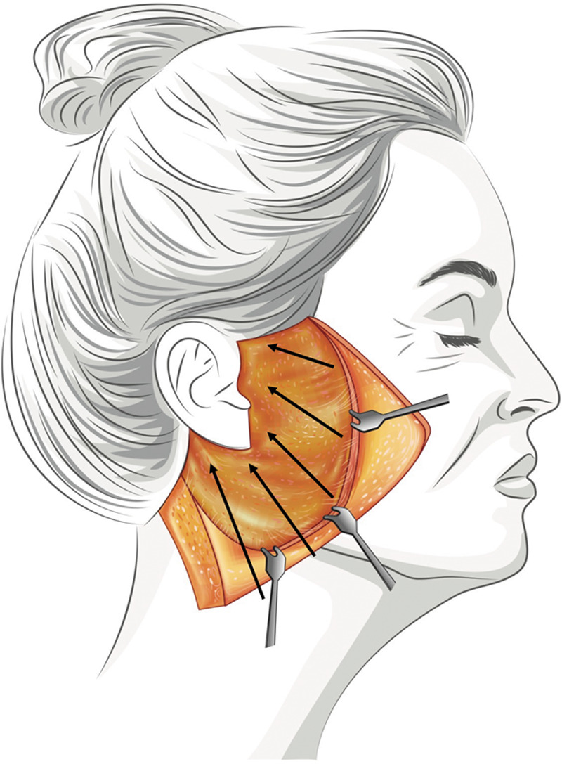
Fig. 3. Areas of proposed SMAS advancement for the first layer of plication and corresponding vectors are represented with black arrows. A minimum of 2–3 cm of tissue is routinely plicated in the first layer of plication. The second and third layers of plication will plicate an additional 0.5–1 cm of tissue. In the malar area and along the jawline, tissue is plicated anterior to the zygomatic osteocutaneous ligament and the masseteric cutaneous ligaments to recruit the mobile SMAS. Vectors are modified based on preoperative assessment and individual patient needs.The triple-C SMAS plication is performed in 3 layers. The vector of the SMAS plication generally follows the 45 degree trajectory of the zygomaticus major, but vectors are modified based on patient needs. Each layer of the plication will address the neck, jawline, oral commissure, nasolabial fold, and midface. Differential vectors and tension can be applied to tailor the degree of SMAS tightening based on the degree of laxity in each corresponding area. The first layer of the triple-C SMAS plication (Fig. 4 ) is undertaken with 5-6 buried, interrupted 2-0 Mersilene sutures (Ethicon Inc.). This layer involves the most aggressive SMAS plication and will invariably lead to areas of bunching and irregularities of the SMAS and deeper soft tissues (Fig. 4 ). The second (Fig. 5 ) and third (Fig. 6 ) layers of the triple-C SMAS plication serve 2 purposes—(1) to bury the sutures of the first layer (the permanent Mersilene sutures will provide long-term durability and reliability but risk palpability and extrusion if placed superficially) and (2) to allow for smoothing, contouring, and refinement of the SMAS and deep soft tissues of the face. The second and third layers are performed with running, locking, horizontal mattress 2-0 Vicryl sutures (Ethicon Inc.). Suturing is continued until the soft tissues have been satisfactorily smoothed ( See Video 3 [online] , which demonstrates Part 1 of the SMAS plication; see Video 4 [online] , which demonstrates Part 2 of the SMAS plication).
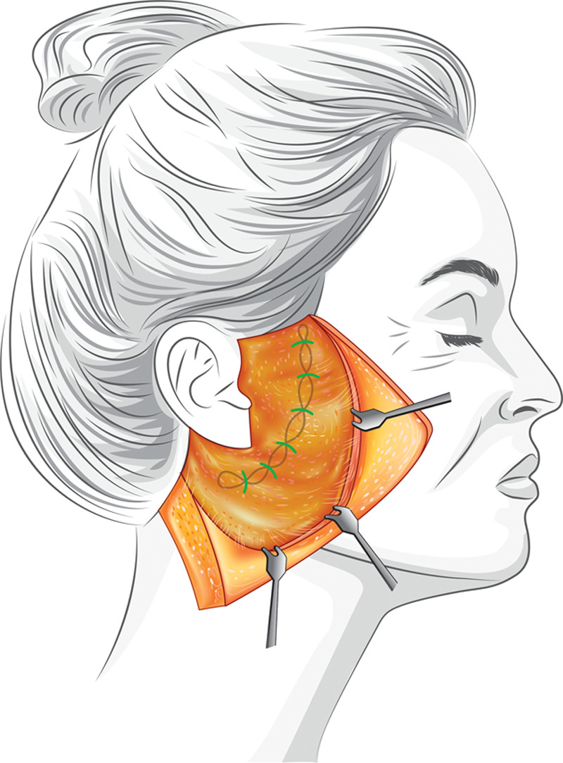
Fig. 4. First layer of SMAS plication, performed with interrupted, buried 2-0 Mersilene sutures. This represents the “load-bearing” layer of SMAS plication.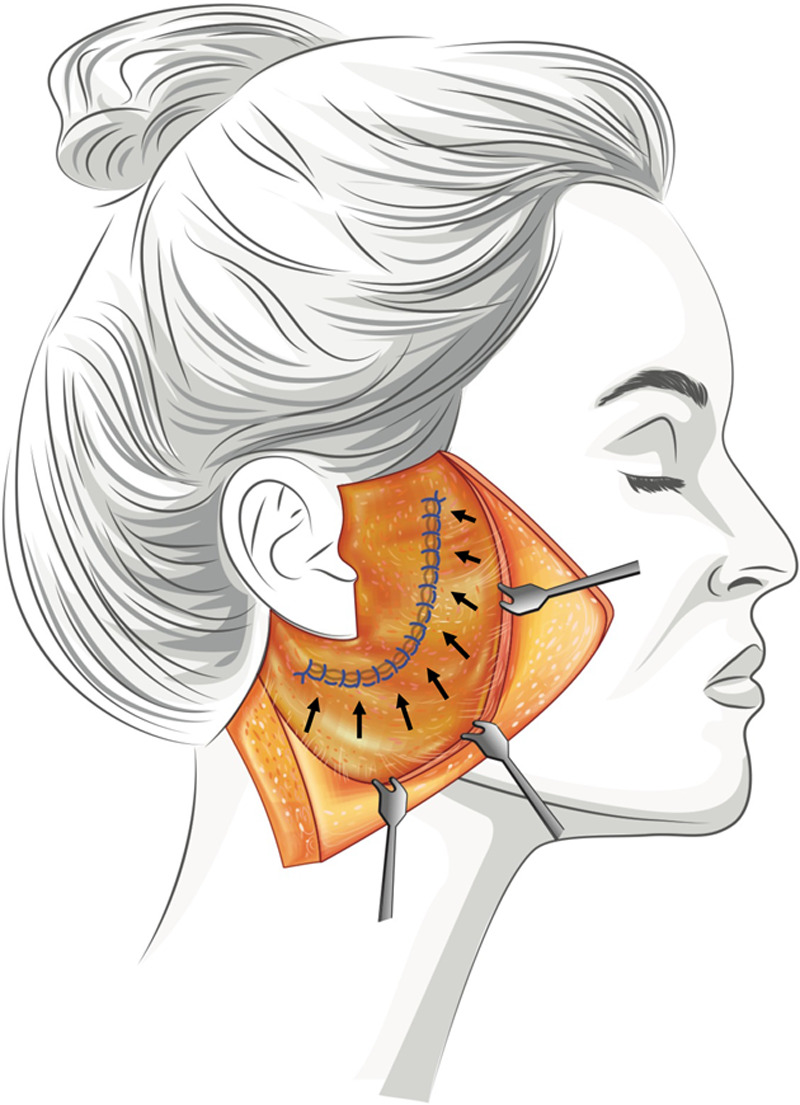
Fig. 5. Second layer of SMAS plication performed with 2-0 Vicryl sutures. This layer of running, locking sutures buries the underlying Mersilene sutures. Black arrows demonstrate corresponding vectors of SMAS plication.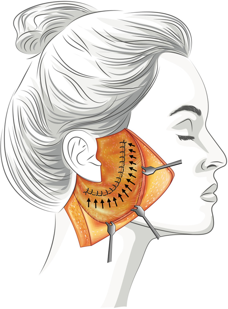
Fig. 6. Third layer of SMAS plication performed with 2-0 Vicryl sutures. This layer of running, locking sutures allows for additional contouring and refinement of the underlying SMAS. Vectors of radial plication are demonstrated with black arrows.Skin is then redraped. The skin redraping and excision follows a primarily posterior trajectory over the face and a posterosuperior trajectory over the neck. Areas of dimpling at the anterior margin of skin flap elevation are released with careful finger dissection at the skin-SMAS interface. Skin is then conservatively trimmed. The skin excision is beveled along the hairline to correspond to the initial incision. Minimal, if any, tension is placed over the skin closure following skin trimming and tailoring. The skin flap is inset with 4-0 Vicryl sutures anteriorly and posteriorly. The lobule is inset with a 3-point suture from skin flap, to lobule, to the underlying SMAS. A 10-French Jackson-Pratt (JP) drain is placed on either side in women. In men, 15-French JP drains are placed. Incisions are then closed with 5-0 Ethilon (Ethicon Inc.) sutures in a running, locking fashion. The incision over the neck is closed with 4-0 Vicryl sutures followed by running, locking 5-0 Ethilon sutures.
Epifoam (Biodermis, Inc., Henderson, NV) is placed to provide uniform compression over the face and neck, followed by a gauze roll and gentle compression with a Coban wrap (3M, Maplewood, MN). Patients are encouraged to ambulate frequently postoperatively and sleep with their head elevated. Patients are seen the following postoperative day (POD). Dressings and drains are removed. Patients are allowed to shower after POD 3. Patients are then seen on POD 7 for suture removal. The majority of patients are able to attend social functions within 1–2 weeks.
补充视频 / 附件(原文未随文提供原图,可于出版方页面查看)gox-7-e2575-s001.mp4Video 1. Neck Liposuction. Video 1 from “Triple-C SMAS Plication Facelift for Natural Facial Rejuvenation”补充视频 / 附件(原文未随文提供原图,可于出版方页面查看)gox-7-e2575-s002.mp4Video 2. Platysmaplasty. Video 2 from “Triple-C SMAS Plication Facelift for Natural Facial Rejuvenation”补充视频 / 附件(原文未随文提供原图,可于出版方页面查看)gox-7-e2575-s003.mp4Video 3. SMAS plication Part 1. Video 3 from “Triple-C SMAS Plication Facelift for Natural Facial Rejuvenation”补充视频 / 附件(原文未随文提供原图,可于出版方页面查看)gox-7-e2575-s004.mp4Video 4. SMAS plication Part 2. Video 4 from “Triple-C SMAS Plication Facelift for Natural Facial Rejuvenation”
The authors performed a retrospective chart review of all patients who underwent triple-C SMAS plication facelift surgery performed by the senior author (K.S.) between January 9, 2017, and January 8, 2018. Data queried included demographic data, operative data, length of follow-up, complication rates, and patient satisfaction rates. Patients with a length of follow-up <100 days were excluded from the study. Patient satisfaction was assessed at patients’ 8-week postoperative visit. Complications assessed included hematoma, seroma, postauricular skin slough >2 cm 2 , facial nerve injury, great auricular nerve injury, hypertrophic scarring, and infection. Hematomas were categorized as major or minor hematomas. Major hematomas were those that required reoperation for treatment. Minor hematomas were hematomas <5 CCs that were treated with aspiration. Patients with any signs of erythematous or elevated scarring at 6 weeks postoperatively were categorized as having hypertrophic scarring and were treated with an injection of a mixture of triamcinolone 40 mg/mL and 5-fluorouracil. Repeat injections were administered at 6-week intervals as indicated. All procedures were in accordance with national and international ethical guidelines for human research and the Helsinki Declaration of 1975.
One hundred ninety-one consecutive facelifts were performed over a 12-month period. One hundred ten patients followed up for at least 100 days and were included in this study. All procedures were performed in the senior author’s outpatient operating facility in Newport Beach, California. Demographic data are summarized in Table 1 . One hundred seven patients were women and 3 were men. Average age was 65.0 years for men and 62.1 years for women. Average BMI was 23.6 kg/m 2 for women and 25.8 kg/m 2 for men. Average follow-up was 404.5 days.
Table 1. Patient Demographics
| n | |
|---|---|
| Men | 3 |
| Women | 107 |
| Nonsmoker | 98 |
| Former smoker | 9 |
| Smoker | 3 |
| Mean | |
| Age (men), y | 65.0 |
| Age (women), y | 62.1 |
| BMI (women), kg/m 2 | 23.6 |
| BMI (men), kg/m 2 | 25.8 |
| Follow-up, d | 404.5 |
The majority of patients underwent a primary facelift (81.8%). Secondary (14.5%) and tertiary (3.6%) facelifts were also performed. A total of 35 isolated facelifts were performed over the study duration. Average operative duration for isolated facelifts was 162.3 minutes. Sixty-five facelift procedures were performed with a total of 143 adjunct procedures. Average operative duration for combined procedures was 195.8 minutes. The most commonly performed adjunct procedures were laser skin resurfacing (35), upper blepharoplasty (33), lower blepharoplasty (15), neurotoxin injections (18), and fat transfer (10). Operative data are summarized in Table 2 .
Table 2. Operative Details
| Operative time | |
|---|---|
| Mean all procedures, min | 185.0 |
| Mean isolated facelift, min | 162.3 |
| Mean facelift + adjunct procedures, min | 195.8 |
| Rhytidectomy | |
| Primary | 90 |
| Secondary | 16 |
| Tertiary | 4 |
| Adjunct procedures | |
| Fat transfer | 10 |
| Upper blepharoplasty | 33 |
| Lower blepharoplasty | 15 |
| Brow lift | 5 |
| Genioplasty | 2 |
| Laser | 33 |
| Hyaluronic acid filler | 7 |
| Neurotoxin | 18 |
| Buccal fat pad excision | 5 |
| Ear lobe reduction | 6 |
| Lesion excision | 3 |
| Chemical peel | 1 |
| Ear lobe reduction | 4 |
| Alar resection | 1 |
| Total | 143 |
Low morbidity was noted. Two major hematomas, 3 minor hematomas, and 5 seromas occurred in the study population. One patient was given a course of oral antibiotics for a mild infection. One neuropraxia involving the zygomatic branch of the facial nerve was noted in a patient undergoing a tertiary facelift. This resolved within 6 months with conservative management. No permanent nerve injuries occurred. Two patients developed postauricular epidermolysis in an area exceeding 2 cm 2 . Twelve patients received steroid and 5-fluorouracil injections for hypertrophic scarring. Three patients developed Vicryl suture extrusion along the incision line; there were no instances of suture extrusion or palpability involving the sutures used for the SMAS plication. Two revision facelift procedures were performed during the study period. Overall patient satisfaction rate was 96.4%. Complications are summarized in Table 3 . Long-term, representative before and after photographs are shown in Figures 7 to 9.
Table 3. Complications
| Minor hematoma | 2.73% |
|---|---|
| Major hematoma | 1.82% |
| Seroma | 4.54% |
| Mild infection | 0.91% |
| Severe infection | 0.00% |
| Hypertrophic scarring | 10.91% |
| Neuropraxia (CN VII) | 0.91% |
| GAN injury | 0.00% |
| Skin slough >2 cm | 1.82% |
| Dehiscence | 2.73% |
| Prolonged edema | 1.82% |
| Prolonged ecchymosis | 0.00% |
| Contour irregularities | 0.91% |
| Pixie ear | 0.00% |
| Revision facelift | 1.82% |
| Scar revision | 3.63% |
| Suture extrusion | 2.73% |
In 2011, the senior author reported his experience with a double-C SMAS plication in 1,532 patients. 12 In the first 614 cases reviewed, the authors utilized a 2-0 Mersilene suture for both layers of the double-C SMAS plication. At this time, the Mersilene suture extrusion rate was 1.9% in women and 3.0% in men. Suture palpability, while not specifically addressed in this case series, was also noted to be an occasional concern. For the subsequent 918 cases in this series, 2-0 Vicryl was utilized for both layers of the SMAS plication, yielding a 0% extrusion rate. Although utilizing Vicryl suture effectively eliminated suture complications, upon reviewing our long-term outcomes, we have felt that Vicryl suture does not offer the long-term reliability of a permanent suture. The triple-C SMAS plication utilizing 2-0 Mersilene for the deeper, load-bearing layer of plication followed by additional plications with 2-0 Vicryl sutures represents an important technical refinement. This technique offers a compromise between the reliability of a permanent Mersilene suture while minimizing suture complications by oversewing the load-bearing layer of the SMAS plication with absorbable Vicryl suture. In this study, although 3 patients developed Vicryl suture extrusion along their incision lines, no suture complications were noted for sutures used for the SMAS plication.
Our data demonstrate that the triple-C SMAS plication can be performed safely and efficiently. Over an average follow-up of 404.5 days, complication rates for this procedure were low. Only 3 major complications were noted in the study period, including 1 facial nerve neuropraxia and 2 major hematomas. The remainder of the complications noted in this study can be categorized as minor complications that resolved with conservative management.
The optimal facelift technique is one that is reliable, reproducible, efficient, and safe. To this end, numerous methods have been formulated that warrant discussion. It has been well established that skin-only techniques that do not utilize the SMAS to restore a youthful contour to the face and neck are inferior to techniques that employ some degree of SMAS modification because they lack reliability and place tension on the skin—a fundamentally elastic structure. 13 Although composite skin-SMAS techniques offer more durability than skin-only techniques, the skin and SMAS must be moved as a single unit in the same direction. These techniques thus lack the versatility of a differential approach to skin and SMAS that addresses these structures separately. 13 , 14 Various strategies within this realm have been discussed, including SMAS plication, SMASectomy, and dual-plane facelift techniques that elevate and translocate separate skin and SMAS flaps.
Although twin studies with long-term follow-up have shown that comparable esthetic outcomes can be achieved with each method, 15 , 16 SMAS plication techniques provide tremendous advantages. Anteriorly based plication techniques elevate sagging fat to soften the nasolabial fold and restore a more youthful contour to the face. 17 When properly planned and performed, SMAS plication procedures can be individually tailored to restructure and sculpt the soft tissues of the face. 18 These techniques offer durability and longevity while avoiding a technically demanding and time-consuming sub-SMAS dissection. Because the SMAS will not be elevated, thicker subcutaneous flaps can be elevated, minimizing the risk of complications such as skin necrosis. Additionally, by avoiding SMAS elevation, the SMAS is not devascularized and potential atrophy of this layer is avoided. Theoretically, plication techniques place the facial nerve at lower risk by avoiding a sub-SMAS dissection. Rates of temporary facial nerve injuries have been estimated to be as high as 3.6% with deep-plane techniques. 20 Indeed, a recent meta-analysis reviewing data from 183 studies demonstrated a lower risk for facial nerve injury, skin necrosis, hematomas, and seromas for SMAS plication techniques. 19
Traditionally, SMAS plication techniques have been described as single-layer, straight-line plications to tighten the face and neck. We feel that these methods lack a degree of versatility. The triple-C SMAS plication offers the flexibility of customizable vectors of plication to independently address laxity and descent of the face and neck with the first layer of the plication in the form of inverted, interrupted sutures. For example, to address significant laxity and descent of the midface, sutures are placed to resuspend the malar fat pad, adapting principles espoused by extended minimal access cranial suspension techniques. 21 The running, locking, curvilinear trajectory of the second and third layers of the triple-C SMAS plication is unique in comparison to traditional, straight-line plications in that it radially gathers, raises, and tightens the deeper structures of the face and neck. This approach better addresses age-related radial expansion of the face when compared with traditional plication techniques.
The double-layer, running, locking sutures offer other important advantages. An experimental study compared the strength of double-layer, running, locking sutures with horizontal mattress sutures in porcine skin specimens. 22 Using a tensometer, the authors demonstrated that the running, locking method required more force to elicit plication failure. These findings suggest that this plication technique may yield more reliable tissue apposition.
It is noteworthy that the neck is routinely opened in the majority of our procedures. In addition to allowing for undermining and skin redraping, the open approach allows liposuction to be performed more safely and precisely. 23 In patients with significant anterior laxity and platysmal banding, the addition of a corset platysmaplasty allows for further refinement. As with the face, the approach to the neck is individualized according to each patient’s needs and goals (Fig. 9 ).
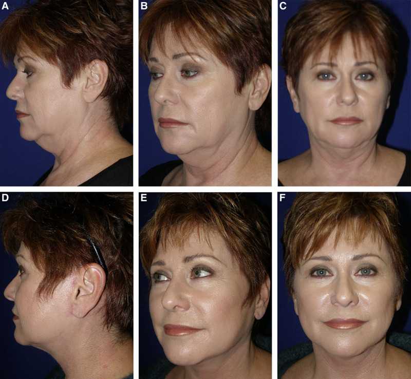
Fig. 7. Pre- and postoperative photographs of a 66-year-old woman who underwent an isolated rhytidectomy at age 54. Photographs are shown preoperatively (A–C) and at 2 months postoperatively (D–F).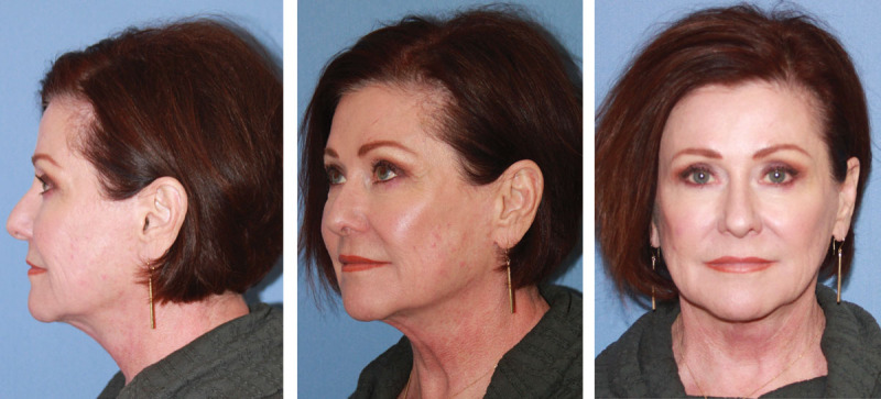
Fig. 8. The patient is shown 12 years postoperatively.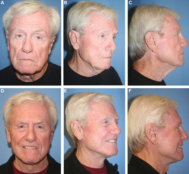
Fig. 9. Pre- and postoperative photographs of an 80-year-old man who underwent rhytidectomy with fat transfer to the face and laser skin resurfacing at age 79. Photographs are shown preoperatively (A–C) and 12 months postoperatively (D–F).Detractors of the SMAS plication technique argue that over time, sutures can tear or “cheese wire” through the SMAS, limiting longevity. However, it is conceivable that this problem could occur with any of the aforementioned techniques. Additionally, SMAS translocation can be limited by the retaining ligaments of the face, which can only be fully released via a sub-SMAS dissection. However, in the senior author’s practice in Newport Beach, California, we have noted that modern facelift patients desire natural and durable facial rejuvenation with minimal down time. Although patients desire an effective and reliable operation, our patients want to soften and highlight the natural, youthful features of the face safely without dramatically altering the architecture of the face.
Since the inception of facelift procedures at the turn of the 20th century, various rhytidectomy techniques have been devised. Long-term data from a cohort of 110 patients demonstrate that the triple-C SMAS plication is a reliable, reproducible, efficient, and safe method for facial rejuvenation. This versatile technique allows for the creation of customizable vectors to lift and radially tighten the deep structures of the face based on preoperative analysis and individual patient needs. This method represents a unique strategy for face lifting by which excellent results can be consistently obtained.
The authors would like to thank Dr. Nicole Rouse, DO, for contributing the illustrations to this work.
声明:中文精读 · 仅供学术参考。内容来自公开文献检索,不代表本人观点,不构成诊疗建议。 医疗美容需在正规医疗机构由执业医师实施。
基于相同主题推荐 · 继续深挖

这项前瞻性、开放标签、随机、多中心(德国11个中心)上市后研究纳入121名受试者,按1:1随机接受 Belotero Volume(CPM-V,n=58)或含利多卡因的 CPM-V
鼻部塑形疗效与安全性:12个月前瞻性研究/PMC13100342_fig1.jpg)
这项单中心前瞻性研究纳入24名中国女性受试者,接受CaHA凝胶鼻部注射(骨膜上/骨膜浅层,单侧单次总量≤2.0 mL,平均0.93±0.22 mL),全部完成12个月随访,无脱落。
研究设计:单中心、单术者回顾性图表复习,2017 年 1 月至 2018 年 1 月连续 191 例除皱手术中 110 例随访满 100 天者纳入分析,平均随访 404.5 天