硅胶下巴假体并发症后的翻修手术:25例并发症队列的术式选择与临床启示
2026年10月12日25 min read
Journal of Clinical Medicine
结论与实践要点
本研究为单中心回顾性病例系列,纳入2019–2025年接受硅胶面部假体植入的98例患者,其中25例(男11例、女14例)发生并发症并接受翻修,21例为外院植入后转诊;最低随访6个月。并发症构成:骨吸收9例(36%)、假体移位3例(12%)、两者并存3例(12%)、患者不满意7例(28%)。骨吸收在固定与未固定假体中均可见到,移位均发生于未行刚性固定者。翻修术式:取出假体+颏部滑行截骨术7例(28%)、定制颏部假体7例(28%)、正颌手术4例(16%)、复位固定1例(4%);翻修后随访期内未再发生并发症。
典型问题提示术前评估的重要性:一例假体上移致骨吸收累及前牙根尖区,取出后行骨增量及正颌手术;一例II类错颌行正颌掩饰治疗加假体,效果不自然;一例假体植入2年后于其上方注射透明质酸,引发生物膜感染仍被迫取出。文献提示未固定假体骨丢失约18.25%,固定者约1%;早期研究报道骨吸收可达3–5 mm(平均30–48个月);总体感染率约0.7%;骨密度较低(如Type 4)者吸收风险或更高。
作者提示:硅胶假体仍有价值,但鉴于骨吸收倾向,使用应审慎;如使用,建议骨膜下放置并以至少一枚钛钉刚性固定、无张力被动放置、尺寸以必要最小为宜;不主张常规用于掩盖II类牙颌面畸形,性别肯定手术中成品假体可靠性亦有限;复杂病例或可采用定制三维假体。局限性:队列异质性大、存在转诊偏倚、随访较短,部分骨吸收发生时间无法确定;证据等级为回顾性病例系列,方案需个体化综合判断。
Background: Silicone chin implants have been widely used is plastic and esthetic surgery of the face being considered as safe and efficient way for chin augmentation. However, complications such as bone resorption, displacement or ectopic bone formation may occur. Methods: The objective of this study was to evaluate complications associated with silicone chin implants and revision surgery protocols. Results: Among 98 patients who received silicone chin implants, 24 (11 males, 13 females) exhibited complications. The most commonly diagnosed issues were displacement ( n = 3), bone resorption ( n = 9), both conditions ( n = 3), and patient dissatisfaction ( n = 7). All patients were qualified for revision surgery, which included silicone implant removal followed by sliding genioplasty ( n = 7), orthognathic surgery ( n = 4), custom-made chin implant placement ( n = 7), and repositioning and fixation ( n = 1). After revision surgery, no complications occurred. Conclusions: Observations from this revision cohort suggest that careful patient selection and consideration of orthognathic or customized implant-based approaches may reduce the risk of dissatisfaction and revision surgery in patients with dentofacial deformities, or those seeking gender confirmation surgeries, compared to stock silicone implants.
Keywords: angle、augmentation、chin、complications、facial implants、implant fixation、malar、silicone、stabilization
Silicone elastomer is a solid, rubber-like polymer composed of polydimethylsiloxane. Silicone facial implants have been established as a versatile tool in contemporary plastic and esthetic facial surgery, as silicone is biocompatible and highly resistant to degradation, with minimal allergic reaction and risk of toxicity [ 1 ]. They have been described as an efficient and less-invasive approach to tissue augmentation than osteotomy-based or bone-grafting techniques, with lower complication rates and morbidity [ 2 , 3 ]. The most commonly used implants are for the chin, mandibular angle, and malar region. Chin implants are used in patients with a receding chin due to aging and/or bone deficiency associated with Class II malocclusion; therefore, they are the most frequently used facial implants, providing an immediate “on-table” effect with a relatively low complication rate. They may also be used as an additive after genioplasty when a deepened labiomental sulcus is diagnosed [ 4 ]. However, bone resorption, implant displacement, and resulting asymmetry have been reported, highlighting the importance of implant stabilization (Niamtu, 2000, 2009) [ 5 , 6 ]. Unstable implants may cause tissue irritation, encapsulation, bone compression and erosion, and facial asymmetry. The current literature provides limited evidence on alloplastic facial implant stabilization and on fixation protocols (sutures, screws, supra- or subperiosteal implant positioning) (Madorsky & Meltzer, 2025) [ 7 ]. Debate continues regarding optimal silicone fixation to prevent these sequelae. This report discusses the clinical manifestations of complications associated with silicone chin implants encountered in the daily practice of plastic and maxillofacial surgeons. It also emphasizes the importance of evaluating malocclusion and considering orthodontic treatment before selecting patients for esthetic surgery.
A retrospective review was conducted on patients who underwent facial reconstruction using silicone facial implants between January 2019 and December 2025.
Eligible patients met all of the following criteria:(1)Facial reconstruction with the use of silicone implants;(2)A minimum follow-up period of 6 months;(3)Availability of complete preoperative and postoperative clinical documentation, including medical records and imaging;(4)Complications were diagnosed.
Before qualification for the operation, facial CT images were obtained using a Carestream 9600 with the following scanning parameters: 120 kV, 5 mAs, and a 300 × 300 × 300 µm voxel matrix (Atlanta, GA, USA). All procedures were carried out in accordance with the Declaration of Helsinki. The study protocol received approval from the Bioethics Committee of the Medical University of Warsaw (AKBE/21/2025). Written informed consent was obtained from all individuals whose clinical photographs are included in this manuscript.
Among 98 patients who attended for primary or secondary procedures with the use of silicone facial implants, 25 of them (11 males—44%, 14 females—56%) exhibited complications and were enrolled into the study ( Table 1 ). Of the analyzed patients, 21 had implants placed elsewhere and were referred for consultation or revision surgery. Indications for chin implant placement were purely esthetic ( n = 10), camouflage of Class II dental deformity ( n = 10), or masculinization of the lower third ( n = 3). The most commonly encountered complications that led to revision surgery were displacement ( n = 3) (12%), bone resorption ( n = 9) (36%) or both ( n = 3) (12%), and patient dissatisfaction ( n = 7) (28%). Displacement was diagnosed in cases where no rigid implant fixation was applied. Bone resorption ranging from mild to severe was seen in both fixed and non-fixed implants ( Figure 1 , Figure 2 , Figure 3 and Figure 4 ). Dissatisfaction was met in the group of patients who underwent chin augmentation with silicone implants due to Class II dentofacial deformity or if they wanted to masculinize their jawline. All patients were qualified for revision surgery: silicone implant removal with subsequent sliding genioplasty ( n = 7) (28%), orthognathic surgery ( n = 4) (16%), custom-made chin implant placement ( n = 7) (28%), and reposition and fixation ( n = 1) (4%) ( Figure 3 and Figure 5 ).
Table 1 Characteristics of the patients enrolled in the study, evaluating indications of the primary surgery, complications, fixation used, and secondary procedures that were performed. Gender: M—male, F—female.
| Patient | Gender | Age (Years) | Indications | Primary | Stabilization with Screws | Complications | Secondary | Plan |
|---|---|---|---|---|---|---|---|---|
| 1 | M | 28 | Masculinization | no | no | Bone resorption | yes | Removal and placement of PEEK custom chin implant |
| 2 | F | 38 | Esthetic | no | no | Displacement and resorption | yes | Removal |
| 3 | M | 25 | Esthetic | no | yes | Bone resorption | yes | Removal |
| 4 | M | 31 | Esthetic | no | no | Dissatisfaction | yes | Removal and placement of PEEK custom chin implant |
| 7 | F | 29 | Class II dentofacial deformity | no | no | Bone resorption | yes | Orthognathic surgery, augmentation |
| 8 | M | 32 | Class II dentofacial deformity | yes | yes | Bone resorption | yes | Orthognathic surgery, bone augmentation |
| 9 | M | 35 | Class II dentofacial deformity | no | yes | Bone resorption | yes | Orthognathic surgery, PEEK angle implants |
| 10 | F | 29 | Class II dentofacial deformity | no | no | Dissatisfaction | yes | Removal and placement of PEEK custom chin implant |
| 11 | F | 45 | Esthetic | no | no | Displacement | yes | Repositioning and stabilization with screws |
| 12 | F | 41 | Esthetic | yes | yes | Infection | yes | Removal |
| 13 | M | 27 | Class II dentofacial deformity | no | yes | Bone resorption | yes | Removal, sliding genioplasty |
| 14 | F | 45 | Esthetic | no | no | Displacement | yes | Removal, sliding genioplasty |
| 15 | M | 30 | Class II dentofacial deformity | no | no | Bone resorption | yes | Removal and placement of PEEK custom chin implant |
| 16 | F | 19 | Class II dentofacial deformity | no | no | Bone resorption | yes | Removal, sliding genioplasty |
| 17 | F | 51 | Esthetic | no | no | Displacement and resorption | yes | Removal, sliding genioplasty |
| 18 | M | 20 | Masculinization | no | yes | Bone resorption | yes | Removal and placement of PEEK custom chin implant |
| 19 | F | 36 | Esthetic | yes | yes | Dissatisfaction | yes | Removal and placement of PEEK custom chin implant |
| 20 | F | 32 | Esthetic | no | no | Displacement | yes | Removal, sliding genioplasty |
| 21 | M | 25 | Class II dentofacial deformity | no | no | Dissatisfaction | yes | Orthognathic surgery |
| 22 | F | 27 | Class II dentofacial deformity | no | no | Dissatisfaction | yes | Removal, sliding genioplasty |
| 23 | F | 36 | Esthetic | no | no | Displacement, resorption | yes | Removal, custom PEEK chin implant |
| 24 | M | 37 | Masculinization | no | yes | Dissatisfaction | yes | Removal, custom PEEK chin implant |
| 25 | M | 40 | Class II dentofacial deformity | no | no | Dissatisfaction | yes | Removal, sliding genioplasty |
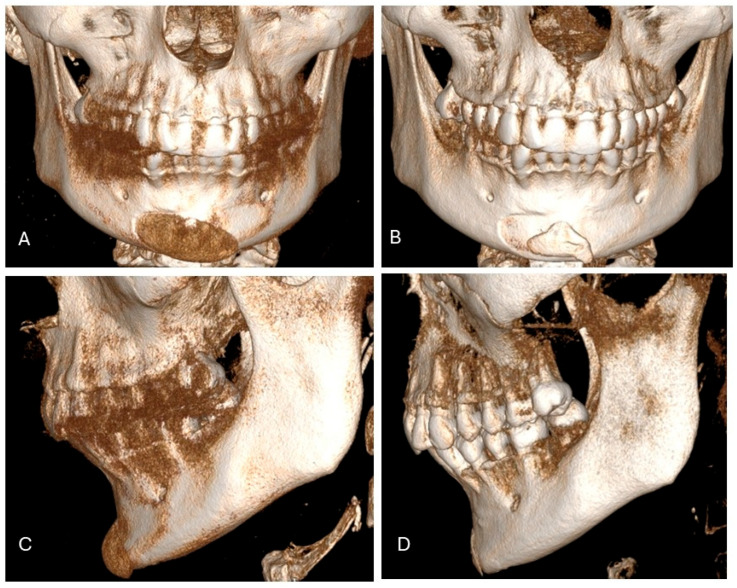
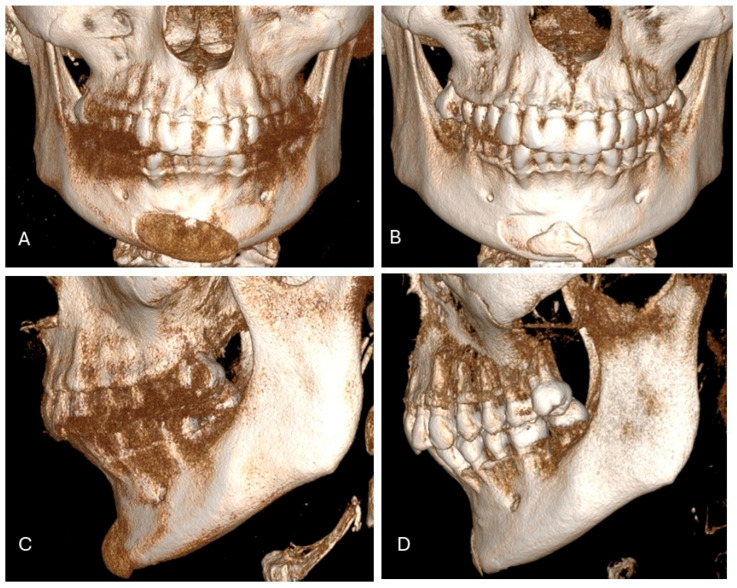
Figure 1 A 42-year-old Caucasian woman presented for consultation because of a mispositioned chin implant placed elsewhere four years earlier (A). She complained of asymmetry, palpable implant mobility, tissue irritation, and mild pain. CT showed implant malposition due to lack of fixation (A,C), with bone resorption and ectopic bone formation (B,D). The patient was scheduled for implant removal with immediate osteoplasty and placement of a custom-made PEEK chin implant.42岁女性:外院植入假体错位4年,CT示骨吸收与异位骨形成;拟取出假体、骨修整后植入定制PEEK颏假体。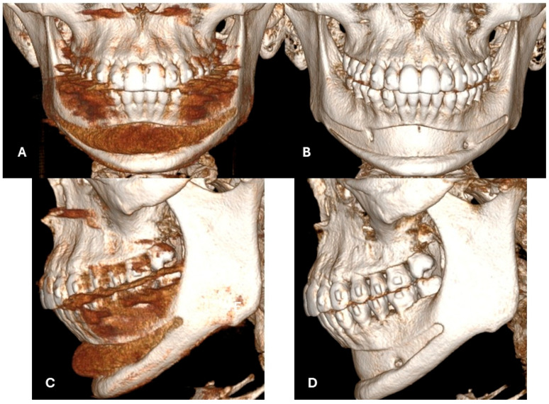
Figure 2 A 21-year-old Caucasian woman who presented for consultation after a failed stock chin-implant placement elsewhere. Clinically, she exhibited Class II malocclusion with vertical overgrowth of the anterior maxilla resulting in a gummy smile, recessed chin, overbite, and retrognathia. She was undergoing orthodontic treatment with removable (camouflage) appliances at the time of consultation. Orthodontic camouflage and implant placement had been attempted to mask the major craniofacial and dental arch relationship anomalies. CT imaging showed the chin implant was positioned inappropriately high relative to the mental protuberance and not on the inferior mandibular border (A–C). This led to implant collapse into the mandibular groove and inadequate soft-tissue projection (B). The implant had been placed passively without stabilization, which caused significant bone resorption along the implant, extending to the apices of the anterior teeth (B,D).21岁女性:II类错颌伴前颌垂直向过长,假体位置过高且无固定,骨吸收扩展至前牙根尖。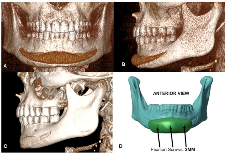
Figure 3 A 30-year-old Caucasian man who presented for consultation after a failed chin implant placed to enhance the lower third’s masculine features. A stock silicone implant had been used and did not provide the expected volumizing effect or masculinization of the mandible (A). CT imaging revealed the implant caused bone resorption along its entire length- anterior view and (B) lateral view. (D). After the digital removal of the silicone implant, reconstructed images confirmed resorption of the bone underneath the implant (C). The surgical plan called for the removal of the silicone implant and immediate placement of a custom-made PEEK chin implant to augment the anterior lower third and create a squarer, more masculine appearance (D).30岁男性:成品硅胶假体男性化效果不足,CT示假体下方全长骨吸收;拟定制PEEK假体。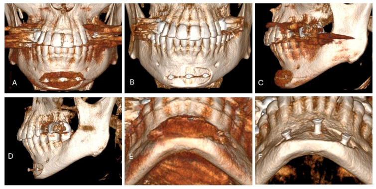
Figure 4 A 31-year-old Caucasian man presented for consultation because he was dissatisfied with a previously placed stock silicone chin implant stabilized with titanium screws (A–F). Although the patient had undergone comprehensive dental and orthodontic treatment, he had a “weak” mandible and jawline from untreated Class II malocclusion. CT showed that, despite bony fixation with titanium screws, the implant had caused mild bone resorption at the implant interface (B,D,E). The treatment plan called for removal of the silicone implant and screws, followed by immediate masculinizing genioplasty and placement of custom mandibular angle implants according to our protocol for lower-third masculinization.31岁男性:钛钉固定假体界面仍见轻度骨吸收;拟男性化颏成形联合定制下颌角假体。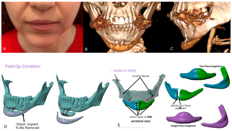
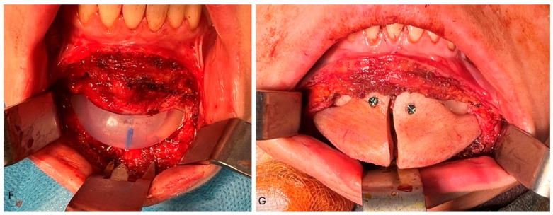
Figure 5 Clinical photographs of a 35-year-old Caucasian woman who presented for consultation because of asymmetry and imbalance of the lower third after prior surgery elsewhere (A). She had a severe Class II malocclusion previously managed with camouflage orthodontics, followed by sliding genioplasty and implant placement one year later (B,C). At the time of consultation, she complained of asymmetry and hollowness at the lateral aspects of the chin resulting from anterior augmentation confined to the central area (A). The treatment plan called for removal of the silicone implant (D,F) and augmentation with a custom two-piece chin implant stabilized with titanium screws (E–G).35岁女性:既往掩饰正畸+颏成形+假体后不对称,取出硅胶假体后以定制两件式假体重建。
Silicone implants have been used in facial surgery since the 1960s and were considered to be one of the safest alloplastic materials, easy to use, and with good results. However, due to increasing reports of complications regarding their longitudinal interference with host tissues, some concerns have been raised regarding their “universal” applications, especially in craniomaxillofacial and esthetic surgery. One of the most commonly discussed issues regarding implants is their displacement due to inadequate fixation, extrusion, bone resorption, and/or spur-like neo-ossification based on subperiosteal bone induction by periosteal distraction [ 1 , 7 , 8 ]. Some studies also describe alteration of facial muscles and lip dysfunction [ 9 ]. This was not observed in the studied cohort.
Implant displacement is associated with a lack of proper stabilization beneath the tissues. In the past, silicone chin implants were routinely placed via submental or intraoral incision and sutured with surgical sutures or wires. Insufficiently stabilized implants may displace, leading to facial asymmetry and/or bone alterations due to micromovements, which was also seen in six presented cases who required revision surgery [ 3 , 10 ]. A chin implant should be placed at the lower border of the mandible and fixed with at least one screw in order to prevent movement. In other cases, it may rotate or be displaced inferiorly or superiorly, leading to resorption of the cortical bone covering the root apices and/or result in inadequate projection, leading to patient dissatisfaction, which was also reported in this manuscript. When not stabilized, it may also result in ectopic bone formation, which consequently also leads to asymmetry and patient dissatisfaction. Also, implants should be placed passively, without any pressure on the bone, as it may induce bone resorption, as was proven by Pearson and Sherris (1999) [ 11 ]. Also, the size of the implant should be as small as needed, as, along with implant size, the risk of bone resorption is increasing according to the study of Wellisz et al. (1995). Also, in the study of Yeung & Wong (2022), it was shown that mandibular bone resorption associated with chin implants highlighted the greatest resorption rates in softer bone types (like Type 4) compared to denser types (Type 1–3) [ 12 ]. Their results indicated a correlation between bone density and resorption outcomes with use of silicone chin implants [ 12 ].
When a too-big implant is used, or a too-small dissection is performed, soft tissue coverage may be insufficient, and implant extrusion may occur. Such scenarios require implant removal with subsequent revision surgery at least a few weeks thereafter. Therefore, silicone chin implants should not be routinely used as camouflage for class II dentofacial deformity, especially in severe cases. This is because they may be insufficient for camouflage, and the effect obtained is unnatural. Also, they may cause bone resorption and the necessity of implant removal with subsequent orthognathic surgery, which is needed in the first place, as seen in the presented cohort. In one presented case, a silicone chin implant was placed onto a bone segment after previously performed genioplasty, both done elsewhere. Such an approach resulted in an unnatural “witch’s chin” deformity. The patient refused orthognathic surgery and thus, a custom extended chin implant was placed in order to restore facial balance ( Figure 5 ).
Bone resorption induced by foreign body response is one of the most discussed issues regarding silicone facial implants. Robinson was the first to describe the process of resorption under silicone and acrylic chin implants, attributed to foreign body giant cell reaction between the implant and the bone or to pressure from the mentalis muscle against the implant (Robinson, 1972) [ 13 ]. The study also revealed that resorption showed up to 3–5 mm at an average of 30–48 months postoperatively, which corresponds to 0.1 mm per month.
Significant resorption poses not only an obvious problem associated with the creation of a bony defect and potential damage to underlying structures like tooth roots, but it also leads to loss of chin projection, which was also seen in the presented cohort [ 9 ]. Hence, a debate about implant positioning and its stabilization has gained attention throughout the last 60 years. Historically, silicone implants have been stabilized by percutaneous temporary sutures as described by von Szalay or Peled, but they did not provide sufficient implant placement, which is still considered one of the factors inducing bone resorption (Peled & Szalay, 1994; Szalay, 1992) [ 14 , 15 ]. Nowadays, subperiosteal implant placement with rigid stabilization with the use of titanium screws is considered a gold standard, yet it still poses some limitations for the use of silicone in the craniofacial area and is associated with the distinct anatomy and physiology of the orofacial region. The mechanism of bone resorption was evaluated by Pearson and Sherris (1999) [ 11 ]. An animal model was used to indicate whether the sub- or supraperiosteal position of the implant promotes osteoclastogenesis. The authors did not observe a significant difference in bone resorption between the sub- or supraperiosteal position of the implants and concluded that some level of resorption is inevitable when silicone implants are applied. The authors concluded that the mechanism of bone resorption under implants that were placed supraperiosteally results from pressure-induced necrosis of the periosteum under the implant. In the case of implants placed directly onto the bone, resorption was a result of periosteal disruption [ 11 ]. From a clinical point of view, subperiosteal placement is more precise, gives better implant alignment with bony curvature, and better mimics natural tissue projection. Also, subperiosteal placed implants are less palpable and tend to migrate in a lesser range than the supraperiosteal position. Also, subperiosteal placement helps to prevent implant exposure.
The placement of chin implants supraperiosteally or without fixation allows for micromovement due to activation of the mentalis. This may lead to erosion into the body of the mandible and/or chronic infection with extrusion [ 9 ]. Bone resorption was also a major causative factor for revision surgery within the presented study, based on secondary sliding genioplasty, adapting 3D custom-made implants or orthognathic surgery. Resorption was diagnosed in both rigid-fixed and non-fixed implants. However, there were no cases with implant migration when rigid fixation was applied. There was one dramatic case, where a superiorly displaced implant induced bone resorption up to the periapical spaces of the teeth. The patient was referred for implant removal with immediate bone augmentation and subsequent orthognathic surgery. This case illustrates clearly when cosmetic procedures such as camouflage orthodontic treatment alongside “minor” esthetic surgery are an inappropriate way of treatment. Overall, non-fixed silicone chin implants are associated with an 18.25% bone loss, versus only 1% in fixated implants, according to the current literature [ 7 , 16 , 17 ]. Hence, in every operated case performed by the Author (primary or revision cases) rigid fixation was applied. According to Madorsky & Meltzer (2025) [ 7 ], subperiosteal placement may, however, induce more complex sequelae of processes that lead to implant “entrapment” under the periosteum, which is induced to promote spur-like neo-ossification based on subperiosteal distraction, which gives a new shade on the conceptualizations based on silicone facial implants. Bone formation between the elevated periosteum and native bone is believed to be induced by distraction osteogenesis mechanisms. This may lead to de novo bone formation above the implant or underneath. New bone forming on top of the implant may induce its compression on the opposing site and bone resorption, leading to implant displacement and asymmetry, which was also seen in one case presented within this study [ 7 ].
Early infections and the necessity of implant removal are rarely seen in silicone chin implants due to the smooth surface being unfavorable for bacterial biofilm formation when compared to other materials. The overall infection rate of silicone chin implants is estimated at 0.7%. However, there were unusual infections of the implants described, secondary to dental procedures. In the study by Danieletto-Zanna et al. (2022) [ 1 ], an infected silicone chin implant had to be removed due to the nonideal placement of dental implants, which developed a bacterial infection [ 1 ]. In another study, an implant had to be removed due to an odontogenic infection spreading into the peri-implant space (Hoffman, 1981) [ 18 ]. In the presented study, there was only one case of implant infection secondary to esthetic procedures performed in the near proximity of the already healed implant, which, in consequence, required removal despite attempts being made to preserve it. These clinical situations clearly show the role of microflora of the oral cavity and risk factors associated with implant contamination from odontogenic or sinus infections (active or developed after implant placement) [ 19 , 20 ].
Among the investigated cohort, there was only one case of implant infection, not related to the surgery itself. It was caused by additional procedures with the use of hyaluronic acid injection at the top of the implant, performed elsewhere two years after implant placement, which caused bacterial biofilm inoculation. Despite efforts being made based on implant debridement and general antibiotic therapy, the implant had to be removed with no procedures performed thereafter. It is evident that any additional procedures based on injectables in the near proximity of the implant may unintentionally be a causative factor if peri-implant infection, and thus, its removal was also seen in the presented cohort [ 21 ].
Patient dissatisfaction was also another cause for revision surgery. This was common in patients with severe Class II dental malocclusion or those who desired masculinization of the lower third. In these cases, either custom-made PEEK implants, genioplasty, or orthognathic surgery was applied. This clearly shows that, along with the development of surgical techniques and 3D modeling, silicone facial implants are gradually being considered less universally applied than before, especially in cases of dentofacial deformities or gender-affirming procedures.
This retrospective study exhibits some limitations. The cohort was heterogeneous; some patients were operated on by different surgeons elsewhere and came for a second opinion and thus, revision surgery. The minimum follow-up for all operated patients (primary or secondary) was six months. However, some patients had implants placed years before the consultation, which resulted in revision surgeries described within this study. In some cases, resorption was asymptomatic and was diagnosed at the time of consultation, making it impossible to assess exactly at which point it began. Additionally, there is currently no information available about the type of bone quality and its tendency for resorption after contact with silicone implants. The current literature does not provide sufficient information on how the age of the patients impacts the tendency for bone resorption after direct contact with silicone implants.
Silicone facial implants still serve as a versatile tool in esthetic facial surgery. However, due to their tendency to induce bone resorption, their use should be judiciously evaluated. They are of great use in minor tissue augmentation in aging faces, and if used, they should be placed in the subperiosteal plane with rigid fixation in order to prevent displacement and bone resorption. These implants should not be routinely used in the camouflage of dentofacial deformities, where sliding genioplasty or orthognathic surgery should be performed in the first place. As stock implants, they are also not found to be a reliable way in gender affirming surgery. In more complex clinical cases or when larger implants are required, 3D solid implants that provide precise alignment within the tissues and better esthetic outcome should be applied.
声明:中文精读 · 仅供学术参考。内容来自公开文献检索,不代表本人观点,不构成诊疗建议。 医疗美容需在正规医疗机构由执业医师实施。
基于相同主题推荐 · 继续深挖

这项前瞻性、开放标签、随机、多中心(德国11个中心)上市后研究纳入121名受试者,按1:1随机接受 Belotero Volume(CPM-V,n=58)或含利多卡因的 CPM-V
回顾性病例系列(2019–2025年):98例硅胶面部假体植入者中25例(男11例44%、女14例56%)发生并发症接受翻修,最低随访6个月。
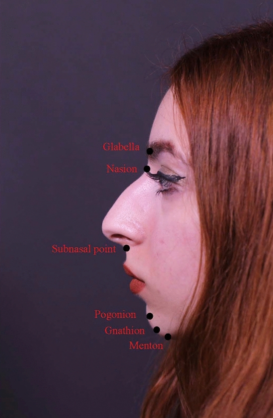
回顾性研究,纳入49例下颌后缩2.5–6 mm的初次隆鼻患者