鼻整形背部轮廓四阶段渐进成形技术:骨性与软骨性操作整合的技术笔记
2026年10月13日22 min read
Plastic and Reconstructive Surgery Global Open
Plastic and Reconstructive Surgery Global Open
结论与实践要点
本文为 Plastic and Reconstructive Surgery Global Open(DOI:10.1097/GOX.0000000000007920)刊登的技术笔记。作者针对鼻整形中最棘手的骨软骨交界区背部轮廓问题,提出 structural contour(结构性轮廓)技术:将骨性与软骨性操作整合为循序渐进的四阶段流程,并在 4 例患者中完成初步描述性应用。需要说明的是,这并非对照研究,文中不包含任何统计量、并发症率或标准化评分数据,其性质是对手术路径与解剖原理的技术性阐述。
四阶段的具体设计为:第一阶段处理骨性框架,依次行 Webster 三角骨切除以缩窄上外侧骨宽、鼻面沟钻孔以界定背外侧轮廓、压电(piezotome)低到低外侧截骨实现精准而创伤较小的骨移动、青枝横向截骨辅助骨穹内侧移,并在存在残余不对称或背高不平时加做旁正中截骨行背部均衡;第二阶段在 keystone 区行骨软骨稳定,以 18G 针在鼻骨钻孔,用 4-0 Monocryl 缝线将鼻骨与上外侧软骨环状固定,恢复骨-软骨界面的受控对合;第三阶段在 W ASA 段行软骨精修,于鼻中隔-上外侧软骨交界(W 点)做约 5 mm 受控松解后,以 5-0 Monocryl 将上外侧软骨下移重置约 2 mm,在改善背线对合的同时保留内鼻阀;第四阶段行软骨背环状缝合,先确认瓣膜通畅,再以连续 5-0 PDS 缝线在距背表面约 2 mm 的后方向 keystone 区走行,稳定鼻侧壁并界定上外侧软骨的休息角,最终形成圆柱形背部构型。
在结果方面,作者报告 4 例患者均按个体解剖特征与美学目标完成了渐进式背部雕刻,各阶段术前-术后正位照片显示背美学线由不连续、不对称逐步转为平行、清晰,且术中确认内鼻阀保持通畅(以上外侧软骨休息角约 ±30 度为参考)。原文未报告任何量化结局、随访时长或不良事件数据,结论停留在技术描述层面,即该流程为背部轮廓管理提供了系统化、可重复、可调整的路径。
对整形外科医生的实操启示有两点。其一,该技术的真正卖点在于「分而治之」的顺序感:先用骨性步骤建立稳定的穹顶基础,再处理骨软骨交界,最后才动软骨背线,避免传统一步到位的背部处理在切皮光线照射下暴露骨-软骨过渡区的不规则。其二,keystone 区钻孔固定与 2 mm 下移重置都属毫米级操作,适合背高基本合适、仅需精修轮廓的病例;对于需要显著降低背高或重塑穹顶的患者,本文技术并非针对性方案。并发症防范方面应关注:截骨序列不当可致骨穹不对称或夹板样移位;keystone 钻孔过深或缝线张力不当可能影响骨-软骨贴合并产生可触及的不规则;W 点松解过度有损中穹支撑与鼻阀功能,故术中反复确认瓣膜通畅这一环节不可省略。
证据边界必须向读者讲清楚:本文为 4 例的技术笔记,没有对照组、没有客观测量(如鼻阀功能检查、NOSE 评分)、没有并发症统计,也没有学习曲线评估;作者本人亦承认多阶段技术存在固有学习曲线,且依赖压电设备、柱形磨头等专门器械。文中所述一切效果均来自术者主观影像观察,任何机构开展此技术前,宜先以小样本、标准化摄影与鼻功能评估逐步验证,不宜将文中美学效果直接外推为普适性结局。
Dorsal definition represents a persistent challenge in rhinoplasty, especially at the osteocartilaginous junction. We present the structural contour technique, a progressive 4-phase surgical approach designed to achieve a more defined and stable nasal dorsum through combined bony and cartilaginous maneuvers. The technique includes bony framework modification through targeted ostectomy and remodeling, osteochondral stabilization at the keystone area using cerclage sutures, controlled inferior reinsertion of the upper lateral cartilages, and final dorsal contour refinement with a continuous absorbable suture to achieve a cylindrical configuration while preserving nasal valve function. In a preliminary descriptive experience involving 4 patients, this technical approach allowed tailored dorsal contour refinement according to individual anatomical and aesthetic requirements. The structural contour technique provides a systematic and reproducible framework for dorsal refinement, integrating bone and cartilage in a controlled and harmonious manner.
Question: How can progressive dorsal refinement be achieved in rhinoplasty while maintaining a stable osteocartilaginous framework and preserving nasal valve function?
Findings: The structural contour technique introduces a 4-phase approach that sequentially integrates osseous and cartilaginous maneuvers, including bony remodeling, osteochondral alignment with cerclage fixation, inferior reinsertion of the upper lateral cartilages, and final dorsal cerclage refinement. This stepwise method allows progressive dorsal definition tailored to each patient’s aesthetic goals.
Meaning: A systematic and reproducible strategy for achieving stable, functional, and harmoniously contoured nasal dorsal lines in rhinoplasty.
The nasal dorsum is a pivotal determinant of facial aesthetics. 1 The osteocartilaginous junction represents a critical transition zone where even subtle irregularities become visible under tangential light. 2 Although dorsal preservation techniques have gained popularity over the last decade, they may not consistently ensure smooth integration between bone and cartilage, especially in patients with preexisting deformities or previously modified dorsums. 3 , 4
To address this limitation, we propose the structural contour technique, a systematic, 4-phase approach that integrates osseous and cartilaginous maneuvers to achieve well-defined, stable, and functional dorsal lines.
The surgical steps are presented for didactic and illustrative purposes, focusing on the technical sequence of the procedure and its anatomical rationale, rather than perioperative management or postoperative care. A detailed operative demonstration of the 4-phase structural contour technique is provided in the Video. ( See Video [online] , which shows the 4-phase approach, including bony framework modification, osteochondral stabilization at the keystone area, cartilaginous refinement, and final dorsal contour definition.)
The 4-phase dorsal approach was applied to adapt surgical planning to each patient’s anatomical characteristics and aesthetic requirements. In cases of a preserved dorsum, either naturally aligned without requiring modification of height or projection, 5 or previously managed using a dorsal preservation technique (excluding the “ballerina maneuver” described by Goksel), 6 the nasal dorsum was systematically treated in 4 sequential phases.
Phase 1 focuses on controlled preparation and equalization of the bony framework to achieve dorsal symmetry and initiate progressive narrowing. The objective of this phase is to establish a stable and balanced osseous foundation that facilitates subsequent osteochondral and cartilaginous refinement.
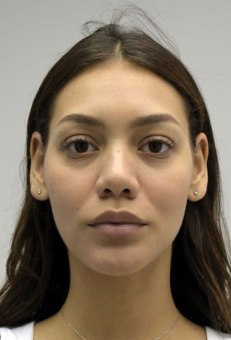
Preoperative views illustrating baseline dorsal morphology before bony framework modification are shown in Figure 1 . A schematic overview of the planned bony framework modifications is provided in Figure 2 . ( See figure, Supplemental Digital Content 1 , which shows phase 1: structural contouring. Preoperative left lateral view, https://links.lww.com/PRSGO/F45.)
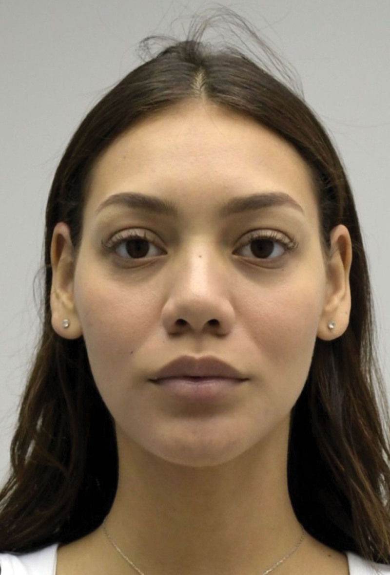
Fig. 1. Phase 1: Structural contouring. Preoperative frontal view.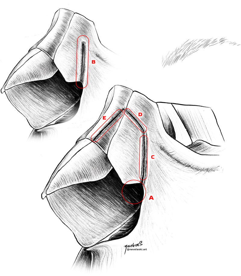
Fig. 2. Phase 1: bony framework. Stepwise illustration of osteotomies and bone modifications: (A) Webster triangle ostectomy; (B) formation of nasofacial groove; (C) low-to-low lateral osteotomy with piezotome; (D) greenstick transverse osteotomy; and (E) paramedian osteotomy for dorsal equalization.The sequence begins with targeted ostectomy of the Webster triangle at the maxillopyriform angle to reduce excessive lateral bony width (Fig. 3 ), followed by creation of the nasofacial groove through controlled bony drilling to define the lateral dorsal contour (Fig. 4 ). A complete low-to-low lateral osteotomy is then performed using a piezotome to allow precise and atraumatic mobilization of the nasal bones (Fig. 5 ). This is followed by a greenstick transverse osteotomy to facilitate controlled medialization of the bony vault (Fig. 6 ). When indicated to correct residual asymmetry or unequal dorsal height, a complete paramedian osteotomy is performed to achieve dorsal equalization (Fig. 7 ).
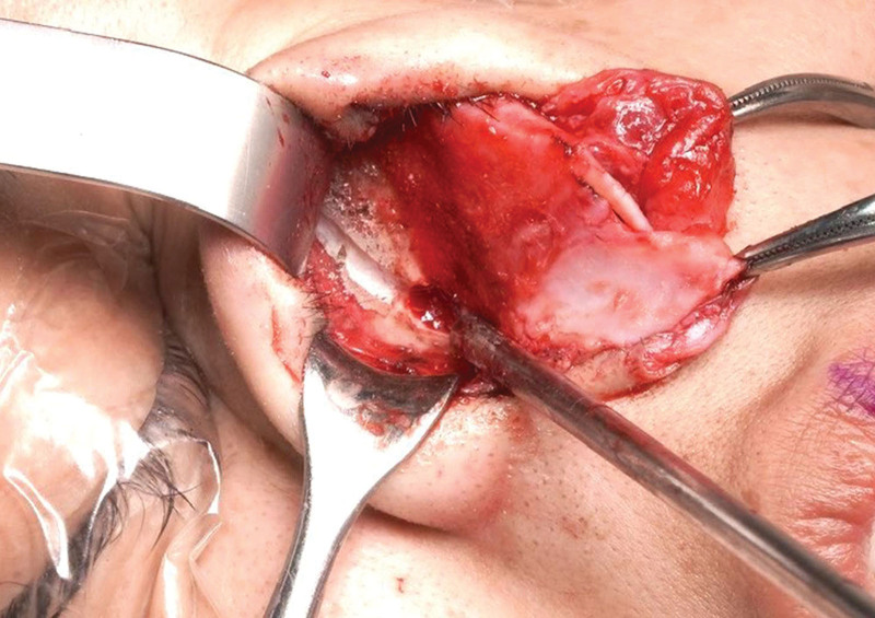
Fig. 3. Webster triangle ostectomy. Intraoperative exposure of the maxillopyriform angle following targeted bony resection, representing the first step of phase 1 in structural contouring.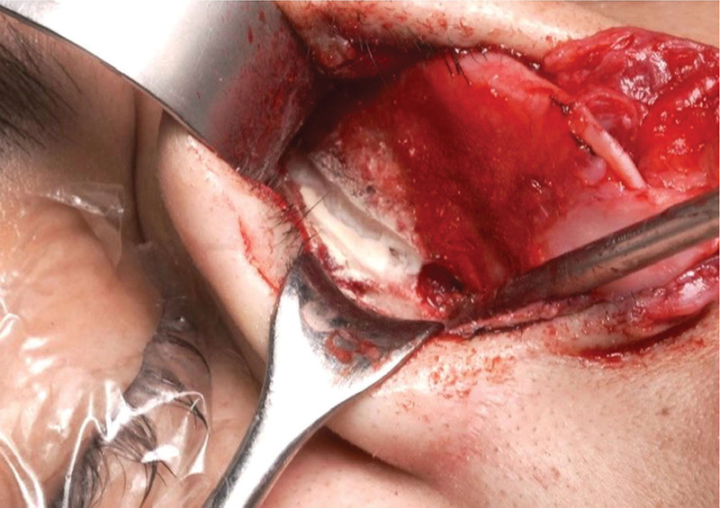
Fig. 4. Nasofacial groove drilling. Intraoperative view showing the creation of a bony channel along the nasofacial groove, corresponding to the second step of phase 1 in structural contouring.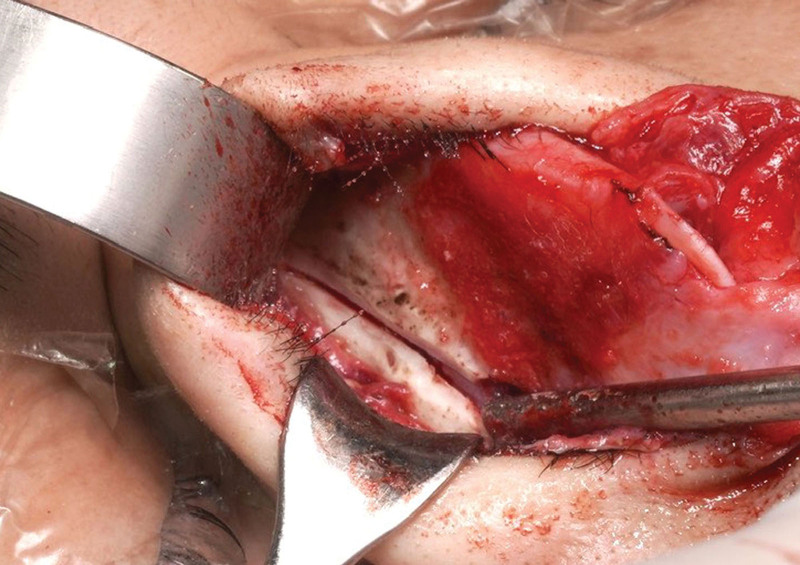
Fig. 5. Complete lateral osteotomy with piezotome. Intraoperative view of the low-to-low lateral osteotomy, representing the third step of phase 1 in structural contouring.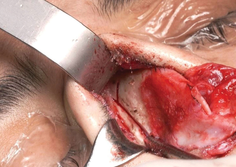
Fig. 6. Greenstick transverse osteotomy. Intraoperative image of the transverse osteotomy, representing the fourth step of phase 1 in structural contouring.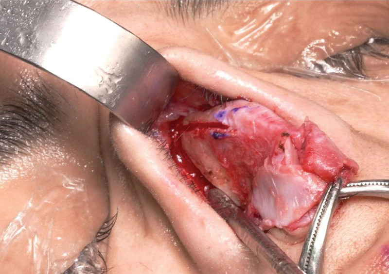
Fig. 7. Complete paramedian osteotomy. Intraoperative exposure of the paramedian osteotomy, representing the fifth step of phase 1 in structural contouring.Postoperative views after completion of phase 1 are presented in Figure 8 . ( See figure, Supplemental Digital Content 2 , which shows phase 1: structural contouring. Postoperative left lateral view, https://links.lww.com/PRSGO/F46.)
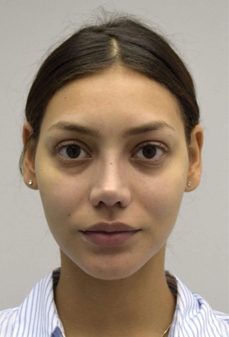
Fig. 8. Phase 1: structural contouring. Postoperative frontal view following completion of bony framework modification.
Phase 2 addresses stabilization of the osteocartilaginous junction at the keystone area, a critical transition zone for dorsal continuity and structural support. The objective of this phase is to restore controlled alignment between the nasal bones and the upper lateral cartilages, ensuring a smooth and stable dorsal contour.
Preoperative views before keystone stabilization are shown in Figure 9 . A schematic representation of the planned osteochondral stabilization strategy is provided in Figure 10 . ( See figure, Supplemental Digital Content 3 , which shows phase 2; structural contouring. Preoperative left lateral view, https://links.lww.com/PRSGO/F47.)
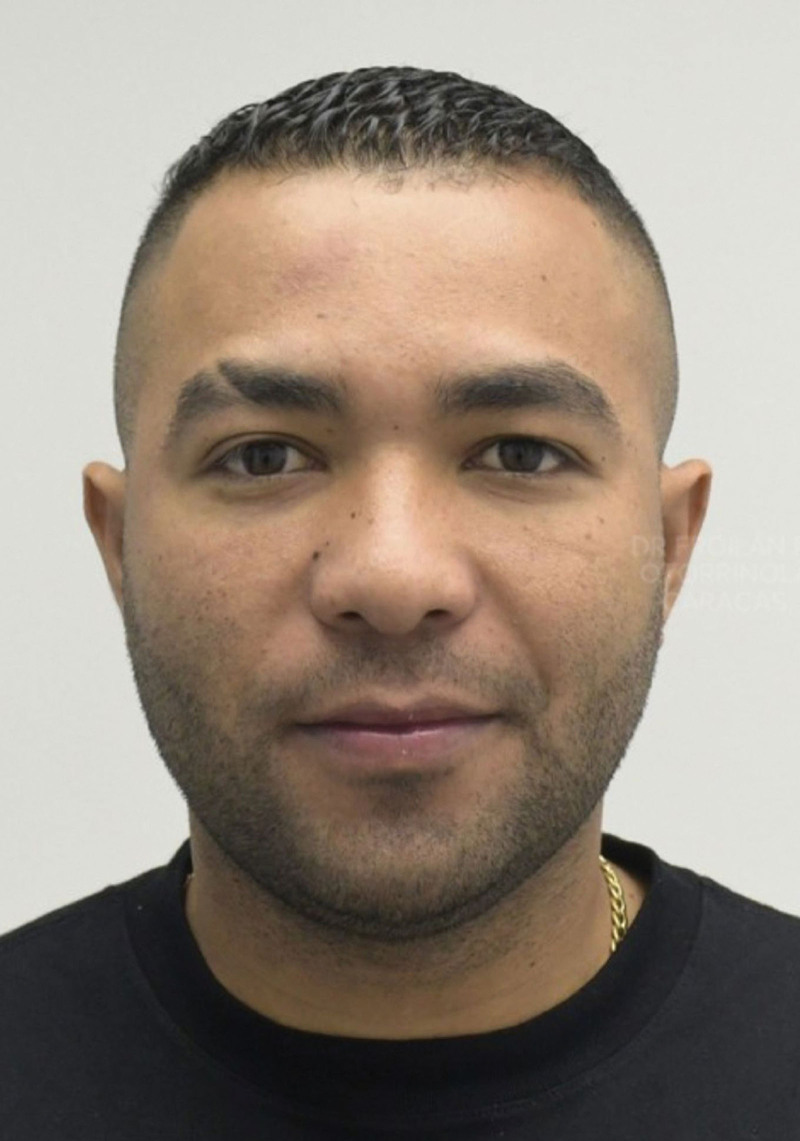
Fig. 9. Phase 2: structural contouring. Preoperative frontal view.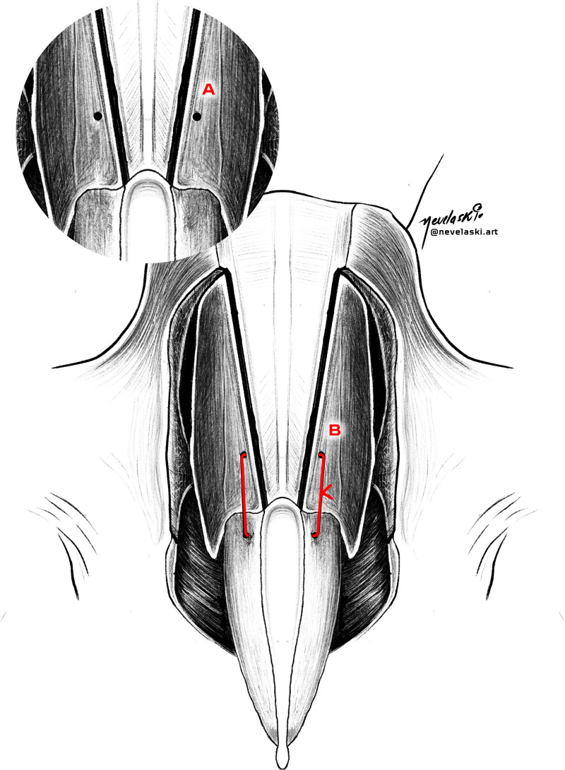
Fig. 10. Phase 2: osteochondral stabilization (keystone area). Illustrative representation of the main steps: (A) Drill hole in the nasal bone and (B) fixation of the nasal bone to the upper lateral cartilages.Intraoperatively, a drill hole is created in the nasal bone using an 18-gauge needle to facilitate precise passage of the fixation suture (Fig. 11 ). The nasal bones are then fixated to the upper lateral cartilages using a 4-0 Monocryl suture, achieving controlled osteochondral alignment and continuity across the keystone area.
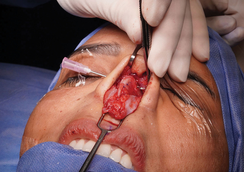
Fig. 11. Osteochondral stabilization (keystone area). Intraoperative image showing creation of a drill hole in the nasal bone and fixation of the nasal bone to the upper lateral cartilage.Postoperative views demonstrating dorsal contour following completion of phase 2 are presented in Figure 12 . ( See figure, Supplemental Digital Content 4 , which shows phase 2: structural contouring. Postoperative left lateral view, https://links.lww.com/PRSGO/F48.)
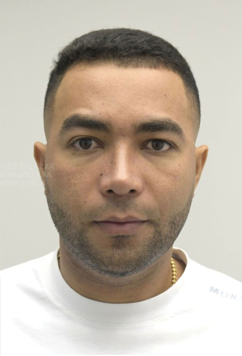
Fig. 12. Phase 2: structural contouring. Postoperative frontal view.
Phase 3 focuses on controlled cartilaginous refinement at the W ASA segment to optimize dorsal alignment while preserving internal nasal valve function. The objective of this phase is to modulate the resting position of the upper lateral cartilages, contributing to dorsal stability and smooth contour transition.
Preoperative views before cartilaginous refinement are shown in Figure 13 . A schematic illustration of the planned cartilaginous modifications at the W ASA segment is provided in Figure 14 . ( See figure, Supplemental Digital Content 5 , which shows phase 3: structural contouring. Preoperative left lateral view, https://links.lww.com/PRSGO/F49.)
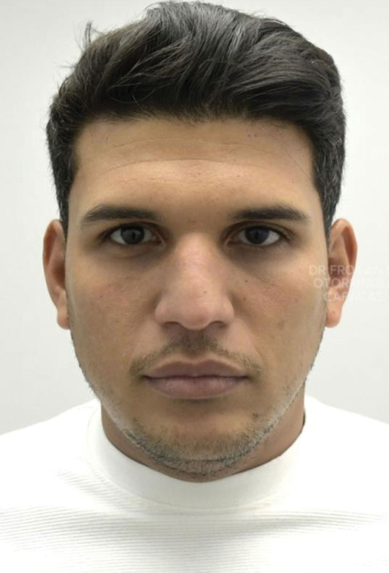
Fig. 13. Phase 3: structural contouring. Preoperative frontal view.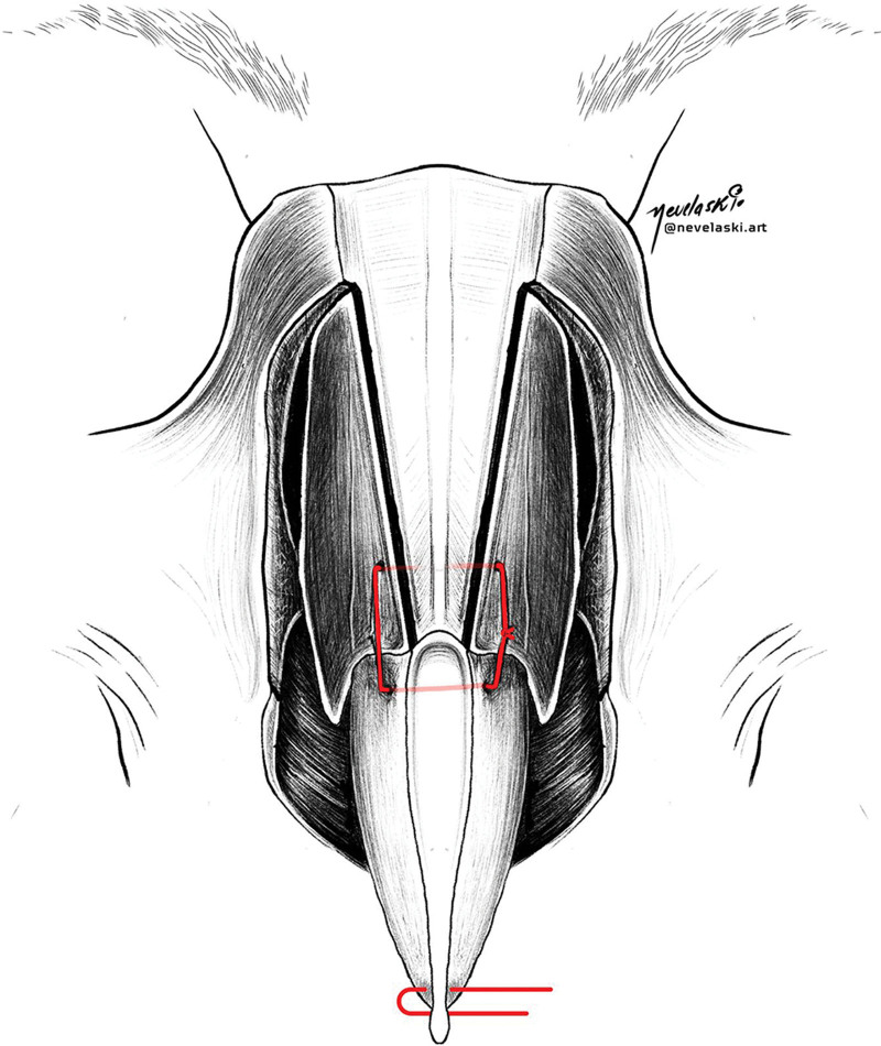
Fig. 14. Phase 3: cartilaginous refinement (W ASA segment). Illustrative image of key steps: 5 mm release at septum–upper lateral cartilage junction; 2 mm inferior reinsertion of upper lateral cartilages; preservation of the internal nasal valve.A controlled release of approximately 5 mm is performed at the septum–upper lateral cartilage junction (W point) to allow mobilization of the cartilaginous framework. Inferior reinsertion of the upper lateral cartilages onto the septum is then performed with a 2-mm advancement using 5-0 Monocryl sutures (Fig. 15 ). This maneuver permits controlled adjustment of the resting angle of the upper lateral cartilages, contributing to improved dorsal alignment and enhanced contour stability while maintaining internal nasal valve patency. Preservation of the internal nasal valve is confirmed during the procedure. ( See figure, Supplemental Digital Content 6 , which shows the maneuver to confirm internal nasal valve angulation ±30 degrees, https://links.lww.com/PRSGO/F50.)
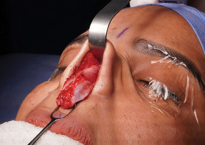
Fig. 15. Cartilaginous refinement (W ASA segment). Intraoperative image demonstrating release at the septum–upper lateral junction (W point), inferior reinsertion of the upper lateral cartilages, and formation of the W ASA segment.Postoperative views demonstrating improved dorsal support and preservation of internal nasal valve function are presented in Figure 16 . ( See figure, Supplemental Digital Content 7 , which shows phase 3: structural contouring. Postoperative left lateral view, https://links.lww.com/PRSGO/F51.)
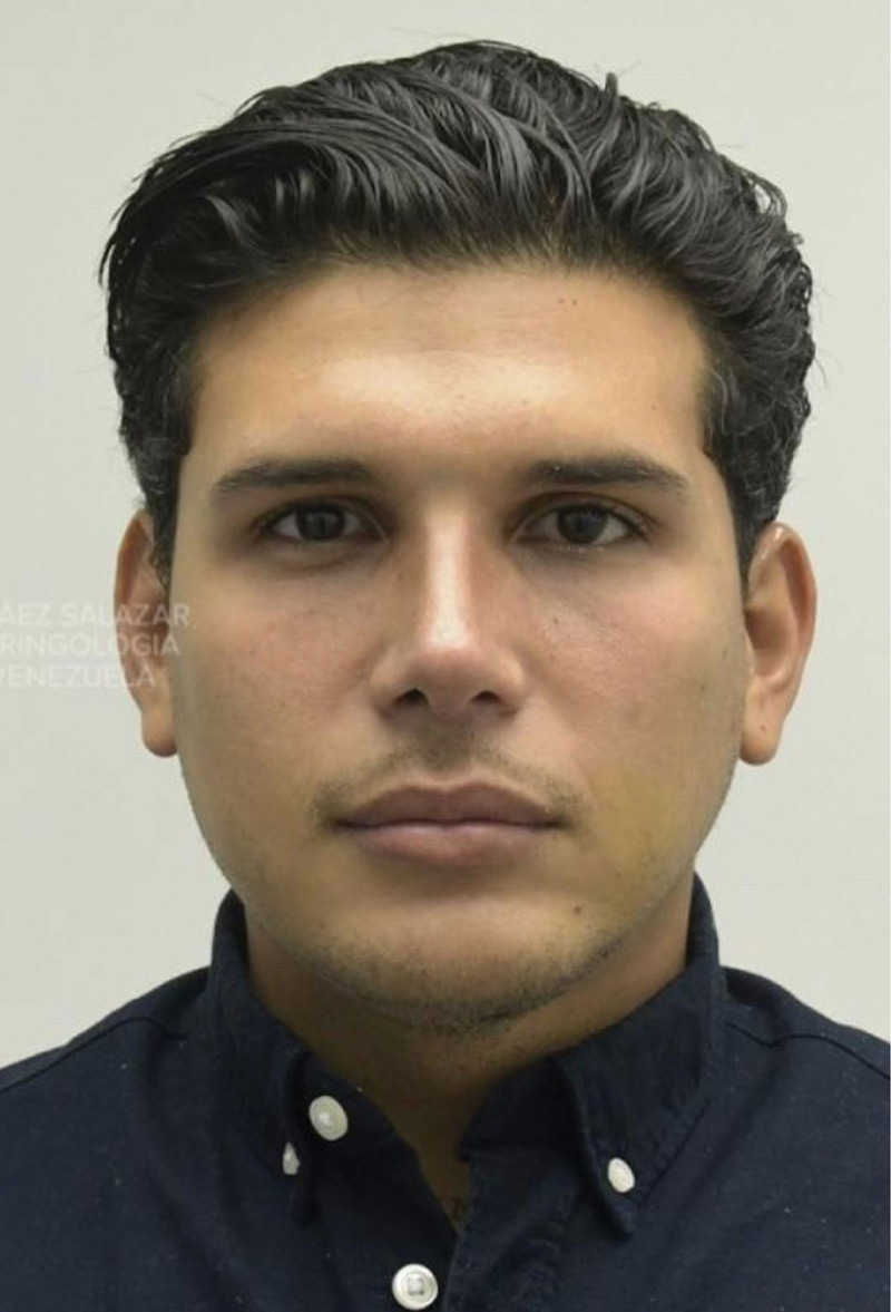
Fig. 16. Phase 3: structural contouring. Postoperative frontal view.
The final phase consists of cartilaginous dorsum cerclage to refine and stabilize the dorsal aesthetic lines. The objective of this phase is to achieve parallel, well-defined, and durable dorsal contours while maintaining nasal valve patency.
Preoperative views before final dorsal refinement are shown in Figure 17 . A schematic overview of the cartilaginous dorsum cerclage sequence is provided in Figure 18 . ( See figure, Supplemental Digital Content 8 , which shows structural contouring. Preoperative left lateral view, https://links.lww.com/PRSGO/F52.)
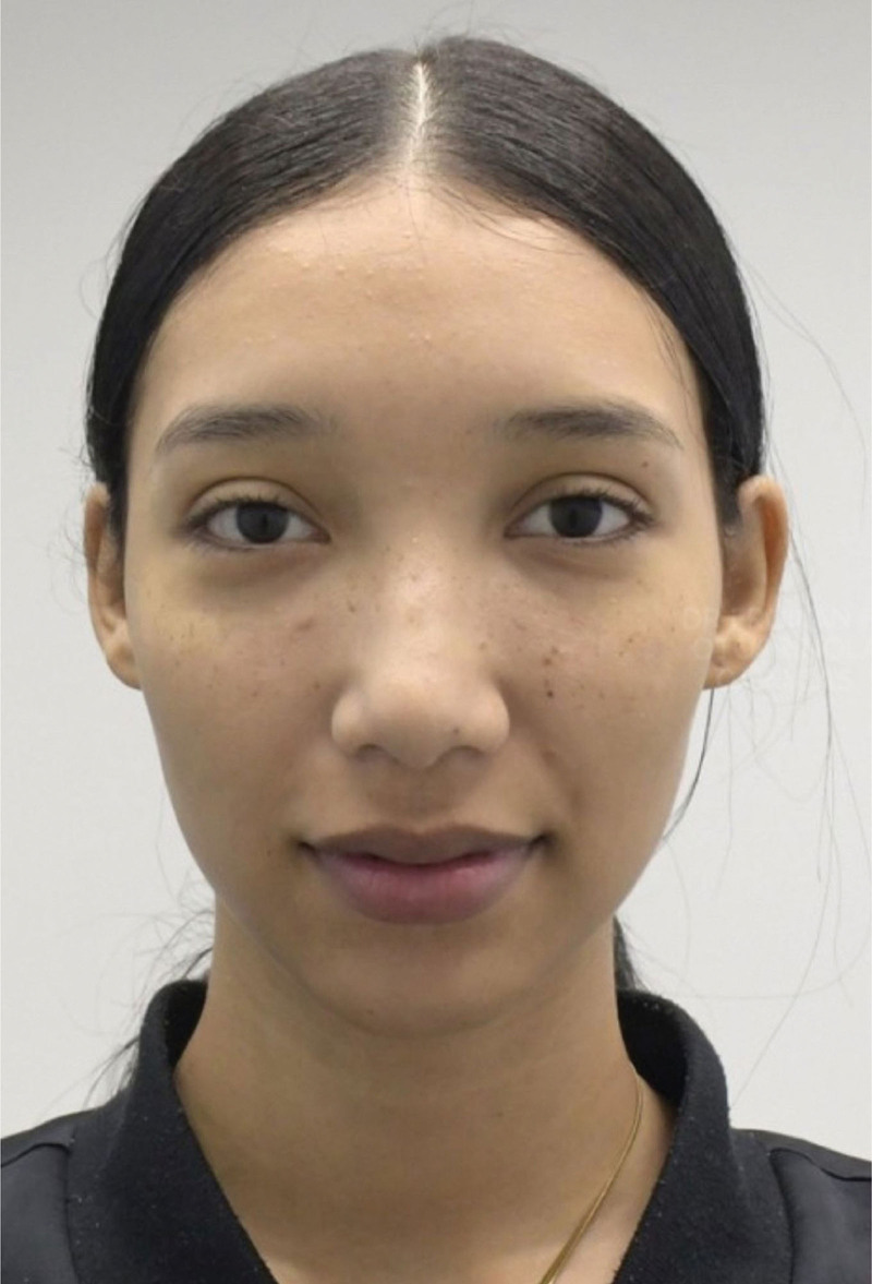
Fig. 17. Phase 4: structural contouring. Preoperative frontal view.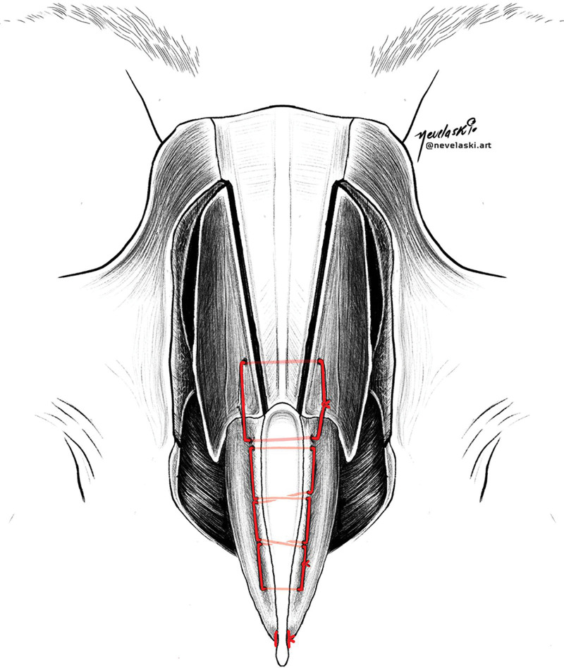
Fig. 18. Phase 4: cartilaginous dorsum cerclage. Illustrative depiction of continuous suture 2 mm posterior to the dorsum to stabilize the lateral wall and create the resting angle of the upper lateral cartilage.Intraoperatively, functional valve patency is first confirmed. A continuous 5-0 PDS suture is then placed approximately 2 mm posterior to the dorsal surface, extending toward the keystone area to stabilize the lateral nasal wall and define the resting angle of the upper lateral cartilage (Fig. 19 ).
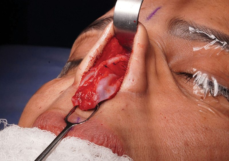
Fig. 19. Cartilaginous dorsum cerclage. Intraoperative image demonstrating continuous cerclage suture from the unobstructed area to the keystone, 2 mm posterior to the dorsum, stabilizing the lateral wall and defining the resting angle of the upper lateral cartilage.Postoperative views demonstrating restoration of parallel and well-defined dorsal aesthetic lines are presented in Figure 20 . ( See figure, Supplemental Digital Content 9, which shows structural contouring. Postoperative left lateral view, https://links.lww.com/PRSGO/F53.)
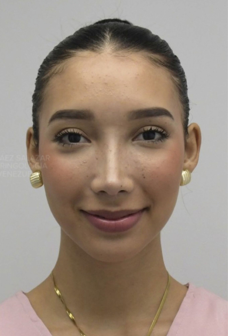
Fig. 20. Phase 4: structural contouring. Postoperative frontal view.Postoperative management followed standard institutional protocols and is not the focus of this technical description.
补充视频 / 附件(原文未随文提供原图,可于出版方页面查看)gox-14-e7920-s001.mp4Video 1. Demonstration of the structural contour technique in rhinoplasty. The video illustrates the four-phase approach, including bony framework modification, osteochondral stabilization at the keystone area, cartilaginous refinement, and final dorsal contour definition.
Dorsal contour refinement remains one of the most challenging aspects of rhinoplasty, particularly at the osteocartilaginous junction, where subtle irregularities may become visually apparent due to the complex interaction between bony and cartilaginous structures. 7 , 8 Achieving stable integration across this transitional zone is essential for maintaining dorsal definition, structural continuity, and long-term aesthetic harmony. 9
The structural contour technique introduces a reproducible 4-phase surgical framework for progressive dorsal refinement. 10 Each phase fulfills a specific technical objective, including controlled modification of the bony vault, osteochondral stabilization at the keystone area, targeted cartilaginous repositioning, and final dorsal contour definition into a cylindrical configuration. 11 , 12 This structured sequencing allows the surgeon to address dorsal contouring in a systematic and adaptable manner according to individual anatomical requirements.
Preservation approaches have emerged as a key strategy in contemporary rhinoplasty by prioritizing maintenance of native dorsal anatomy and minimizing disruption of osteocartilaginous relationships. 13 However, in patients presenting with preexisting asymmetries, inherent dorsal irregularities, or prior nasal surgery, these strategies may not consistently provide adequate contour integration or predictable dorsal alignment. 14 – 16 In this context, structured techniques that permit controlled reshaping and stabilization of both osseous and cartilaginous components may contribute to improved dorsal aesthetic continuity and surgical predictability.
The proposed approach also builds upon established biomechanical principles of cartilage behavior. The concept of a “resting angle,” previously described for the lower lateral cartilages, is extended in this technique to the upper lateral cartilages, providing a rationale for controlled modulation of middle vault architecture and dorsal contour alignment. This contributes to enhanced structural stability while preserving internal nasal valve function and avoiding excessive tension on surrounding tissues. 17
Recent reports have highlighted both structural strategies for dorsal framework stabilization and the emerging role of biologic approaches aimed at improving cartilage regeneration and long-term contour predictability in rhinoplasty. Techniques involving autologous grafting and tissue engineering concepts have been explored to enhance soft tissue adaptation and functional reliability of nasal reconstruction. In this context, mechanically driven dorsal contouring techniques such as the structural contour framework described in this report, may provide a structured platform for the future integration of regenerative adjuncts designed to optimize osteocartilaginous healing and aesthetic durability. 18 – 20
From a technical standpoint, the structural contour technique offers a structured and reproducible operative framework characterized by progressive intraoperative adaptability and integrated management of osseous and cartilaginous components within a single surgical strategy. Rather than promoting a specific aesthetic paradigm, the technique is intended to function as a versatile tool that can be tailored according to individual anatomical characteristics and aesthetic objectives through careful surgical planning.
Limitations include the inherent learning curve associated with multiphase structural techniques and the requirement for specialized instrumentation such as piezoelectric devices and cylindrical burrs. 21 – 23 Given the descriptive nature of this technical note and the preliminary clinical experience presented, formal assessment of reproducibility, complication rates, functional outcomes, standardized NOSE scoring, and learning curve dynamics was beyond the scope of the present report.
Future prospective studies involving larger patient cohorts, longer follow-up periods, and standardized outcome measures will be necessary to further validate the clinical applicability, reproducibility, and functional impact of this technique.
This report is intended to describe the technical rationale and stepwise execution of the proposed approach, rather than to assess comparative clinical outcomes, in keeping with the scope of a technical note.
The structural contour technique provides a systematic, stepwise approach for dorsal refinement that integrates bony, osteochondral, and cartilaginous maneuvers within a single surgical framework. By addressing the nasal dorsum as a continuous structural unit, this technique allows controlled shaping of dorsal aesthetic lines while preserving nasal valve function. As a technical note, this report aims to describe the surgical rationale and execution of this approach, offering a reproducible and adaptable method for dorsal contour management in selected rhinoplasty cases. Further prospective studies with larger patient cohorts and longer follow-up will be required to validate clinical outcomes and reproducibility of the described maneuvers.
The authors have no financial interest to declare in relation to the content of this article.
The patients provided written consent for the use of their images.
This technical note was conducted in accordance with the ethical standards of the institutional and national research committee and with the 1964 Declaration of Helsinki and its later amendments. Written informed consent was obtained from all patients for the surgical procedures, clinical photography, and the publication of anonymized clinical data.
声明:中文精读 · 仅供学术参考。内容来自公开文献检索,不代表本人观点,不构成诊疗建议。 医疗美容需在正规医疗机构由执业医师实施。
基于相同主题推荐 · 继续深挖
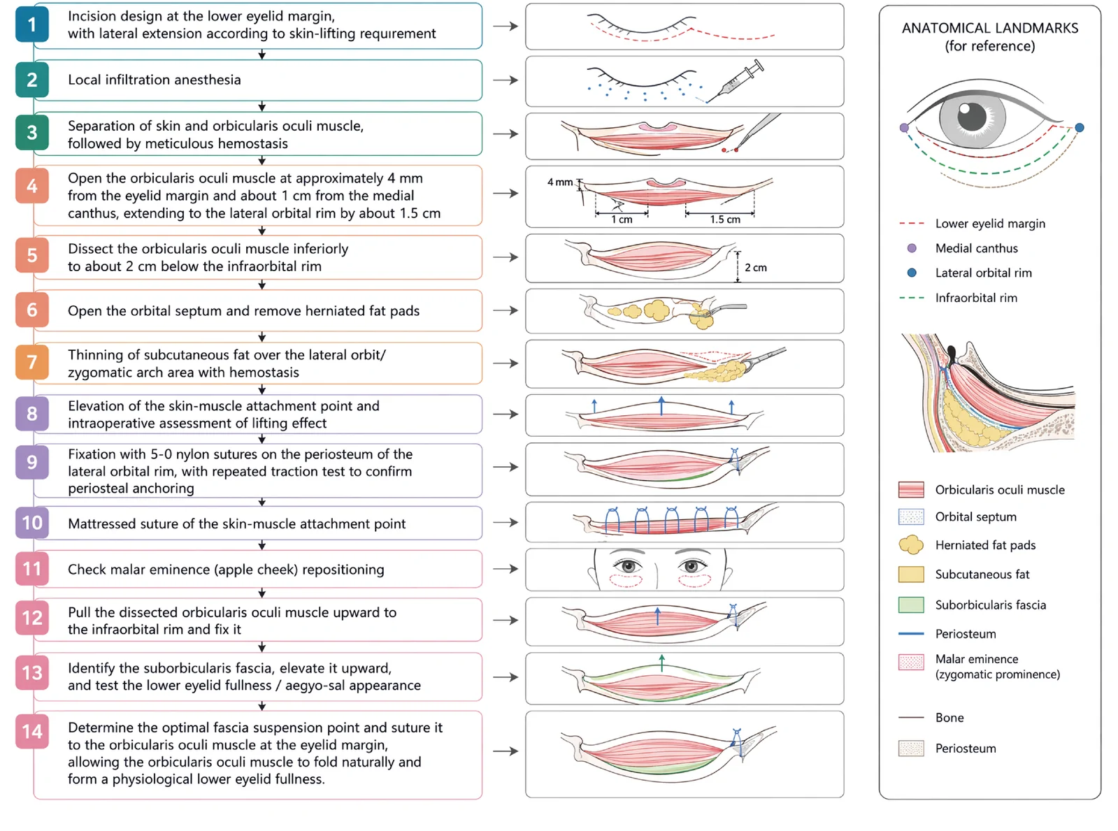
V3中面部提升经睫毛下切口,沿内眦线、外眦线、颧脂肪垫下界三个向量悬吊,可同时改善中面部下垂、下睑袋及睑颊轮廓不连续。
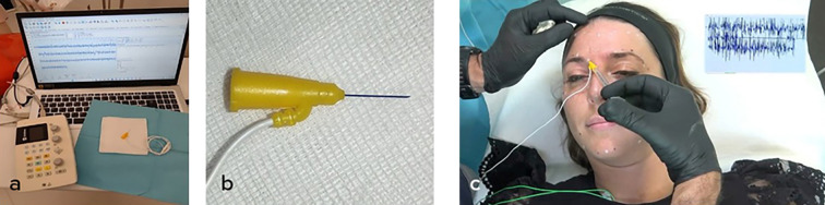
由6名意大利多学科专家(皮肤科、整形外科、神经科)基于文献回顾(PubMed 1973–2025)与临床经验形成的专家共识,针对incobotulinumtoxin A(Bocou
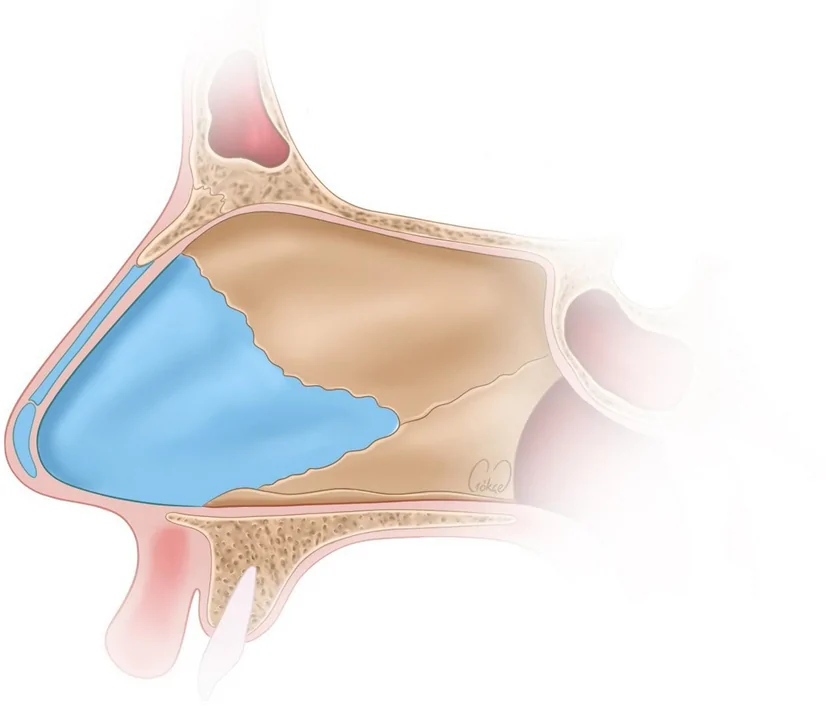
回顾性队列研究(循证等级IV),50例轴向偏曲(C形28例、反C形22例)歪鼻患者,由单一术者于2021至2023年行开放鼻整形,平均随访2年(1至3年)。