694nm红宝石点阵激光治疗黄褐斑:东亚人群临床研究与AI影像量化评估
2026年10月13日37 min read
Journal of Cosmetic Dermatology
结论与实践要点
本研究为发表于 Journal of Cosmetic Dermatology 的回顾性真实世界研究(DOI: 10.1111/jocd.71071),数据来自台湾高雄一家皮肤科医美机构2022年5月至2023年5月的病历数据库,设有双重目的:一是评估低能量点阵694 nm Q开关红宝石激光(QSRL)在东亚黄褐斑患者中的疗效与安全性,二是验证一套AI辅助的ROI(感兴趣区)色彩量化分析框架。原文纳入标准为病程超过10年的混合型黄褐斑、入组前3个月未接受激光或外用漂白剂治疗,共18例入组、16例完成治疗并纳入分析;平均年龄49岁(范围40–66岁),全部为女性,Fitzpatrick分型以IV型为主(17/18例,94.4%),1例为III型。
治疗方面,原文使用694 nm红宝石激光(Q Plus R,Quanta System)配备9 mm High Coverage(9 HC)点阵手具,能量密度0.77–0.9 J/cm²,Q开关脉宽6 ns,单遍非重叠扫描,治疗终点为轻度红斑(数分钟内消退);治疗间隔3–8周,每人至少4次、最多5次,治疗前以利多卡因-丙胺卡因(EMLA)封包全脸麻醉1小时,18例患者均未口服氨甲环酸。疗效评估以Nikon D5200标准化摄影结合VISIA系统,选取颧骨最暗区域为ROI,通过SURF特征对齐与RANSAC算法实现跨次访视的像素级配准,再以灰度共生矩阵(GLCM)纹理分析与亮度系数量化色素强度;其色素分型系统经500张专家标注图像训练、100张独立图像验证,准确率93.2%、F1得分>0.90。统计分析采用GraphPad Prism配对t检验(单尾),以p<0.05为差异有统计学意义。
原文将ROI色素分为三型:III型为细点状或团块状(较浅表)、IV型为弥漫斑片状褐色、V型为均质深褐色(常伴真皮沉积),以面积占比量化。主要结果如下:右颊V型色素占比由基线(第1次观察)8.31%降至第5次观察5.18%(配对t检验p=0.036),第2次与第3次(p=0.044)、第2次与第5次(p=0.045)比较亦呈显著;右颊浅表色素(III+IV型)合计占比由91.80%升至94.87%,且个体间变异度显著缩小(标准差由5.24%降至1.96%,p=0.024)。左颊V型由9.89%降至7.61%(p=0.235)、总体V型由9.10%降至6.39%(p=0.100),均未达统计学显著;总体浅表色素由90.91%升至93.87%(p=0.051,边缘显著)。中文解读:改善主要体现为深色均质色素(V型)的减少与色素分布的均质化,而非全局色素面积的简单缩小;左右侧显著性不一致(右p=0.036对左p=0.235)提示疗效可能存在部位依赖性,原作者推测与不对称紫外线暴露有关。
安全性方面,原文报告随访期内未见PIH、色素减退、色素反跳、皮损扩大或皮肤敏感;正文表述为末次治疗后至少6个月随访未见上述情况,摘要另提及1年随访未见PIH与色素反跳,两处随访时长表述不一致,宜以正文的至少6个月为准。图7展示了一例40岁Fitzpatrick III型女性患者5次治疗前后的VISIA图像对比,可见色素与黄褐斑减轻。
临床落地解读(参数与疗程):该研究为东亚III–V型皮肤提供了一个具体参数参考——0.77–0.9 J/cm²、6 ns脉宽、9 HC手具单遍扫描、间隔3–8周、3–5次治疗。原文讨论强调,与1064 nm Nd:YAG「laser toning」常需数千击、多遍扫描相比,单遍方案可降低累积热负荷,从而减少高黑色素吸收波长(694 nm、532 nm)相关的PIH风险。需要提醒的是,上述能量密度为特定设备与手具下的设置,更换设备时不宜直接照搬数值,宜以终点反应(轻度红斑)作为滴定依据;本研究未联合口服氨甲环酸或外用漂白剂,尚不能回答与标准三联外用药联合能否进一步增效的问题。
临床落地解读(评估工具与机制推测):本研究方法学上示范了ROI级AI影像量化(SURF+RANSAC配准联合GLCM)对亚临床色素改变的捕捉能力——即使肉眼与MASI评分判为「无变化」,纹理参数仍可识别黑色素簇的碎裂与分布均质化。在有标准化摄影条件的机构,可借鉴此类纵向同ROI对比用于疗效随访;在常规门诊中,MASI/mMASI等成熟量表仍是可及性更高的选择。原文讨论还提出694 nm波长可能兼具影响真皮血管成分的「双靶点」假说,其依据是离体与文献证据(VEGF在黄褐斑皮损高表达、黑素细胞表达VEGFR-1/VEGFR-2),并未在临床终点中直接验证,属于机制推测,不能作为疗效承诺的依据。
局限性与证据边界:(1)回顾性单组设计,无对照组,无法排除自然波动、季节与护理习惯等混杂;(2)样本量小(16例完成分析),且以IV型皮肤为主,III型与V型代表性有限;(3)随访约6个月,长期复发率未知;(4)色素III/IV/V分型是基于照片校准的操作性定义,并非标准Fitzpatrick灼伤/晒黑反应定义,解读时应注意;(5)部分作者受雇于设备厂商(El.En. Group),存在潜在利益相关性。综合而言,在该回顾性、小样本条件下,低能量点阵694 nm红宝石激光在东亚人群中显示出以V型色素减少为主的改善倾向与可接受的安全性,其确切效应尚需前瞻性对照研究确认。
BackgroundMelasma is a chronic, relapsing pigmentary disorder with a significant psychosocial impact, particularly in darker skin types. Despite various therapeutic options, its resistance to treatment necessitates optimized laser protocols and more precise evaluation tools.
AimsThis study follows a dual objective: to evaluate the efficacy and safety of a low‐fluence fractional 694‐nm Q‐switched Ruby laser (QSRL) protocol in Asian patients, and to validate a novel AI‐assisted region‐of‐interest (ROI) chromatic analysis for objective clinical assessment.
MethodsA retrospective analysis was conducted on patients (Fitzpatrick III–V) treated with at least four treatments using a 694 nm (QSRL) at 3–8 weeks intervals (0.77 and 0.94 J/cm 2 ). Clinical outcomes were assessed using a digital imaging framework incorporating advanced feature‐alignment algorithms (SURF and RANSAC) for precise longitudinal ROI tracking. Objective improvement was quantified via Gray‐Level Co‐occurrence Matrix (GLCM) texture analysis and luminosity coefficients.
ResultsSixteen patients completed the study. Significant reductions in hyperpigmentation were recorded in midfacial regions. The AI‐assisted analysis demonstrated high sensitivity in capturing localized improvements and textural shifts often missed by global clinical impressions. Notably, no cases of PIH or rebound pigmentation occurred during the 1‐year follow‐up. High patient satisfaction correlated strongly with quantitative pigmentary transitions.
ConclusionLow‐fluence fractional 694‐nm QSRL provides a safe, effective treatment option for melasma in Asian skin. Moreover the study validates the AI‐assisted ROI analysis provides a superior assessment tool. This dual approach offers a promising paradigm for long‐term melasma management, combining an optimized laser protocol with a robust, reproducible benchmark for precision dermatology.
Keywords: fractional laser、melasma、Q‐switched ruby laser (694 nm)
MTZmicroscopic thermal zones
PIHPost‐Inflammatory Hyperpigmentation
ROIRegion of Interest
1
Melasma is a chronic pigmentary disorder characterized by symmetric hyperpigmented patches on the face, predominantly affecting women and individuals with darker skin tones (Fitzpatrick types III‐VI) [ 1 ]. Beyond its clinical presentation, its relapsing nature exerts a profound psychosocial impact, significantly impairing self‐esteem and quality of life. The pathogenesis is complex and multifactorial, involving an interplay of UV radiation, hormonal shifts, and chronic inflammation linked to photoaging, characterized by increased vascularity and the activation of pro‐inflammatory mediators. Despite various therapeutic approaches focusing on sun protection and topical agents, its resistance to treatment poses ongoing challenges [ 2 ].
Laser‐based therapies have emerged as a promising alternative, yet the use of traditional, high‐fluence lasers in darker skin types has historically been limited by the significant risk of post‐inflammatory hyperpigmentation (PIH).
Recently, the development of low‐fluence, fractional Q‐switched laser technologies has shifted the treatment paradigm. These methods deliver energy in a pixelated pattern, creating micro‐zones of damage that promote faster healing and minimize downtime. Specifically, the 694‐nm Q‐switched Ruby Laser (QSRL) may offer a unique therapeutic window due to its high selectivity for melanin and minimal absorption by competing chromophores, effectively minimizing thermal damage [ 3 , 4 , 5 ].
However, evaluating the efficacy of these technological advancements remains a significant hurdle. Current “gold standard” assessment tools, such as the Melasma Area and Severity Index (MASI), introduced in 1994 by Kimbrough‐Green et al. [ 6 ], rely on global visual impressions and are prone to inter‐observer variability. These traditional methods fail to capture localized chromatic shifts or subtle changes in spatial distribution, and they often overlook the crucial contrast between lesional and adjacent normal skin. Because clinical response to laser therapy is often region‐specific and heterogeneous, conventional scales may even fail to detect paradoxical aggravation or lesion enlargement.
The quest for more sensitive and objective assessment tools in melasma has led to the proposal of several alternatives to the traditional MASI score. Recent efforts have focused on enhancing the weight of pigment intensity, as seen in the Melasma Severity Index (MSI), which utilizes a squared parameter for darkness to better reflect subtle chromatic improvements [ 7 ].
Other researchers have explored quasi‐quantitative methods such as the Point Counting‐Serial Image Index (PCSI) [ 5 ], designed to track the spatial fragmentation of lesions, a common occurrence following fractional laser therapy that global scales often fail to quantify [ 8 ]. Furthermore, the push toward Digital Image Analysis (DIA), utilizing metrics like the Weighted Skin Intensity (WSI) ratio and the Lab* color space, aims to eliminate inter‐observer subjectivity by providing a purely mathematical evaluation of the contrast between lesional and perilesional skin.
Despite these advancements, most of these emerging scales are still undergoing validation or require specialized equipment. Consequently, the present study is designed with a dual objective: first, to evaluate the clinical efficacy of a specific low‐fluence fractional 694 nm QSRL protocol in Asian patients (skin Type III, IV and V); and second to validate a more adequate AI‐assisted quantitative imaging framework for monitoring such improvements.
By employing a two‐stage alignment process based on SURF‐feature [ 9 ] alignment and RANSAC [ 10 ], we ensured that identical midfacial Regions of Interest (ROIs) were analyzed with high reproducibility. Unlike methods that rely solely on global visual impressions, our approach integrates Gray‐Level Co‐occurrence Matrix (GLCM) texture analysis and luminosity coefficients to track the evolution of both pigment intensity and textural uniformity. This dual‐purpose methodology not only assesses the therapeutic performance of the ruby laser but also addresses the diagnostic gap in melasma, offering a granular, objective benchmark that captures treatment‐induced changes often missed by traditional scoring systems.
2
2.1
This study is based on a real‐world data analysis, utilizing data from a retrospective chart review from the database of theHuang PH Dermatology and Aesthetics, Kaohsiung, Taiwan. Data were collected from May 2022 to May 2023.
The analysis encompasses patients who were diagnosed with mixed melasma, persisting for over 10 years, who had not undergone laser treatment or used topical bleaching agents within three months prior to the initial treatment. All included patients received at least four treatment sessions, spaced 3–8 weeks apart, using the solid‐state fractionated 694 nm ruby laser (Q Plus R, Quanta System S.p.A., Samarate, Italy), up to max five sessions.
Patients were excluded if they presented with any of the following: concomitant use of photosensitizing agents, retinoids, isotretinoin, anticoagulants or immunosuppressants, a history of epilepsy or suspected epileptic reactions during treatment; pregnancy or breastfeeding women; presence of cancerous or precancerous lesions, Herpes simplex, or wounded skin in the treatment areas; high skin tanning.
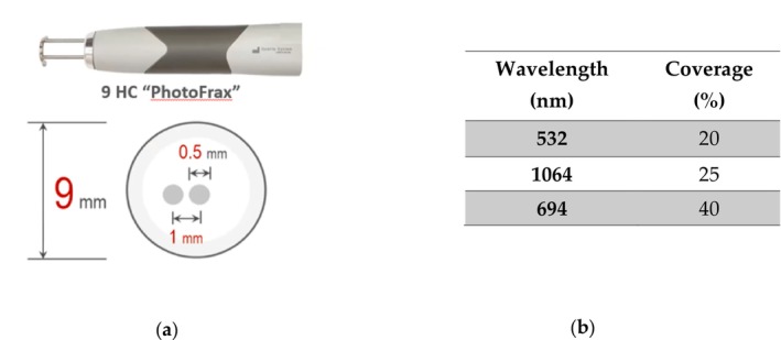
The treatment was performed with 694 nm ruby laser at fractional 9 mm High Coverage (9 HC) handpiece (Figure 1 ). The parameters used were fluence of 0.77 to 0.9 J/cm 2 , Q‐Switched pulse duration of 6 ns. Each patient was scanned with non‐overlapped sweeping.
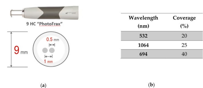
FIGURE 1 Representative image of 9 mm High Coverage handpiece (9 HC).9 mm High Coverage(9 HC)点阵手具的代表性图像,本研究治疗即使用该手具。The laser energy is delivered with a fractionated handpiece to control collateral thermal damage of the skin tissue when longer pulse duration is used. The scanner delivers each energy pulse in a round of 63,62 mm 2 area and fractionalizes the energy in circular dots of 0,5 mm in diameter with a pitch of 1 mm for the 694 nm Ruby laser the coverage is up to 40% of 63,62 mm 2 (Figure 1 ). Contributing to a good uniformity of energy distribution, representing a good compromise compared to previously developed versions or spot sizes (Figure 2 ). The endpoints were mild redness, which quickly faded in a few minutes.
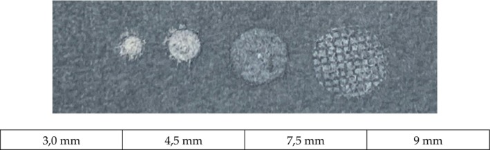
FIGURE 2 Representative image of 9 mm High Coverage handpiece (9 HC) energy distribution.9 HC手具能量分布的代表性图像:扫描单次覆盖63.62 mm²面积,能量被分割为直径0.5 mm的圆形光点、间距1 mm,694 nm红宝石激光的覆盖率可达该面积的40%,能量分布均匀,是相较既往版本或光斑大小的一种折中设计。2.2
Topical anesthesia with lidocaine‐prilocaine (EMLA) applied to the whole face under occlusion for 1 h. The anesthetic cream was removed immediately before laser surgery using wet gauze.
Oral tranexamic acid was not prescribed to any of the 18 included patients.
2.3
Objective improvement parameters were assessed via clinical examination and standardized digital photography. Image acquisition was performed using a 2D digital imaging camera (Nikon D5200, Tokyo, Japan) under controlled lighting and standardized geometric registration.
These images served as the primary data source for a quantitative, automated evaluation system designed to analyze the intensity of pigmentation within a specific region of interest (ROI), located at the darkest area of the cheekbones.
The intensity of pigmentation was quantified by categorizing the skin within the ROI into three specific phototypes (III‐V), representing the most prevalent tones in the Asian population [ 11 ].
Although the Fitzpatrick system is formally defined by skin response to ultraviolet exposure (burning or tanning), in clinical practice dermatologists usually infer these responses directly from baseline skin color, which is more practical and widely understood. Therefore, a representative phototype III skin tone was first selected as the reference baseline. Darker pigmentation patterns observed within melasma lesions were subsequently categorized as Type IV and Type V, with Type V operationally defined as the darkest homogeneous pigmentation observed within the lesion area.
During image analysis, representative regions corresponding to these three pigmentation types were used to establish thresholds based on color and lighting proxies derived from digital image channels. Type III generally corresponded to lighter brown areas with higher luminance, Type IV to intermediate brown pigmentation, and Type V to darker, more homogeneous brown regions.
Pigmentation intensity was defined quantitatively from the registered region of interest (ROI) as a measure of skin darkness, computed from standardized color values under controlled imaging conditions. This was derived from the Value (V) channel in HSV and luminance‐weighted brightness in RGB, where lower V or lower luminance indicates higher pigmentation intensity. Reference samples were established through camera‐calibrated, within‐image color anchoring rather than comparisons across ethnic groups. In this study, the clinically recognized skin tones were translated into quantifiable image‐derived signals so that pigmentation classification could be objectively identified from optical properties within a fixed cheekbone ROI.
Under standardized acquisition and geometric registration, pigmentation intensity was calculated from the ROI's lightness/luminance after masking invalid pixels. Severity was quantified using two complementary metrics: (1) a continuous darkness measure (ΔV or Δluminance relative to baseline) and (2) an area‐based metric representing the proportion of the ROI assigned to darker calibrated bins (Types III–V), where a higher Type V fraction indicates greater pigmentation burden. The ROI was positioned at the darkest cheekbone region so that measurements reflected lesion‐dominant pigmentation rather than overall facial skin tone.
For pre‐ and post‐treatment analysis, follow‐up images were acquired using a digital imaging system (VISIA, CANFIELD Imaging System, Fairfield, NJ, USA).
This data, as well as all other medical information, was collected in full compliance with ethical standards: all patients signed photographs for future research purposes. The management of this data was carried out in accordance with institutional guidelines, and the study received approval from the Mackay Memorial Hospital Institutional Review Board (IRB), Approval certificate for the clinical trial “Application of AI‐assisted analysis and diagnostic system for pigmentary skin disorders.” IRB No. 21MMHIS317e. Taipei (Taiwan): Mackay Memorial Hospital; 2022 May 19.
2.4
The automatic system processes standardized facial photographs taken under consistent lighting conditions using a 2D camera (Nikon D5200, Tokyo, Japan). Images are captured at a resolution of 4928 × 3264 pixels. All photographs are taken perpendicular to the facial surface, with subjects positioned using a standardized chin rest and forehead support to maintain consistent alignment between sessions.
The system aligns facial images across sessions via a two‐stage approach. First, an operator defines a baseline ROI, selecting an area of at least 100 × 100 pixels. In the second stage, a SURF‐based feature alignment [ 9 ] automatically aligns follow‐up ROIs using a holography matrix estimated with Lowe's ratio test [ 12 ] and RANSAC [ 10 ]. This process robustly maps points from the follow‐up image to the baseline image by minimizing the effect of outliers, ensuring that identical skin areas are analyzed over time and enhancing the reliability of our results.
Quality metrics (Q1, Q2, Q3)are then calculated across non‐overlapping 100 × 100 pixel windows in the ROI containing at least 80% valid skin pixels. Pixels are considered valid if their color values fall within the typical range of human skin tones and are not saturated or underexposed. For Q1, we calculate the signal‐to‐noise ratio in the HSV space: Q1=ηHSDH+ηSSDS+ηVSDV, where ηH,ηS,ηV, are the median values of the hue, saturation, and value channels, respectively; SDH,SDS,SDV are the corresponding standard deviations of these channels.
For Q2, we use luminosity coefficients derived from the human vision luminosity model to weight the contributions of each color channel in RGB color space, emphasizing the green channel: Q2=0.2989ηRSDR+0.5870ηGSDG+0.1140ηBSDB.
Q3 leverages texture analysis via Gray‐Level Co‐occurrence Matrix (GLCM) features, summarizing contrast, homogeneity, energy, and correlation [ 13 ]. Let Gi,j be the normalized GLCM, N be the number of gray levels, and μi,,μj,σi,σj be the means and standard deviations of the marginal distributions. Contrast Q3c emphasizes the difference between gray levels of neighboring pixels: Q3c=∑i=0N−1∑j=0N−1i−j2Gi,j. Homogeneity Q3h measures the closeness of the distribution of elements in the GLCM to the diagonal: Q3h=∑i=0N−1∑j=0N−1Gi,j1+i−j. Energy Q3e represents the textural uniformity of the image: Q3e=∑i=0N−1∑j=0N−1Gi,j2. Correlation Q3r evaluates how much a pixel is correlated to its neighbor in terms of gray levels: Q3r=∑i=0N−1∑j=0N−1i−μij−μjGi,jσiσj We combine the GLCM features into a single metric Q3, by computing their mean. The composite metric CM, derived from Q1,Q2 and Q3 quality metrics, is compared with thresholds to provide a quantitative skin type breakdown for the ROI. Classification was calibrated using a training set of 500 expert‐labeled images, encompassing all Fitzpatrick skin types. Validation was performed on an independent test set of 100 images, achieving 93.2% accuracy and an F1 score above 0.90 Figure 3 .
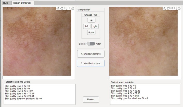
FIGURE 3 User interface of the skin color and quality measurement system, displaying the analysis of a region of interest (ROI) before and after processing. The system quantifies different skin quality types and handles shadow removal for accurate classification, as detailed in the pre and post‐processing statistics.皮肤颜色与质量测量系统的用户界面截图,显示一个ROI处理前后的分析结果;系统可量化不同皮肤质量类型并处理阴影去除,处理前后的统计信息用于保障分类准确性。2.5
Statistical analysis was performed using GraphPad Prism (v10.3 for Windows, GraphPad Software, www.graphpad.com). Based on clinical rationale, we hypothesized an increase in Type III and IV, and a decrease in Type V over time. A paired t‐ test was employed to specifically test these hypotheses and to compare the skin improvement among different treatment sessions. A p‐value < 0.05 was considered statistically significant.
3
In this study, a total of 18 patients met the inclusion and exclusion criteria, while 16 patients completed the treatment session and were included in the analysis. The mean age was 49 years (range: 40–66 years). Fitzpatrick skin types were predominantly type IV (17/18 patients, 94.4%), with one patient presenting with type III skin type (5.6%) (Table 1 ).
TABLE 1 Further breakdown of melasma before treatment: skin color distribution.
| Melasma | Gender M/F | Skin type III/IV | Age (mean, max, min) |
|---|---|---|---|
| Mixed‐type | 0%/100% | 5.6%/94.4% | 49, 66, 40 |
All cases were clinically classified as mixed‐type hyperpigmentation, involving both epidermal and dermal components.
Pigmentation analysis was conducted on a cohort of 16 female patients, the vast majority of whom presented with Fitzpatrick skin type IV. As a result, the pigmentation assessment focused on Types III, IV, and V, given the absence of cases corresponding to other classifications.
Each patient underwent a minimum of three treatment sessions, with 11 out of 16 patients completing a fourth observation point (Obs4) and 10 out of 16 patients completing a fifth observation point (Obs5), enabling longitudinal analysis over time.
Pigmentation was classified into three distinct types based on clinical photographs: Type III, characterized by fine dots or globules (typically more superficial); Type IV, consisting of diffuse, patchy brown pigmentation; and Type V, marked by homogenous dark brown areas often associated with deeper dermal deposition.
Quantitative assessments were performed by calculating the percentage area involved for each pigmentation type, separately for the right (R) and left (L) sides of the face, as well as an overall (O) average derived from both regions. These percentages were recorded at five observation points (Obs1 to Obs5), corresponding to the sequential treatment sessions. Data are reported as mean ± standard deviation, and temporal changes were evaluated statistically using paired t ‐test (1‐tailed), with Obs1 serving as the baseline.
This analytical approach allowed for a comprehensive evaluation of how each pigmentation subtype responded to treatment over time, with attention to spatial distribution (right, left, overall) and the persistence or resolution of pigmentary features. The results are structured into two parts: first, a detailed analysis of Types III, IV, and V individually; and second, a combined assessment comparing the overall evolution of superficial pigmentation (Type III + IV) against deeper pigmentation (Type V).
On the right side (R), for Type III pigmentation, values ranged from 10.47% to 15.99%, with no statistically significant variation over time ( t ‐test, p1vs5 = 0,172). Type IV pigmentation showed stable values across sessions (range: 76.74%–80.61%), with no significant differences ( t ‐test, p1vs5 = 0,428). Notably, Type V showed a decrease in mean pigmentation from 9.10% (Obs2) to 5.18% (Obs5), with significant differences between Obs1 and Obs5 ( t ‐test, p = 0,036). Significance on Type V seems evident also comparing different sessions performed on the right cheek, starting from the second session ( t ‐test, p2vsp3 = 0,044; p2vsp5 = 0,045), as indicate in Table 2 and Figure 4 .
TABLE 2 Comparison between sessions for single side of treatment (right or left cheek) and overall (right + left cheek).
| Obs. n.1 | Obs. n.2 | Obs. n.3 | Obs. n.4 | Obs. n.5 | p (obs. I vs. V) | |
|---|---|---|---|---|---|---|
| R:Type III (Mean ± SD) | 15,06 ± 13,81 | 10,47 ± 8,94 | 13,72 ± 9,22 | 14,61 ± 13,13 | 15,99 ± 11,51 | 0,172 |
| R:Type IV (Mean ± SD) | 76,74 ± 11,96 | 80,61 ± 7,69 | 79,54 ± 7,99 | 78,06 ± 12,73 | 78,87 ± 11,13 | 0,428 |
| R:TypeV (Mean ± SD) | 8,31 ± 5,40 | 9,10 ± 3,95 | 5,80 ± 2,95 | 7,17 ± 3,44 | 5,18 ± 1,90 | 0.036 * |
| L:Type III (Mean ± SD) | 11,48 ± 11,71 | 11,24 ± 9,45 | 13,10 ± 10,94 | 14,04 ± 13,7 | 13,64 ± 9,82 | 0,258 |
| L:Type IV (Mean ± SD) | 78,55 ± 10,72 | 79,78 ± 10,01 | 76,31 ± 13,10 | 77,23 ± 13,03 | 79,24 ± 9,21 | 0,445 |
| L:Type V (Mean ± SD) | 9,89 ± 6,07 | 9,00 ± 6,04 | 10,62 ± 9,72 | 8,73 ± 7,82 | 7,61 ± 6,21 | 0,235 |
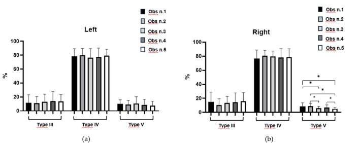
FIGURE 4 Comparative test between different sessions and different kind of pigmentation (III, IV and V). (a) Measures in the left cheek; (b) Measures of the right cheek. Statistical significance: *p < 0.05.不同治疗次数与不同色素类型(III、IV、V型)间比较的统计图:(a) 左颊测量值;(b) 右颊测量值;标注 *p<0.05。On the left side of the face, values for Type III ranged between 11.24% and 14.04% with no statistical significance across timepoints. Type IV values remained consistent (78.55%–79.78%), and no significant differences were detected. Type V values declined slightly (from 9.89% to 7.61%) with no significant changes ( t ‐test, p1vs5 = 0,235).
On the right side of the face, for type III + IV combined values increased slightly from 91.80% (Obs1) to 94.87% (Obs5), with a significant reduction in variability (std: 5.24% to 1.96%; t ‐test p = 0.024). Notably, Type V showed a decrease in mean pigmentation from 9.10% (Obs2) to 5.18% (Obs5), with significant differences between Obs1 and Obs5 ( t ‐test, p = 0.036). On the other hand, the left side of the face presented for Type III + IV, a gradual increase (from 90.02% to 92.87%), but this was not statistically significant ( t ‐test p1vs5 = 0.130). Type V values declined slightly (from 9.89% to 7.61%) with no significant changes ( t ‐test p1vs5 = 0.235) (Table 3 and Figure 5 ).
TABLE 3 Comparison between the session's mean for single side of treatment (right or left cheek) and overall (right + left cheek).
| Session n.1 | Session n.2 | Session n.3 | Session n.4 | Session n.5 | p (session I vs. V) | |
|---|---|---|---|---|---|---|
| R:Type III + IV (Mean ± SD) | 91,80 ± 5,24 | 91,08 ± 4,86 | 93,26 ± 2,62 | 92,67 ± 3,37 | 94,87 ± 1,96 | 0,024* |
| R:TypeV (Mean ± SD) | 8,31 ± 5,40 | 9,10 ± 3,95 | 5,80 ± 2,95 | 7,17 ± 3,44 | 5,18 ± 1,90 | 0,036* |
| L:Type III + IV (Mean ± SD) | 90,02 ± 6,23 | 91,02 ± 6,04 | 89,41 ± 9,86 | 91,27 ± 7,82 | 92,87 ± 5,62 | 0,130 |
| L:Type V (Mean ± SD) | 9,89 ± 6,07 | 9,00 ± 6,04 | 10,62 ± 9,72 | 8,73 ± 7,82 | 7,61 ± 6,21 | 0,235 |
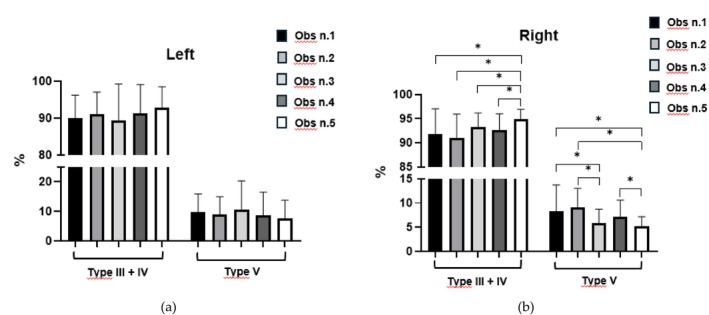
FIGURE 5 Comparative test between different sessions and different kinds of pigmentation (III + IV and V). (a) Measures in the left cheek; (b) Measures of the right cheek. Statistical significance: *p < 0.05.不同治疗次数与不同色素类型(III+IV型对V型)间比较的统计图:(a) 左颊测量值;(b) 右颊测量值;标注 *p<0.05。Overall pigmentation trends mirrored the side‐specific values. Type III + IV increased from 90.91% to 93.87% with borderline statistical significance ( t ‐testp1vs5 = 0,051).
Type V decreased from 9.10% to 6.39%, with a near‐significant p ‐value ( t ‐test p = 0, 100) (Table 4 and Figure 6 ).
TABLE 4 Comparison between sessions for overall (right + left cheek).
| Session n.1 | Session n.2 | Session n.3 | Session n.4 | Session n.5 | p (session I vs. V) | |
|---|---|---|---|---|---|---|
| O:Type III (Mean ± SD) | 13,27 ± 11,92 | 10,85 ± 8,64 | 10,62 ± 9,72 | 14,33 ± 12,24 | 14,81 ± 10,21 | 0,198 |
| O:Type IV (Mean ± SD) | 77,64 ± 10,86 | 80,20 ± 7,66 | 77,92 ± 10,18 | 77,65 ± 11,06 | 79,05 ± 9,76 | 0,427 |
| O:Type V (Mean ± SD) | 9,10 ± 5,35 | 9,05 ± 3,73 | 8,21 ± 5,62 | 7,95 ± 4,70 | 6,39 ± 3,65 | 0,100 |
| O:Type III + IV (Mean ± SD) | 90,91 ± 5,34 | 92,05 ± 3,91 | 91,33 ± 5,24 | 91,97 ± 4,62 | 93,87 ± 3,45 | 0,051 |
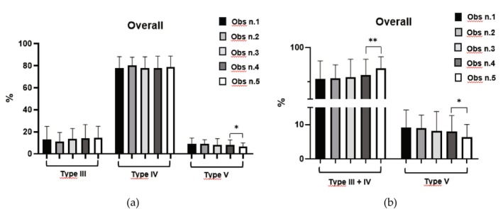
FIGURE 6 Overall comparative test between different sessions and different kinds of pigmentation (a) Overall measures of III, IV, and V; (b) Overall measures of III + IV vs. V.Statistical significance: *p < 0.05; **p < 0.01.总体比较统计图:(a) III、IV、V型的总体测量值;(b) III+IV型对V型的总体测量值;标注 *p<0.05、**p<0.01。There was no rebound pigmentation, expanded melasma areas, sensitive skin, or hypopigmentation at least 6 months following the final treatment session.
A clinical case with a visible improvement after laser treatment are shown in Figure 7 .
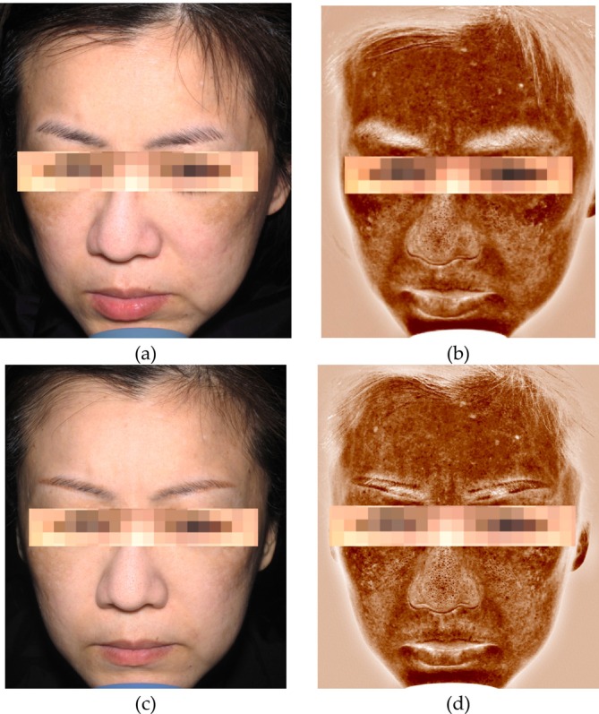
FIGURE 7 A decrease in pigmentation and melasma in a 40‐year‐old female patient (FST III) at baseline and after 5 treatment sessions. (a‐b) VISIA image before treatment; (c‐d) VISIA image after treatment.一例40岁女性患者(Fitzpatrick III型)治疗前与5次治疗后的临床图像对比,显示色素与黄褐斑减轻:(a-b) 治疗前VISIA图像;(c-d) 治疗后VISIA图像。4
The management of melasma remains a significant clinical challenge due to its chronic nature and the high risk of post‐inflammatory hyperpigmentation (PIH), particularly in patients with darker skin tones (Fitzpatrick types III‐V).
A central hurdle in melasma research is the lack of spatial reproducibility in follow‐up photography, as traditional “gold standard” tools like the MASI score are prone to inter‐observer variability and fail to capture localized chromatic shifts. To overcome this, our study employed a two‐stage alignment process based on SURF‐feature alignment and RANSAC, ensuring that identical midfacial Regions of Interest (ROIs) were analyzed with pixel‐perfect reproducibility.
By locking the coordinate system of the face, we eliminated positional noise and enabled an approach analogous to a “digital biopsy” of the lesion. This level of spatial precision is particularly fundamental when evaluating the Fractional Ruby Laser (694 nm); since this wavelength possesses the highest selectivity for melanin, its therapeutic impact often manifests as a complex spatial reorganization of pigment rather than a simple decrease in global intensity.
The most innovative aspect of our validation framework lies in its ability to capture subclinical pigmentary shifts through GLCM texture analysis. While conventional scales only record when a lesion appears lighter to the naked eye, GLCM tracks the fragmentation of melanin clusters, a phenomenon directly linked to the “shattering” effect of the Ruby laser on dermal pigment.
Our results suggest that the fractional ruby laser induces a transition toward textural uniformity. Even in cases where overall luminosity showed subtle changes, GLCM parameters identified the disruption of melanin‐laden macrophages and the breakdown of dense pigmentary aggregates. By providing an objective benchmark that captures treatment‐induced changes often missed by traditional scoring systems, this framework offers a granular view of how the laser alters the optical properties of the dermis, effectively mapping the transition from pathological clustering to a restored, homogenous skin texture.
Beyond direct pigmentary ablation, it is worth considering whether the efficacy of the 694 nm wavelength might also be partially linked to its potential impact on the vascular component of melasma. Melasma is increasingly hypothesized to be a multi‐factorial “photoaging” disorder in which dermal angiogenesis could play a significant role [ 14 ]. Supporting this view, diagnostic studies have reported that Vascular Endothelial Growth Factor (VEGF), a potent mediator of vascular endothelial cell proliferation and migration, is significantly upregulated in melasma lesional skin [ 15 ]. Crucially, the interaction between angiogenesis and pigmentation appears to be mediated by specific receptors; research has demonstrated that melanocytes express numerous VEGF receptors in vitro, including VEGFR‐1 and VEGFR‐2. The finding that VEGFR‐2 expression is upregulated after UV irradiation suggests a mechanism by which sun exposure simultaneously triggers both vascular expansion and melanocyte activation, potentially creating a pro‐melanogenic feedback loop [ 16 ].
In this context, our findings while primarily focused on textural and pigmentary changes do not exclude the possibility that the fractional Ruby laser might modulate the dermal microenvironment by tentatively disrupting this angiogenic‐melanogenic crosstalk.
While traditional vascular lasers like the Pulsed Dye Laser (PDL) and copper bromide (CuBr) are highly effective at targeting oxyhemoglobin, they often carry a significant risk of purpura or PIH in darker skin types [ 17 , 18 , 19 ]. In contrast, the 694 nm wavelength may offer a “dual‐target” potential: it penetrates deeply enough to reach the dilated dermal plexus, potentially reducing the chronic telangiectasia and the subsequent release of inflammatory mediators that drive melanocyte hyperactivity. This secondary vascular effect likely contributes to the reduction of the “muddy” erythematous‐pigmentary appearance of mixed melasma, providing a more stable clinical result.
Our findings align with and expand upon the existing literature, confirming that multiple sessions of low‐dose fractional Q‐switched Ruby Laser (QSRL) significantly reduce pigmentation while maintaining an excellent safety profile.
These results are consistent with the systematic review by Lee et al. (2022) [ 20 ], which emphasized that low‐fluence Q‐switched strategies specifically for the Nd:YAG laser minimize adverse events like mottled hypopigmentation. However, our study suggests that the 694 nm wavelength offers a strategic advantage; its higher absorption peak for melanin compared to the 1064 nm Nd:YAG allows for more precise targeting of chromophores with lower overall energy delivery, thereby reducing the cumulative thermal burden on the dermis.
The clinical efficacy of this approach was previously demonstrated by Jang et al. (2011) [ 3 ] in Korean patients, where six sessions led to a significant MASI reduction (from 15.1 ± 3.3 to 10.6 ± 3.9) and a notable increase in skin lightness (L‐value).
Our results further corroborate these findings, proving that substantial improvements in melasma can be achieved in Asian cohorts (Fitzpatrick III‐IV) with as few as three to five sessions [ 3 ]. This is particularly significant when compared to the work of Hilton et al. (2013) [ 4 ]; while their study reported high efficacy in Caucasian patients (Fitzpatrick I‐III), our data demonstrates equivalent safety in a population far more predisposed to Post‐Inflammatory Hyperpigmentation (PIH). The consistency of these results across different ethnicities highlights the robustness of the fractional approach: by creating Microscopic Thermal Zones (MTZs), we promote rapid epidermal repair and mitigate the stimulation of inflammatory mediators.
A critical factor in our observed safety profile was the implementation of a single‐pass protocol using a 9 mm High Coverage (HC) handpiece. Unlike traditional “laser toning” with 1064 nm Nd:YAG, which often requires thousands of shots and multiple passes potentially leading to cumulative thermal injury or guttate hypomelanosis our findings demonstrate that a single pass is sufficient to achieve therapeutic goals. This minimizes the risk of hyperpigmentation, which is particularly high for wavelengths with intense melanin absorption like the 694 nm or 532 nm. By reducing the number of passes, we decreased the cumulative thermal burden, effectively lowering the likelihood of PIH and shortening post‐treatment erythema [ 21 ].
The sensitivity of our ROI‐based digital analysis further elucidated these dynamics, revealing a statistically significant reduction in Type V skin color areas on the right side of the face ( p = 0.036) that was not mirrored on the left ( p = 0.235).
This discrepancy, likely linked to asymmetric UV exposure, underscores the ability of our framework to detect localized variations that global scales often average out. Ultimately, the integration of fractional 694 nm technology with an objective imaging framework moves melasma management away from subjective evaluation toward a reproducible, data‐driven science, offering a promising approach for the long‐term management of this challenging condition even in high‐risk skin phototypes.
Despite the positive outcomes, the retrospective nature of this study and the six‐month follow‐up period limit insights into long‐term recurrence rates. However, the integration of fractional 694 nm technology with an AI‐assisted quantitative imaging framework moves melasma management away from subjective evaluation toward a reproducible, data‐driven science. This methodology not only validates the ruby laser's performance but also provides a more sensitive tool for the early detection of therapeutic response, ultimately offering a promising approach for the long‐term management of this challenging condition.
5
This study confirms that a low‐fluence fractional 694‐nm Q‐switched Ruby laser protocol is a safe and highly effective treatment for melasma in Asian patients with Fitzpatrick skin types III–V. By targeting melanin with high specificity and utilizing a fractional delivery system, we achieved significant clearing of hyperpigmented lesions while maintaining an excellent safety profile, notably avoiding common complications such as post‐inflammatory hyperpigmentation or leukoderma.
Beyond clinical outcomes, our work highlights the critical role of AI‐assisted imaging in modern dermatology. The transition from subjective visual scales to an objective framework—integrating SURF‐RANSAC alignment and GLCM texture analysis—allows for the detection of subclinical melanin fragmentation and textural shifts. This “digital biopsy” approach provides a granular benchmark for therapeutic response that traditional scoring systems fail to capture. Ultimately, the synergy between advanced laser technology and objective digital validation offers a more precise, reproducible, and data‐driven path for the long‐term management of melasma.
Patrick Po‐Han Huang: conceptualization, methodology, supervision, writing, data curation. Ilia Tetin: conceptualization, methodology, writing. Chih‐Yu Wang: conceptualization, methodology. Sherry Yunshan OuYang: investigation. Pei‐Ling Chih: investigation. Daniela Martinelli, Alessandra Zevini and Riccardo Barini: data analysis, supervision, writing – review and editing. All authors approved the final version of the manuscript.
Approval by the Mackay Memorial Hospital Institutional Review Board, pursuant to Protocol No.: 21MMHIS317e dated May 19, 2022, issued by the Taiwan Christian Foundation for the Elderly, Mackay Memorial Hospital Institutional Review Board. The study was conducted in accordance with the Declaration of Helsinki. The device has been CE Marked since 2014 and registered for Taiwan market since 2017.
All participants in the study provided their informed consent to the publication of their images.
Daniela Martinelli, Alessandra Zevini, and Riccardo Barini are employed at El.En. Group. The other authors declare that the research was conducted in the absence of any commercial or financial relationships that could be construed as potential conflicts of interest.
声明:中文精读 · 仅供学术参考。内容来自公开文献检索,不代表本人观点,不构成诊疗建议。 医疗美容需在正规医疗机构由执业医师实施。
基于相同主题推荐 · 继续深挖
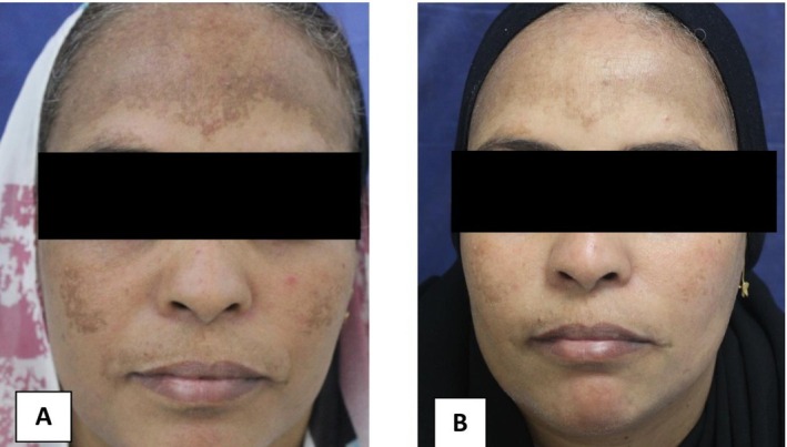
随机对照临床试验(埃及 Aswan 大学,NCT06522984):45 例难治性黄褐斑(标准外用治疗至少 3 个月应答不佳)按 1:1:1 分三组,每组 15 例,结局评估者与数
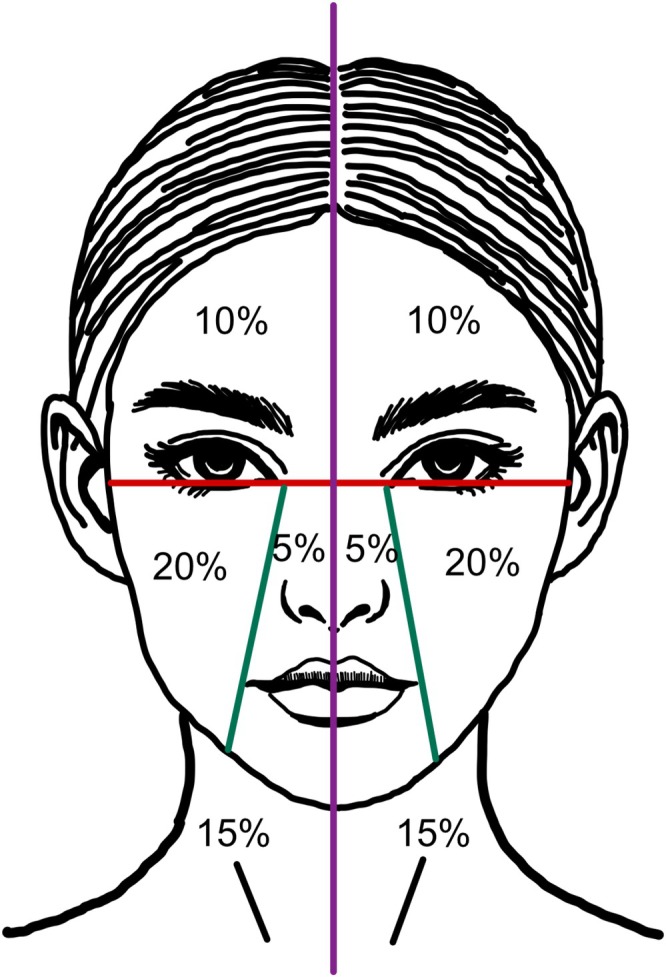
前瞻性随机分面对照、评估者盲法试验(泰国 Ramathibodi 医院,2022-11 至 2025-02):12 例活检确诊 LPP(平均 55±11.1 岁,81.8% 女性,

回顾性队列研究:2015年1月至2024年9月广州某医院130例纹身患者,皮秒(PS)1064nm组73例、Q开关(QS)1064nm组57例,分组依设备可用性与医师裁量而非随机;