皮秒与Q开关1064nm激光洗纹身对比:总体疗效相当,皮秒激光优等清除率更高、所需次数更少
2026年10月13日29 min read
Journal of Cosmetic Dermatology
Journal of Cosmetic Dermatology

结论与实践要点
本研究为发表于 Journal of Cosmetic Dermatology 的回顾性队列研究(DOI: 10.1111/jocd.71098),纳入2015年1月至2024年9月广州某医院收治的130例纹身患者,其中皮秒(PS)1064 nm激光组73例、Q开关(QS)1064 nm激光组57例,分组由设备可用性与临床医师裁量决定而非随机分组,旨在比较两种1064 nm激光在纹身清除中的疗效与安全性。原文报告两组在年龄、性别、Fitzpatrick分型、纹身部位、颜色、墨密度与纹身类型方面的基线差异均无统计学意义(p>0.05);队列平均年龄25.79岁,女性76例(58.46%)、男性54例(41.54%),四肢纹身占60.77%,蓝/黑色纹身占83.08%,皮肤分型为III型31.54%、IV型68.46%。
治疗方案上,QS组使用MedLite C6设备(脉宽5–10 ns),PS组使用Syneron-Candela皮秒设备(脉宽450 ps);两组能量密度均为1.0–6.0 J/cm²、光斑3–5 mm、频率3–5 Hz,治疗前利多卡因乳膏封包1小时,临床终点为纹身表面变白(frosting),治疗间隔8–12周,于末次治疗后3个月随访评估清除情况与不良反应。疗效评估由3名对激光类型设盲的皮肤科医师依据标准化照片按四分位量表评定:差(清除0–25%)、中(26–50%)、好(51–75%)、优(76%及以上);评分者间信度Fleiss' κ=0.784(95%CI 0.709–0.859,p<0.01),总有效率定义为清除超过50%的患者比例。
主要疗效结果:PS组优等清除率为78.08%(57/73),显著高于QS组的54.39%(31/57)(p=0.029);达到优等清除所需中位治疗次数PS组为3次(IQR 3.00–4.00),QS组为4次(IQR 3.00–5.00)(p=0.020)。总有效率(>50%清除)PS组94.52%(69/73)对QS组85.96%(49/57),差异未达统计学意义(p=0.094);达到有效清除的中位次数PS组3次(IQR 2.00–4.00)少于QS组4次(IQR 2.00–5.00)(p=0.034)。由于基线纹身颜色分布存在近显著不平衡(蓝/黑色占比PS组89.04%对QS组75.44%,p=0.061),作者对108例蓝/黑色纹身行亚组分析:优等清除率PS组80%(52/65)对QS组55.81%(24/43)(p=0.007),总有效率93.85%对88.37%(p=0.314)。
安全性方面,原文报告两种设备的安全性相当:短暂红斑与水肿见于PS组10.96%(8/73)与QS组12.3%(7/57),均于48–72小时内自行消退;两组各有2例发生PIH;未观察到水疱、瘢痕或持久性色素异常,组间安全性差异无统计学意义(p>0.05)。
中文解读(对临床的含义):本队列中皮秒设备的优势集中在「高分段」清除(优等清除率)与减少治疗次数,而在「是否达到半数以上清除」这一总体终点上两组相当——这与原文标题「overall effectiveness comparable」的表述一致。对日常门诊的含义是:若患者治疗目标为尽可能清除且希望减少往返次数,皮秒1064 nm可作为考虑方向(本数据中中位3次对4次);若仅期望部分淡化,两种设备的总体把握相近。原作者在讨论中指出,既往文献结论相互矛盾可能与治疗次数及能量设置有关:报告皮秒占优的研究(Ross、Lorgeou、Choi等)治疗次数较多(4–7次),而报告两组无差异的研究(如Pinto等)最多仅2次治疗;本队列QS组优等清除率(54.39%)低于部分既往报告,原作者归因于所用能量偏低(1–6 J/cm²对既往研究的5–10 J/cm²)。解读:参数(能量、次数)对结局的影响可能不亚于设备代际本身,跨研究比较数值时应保持谨慎。
证据边界提示:讨论中引用的Du等研究为大鼠单次治疗实验、Choi等研究为豚鼠实验,均属动物实验证据,其结论(等能量条件下皮秒优于纳秒)不能直接外推为人体在体临床结论;本研究的人体数据本身亦受回顾性设计限制。此外,本队列皮肤分型集中于III–IV型,对深肤色(V–VI型)患者的适用性数据不足,此类患者通常需要更多次数、更长波长并面临更高的色素异常风险。
局限性与证据边界:原作者明确列出四项局限:(1)回顾性、非随机设计,治疗分配基于设备可用性,存在选择偏倚;纹身墨成分、颗粒大小与深度等未测量因素可能影响结局;(2)蓝/黑色纹身占83.08%,结论向多彩色纹身外推受限;(3)9年研究跨度(2015–2024)可能存在平台与参数漂移,包括手具性能、能量校准与操作者技术的演变;(4)3个月随访可能不足以捕捉长期复发与远期不良反应。总有效率差异未达显著(p=0.094)进一步提示,皮秒设备在优等清除上的优势不宜被夸大;作者认为本研究发现应被视为「产生假设」的证据,需前瞻性随机对照试验验证。中文解读:向患者介绍两种方案时,宜表述为「总体清除比例相近,皮秒在高度清除与减少治疗次数上可能有优势」,并结合个体纹身颜色、深度与皮肤分型个体化选择。
BackgroundLaser tattoo removal is becoming more popular, with the Q‐switched (QS) 1064‐nm nanosecond laser considered the traditional standard. While the picosecond (PS) 1064‐nm laser offers an alternative for optimizing treatment protocols, comparative data regarding its relative efficacy remain limited and inconsistent.
AimThis study aims to evaluate and compare the clinical efficacy and safety profiles of QS 1064‐nm laser and PS 1064‐nm laser for tattoo removal.
MethodsA retrospective analysis was conducted on 130 patients with tattoos between January 2015 and September 2024 in a hospital. Participants received either QS 1064‐nm laser ( n = 57) or PS 1064‐nm laser ( n = 73) therapy. Efficacy was measured utilizing a four‐grade clearance scale (poor to excellent), and adverse effects were recorded.
ResultsThe PS 1064‐nm laser achieved significantly higher excellent clearance (78.1% vs. 54.4%, p = 0.029) and required fewer median sessions (3 vs. 4, p = 0.020). Although the overall effectiveness rate (> 50% clearance) was higher for the PS 1064‐nm laser (94.52%) than for the QS 1064‐nm laser (85.96%), this trend did not reach statistical significance ( p = 0.094). After adjusting for baseline color imbalance ( p = 0.061), a subgroup analysis of blue/black tattoos ( n = 108) showed PS 1064‐nm laser had significantly greater excellent clearance (80% vs. 55.8%, p = 0.007), with comparable overall effectiveness (93.9% vs. 88.4%, p = 0.314). Both treatments maintained excellent safety performance; reported side effects were limited to transient erythema/edema, with no severe complications reported.
ConclusionThe PS 1064‐nm laser showed comparable overall effectiveness to the QS 1064‐nm laser, with advantages in excellent clearance and fewer sessions. Limitations include predominantly blue/black tattoos and a 3‐month follow‐up, necessitating prospective validation.
Keywords: efficacy、picosecond laser、Q‐switched laser、safety、tattoo removal
1
Tattoos, once primarily symbols of cultural identity, have evolved into a prevalent form of contemporary self‐expression. As their popularity has grown, so too has the demand for tattoo removal, driven by shifting aesthetic preferences, workplace requirements, and social pressures [ 1 ]. Traditional interventions, such as surgical excision and chemical treatments, have largely been replaced by laser techniques, which offer greater precision, reduced invasiveness, and lower the risk of scarring [ 2 ]. For several decades, the Q‐switched (QS) Nd:YAG laser operating at 1064‐nm has served as the clinical standard. This technology utilizes nanosecond (ns) pulses to fragment tattoo pigments through photomechanical disruption [ 3 , 4 ]. Recently, the advent of the 1064 nm picosecond (PS) laser has further transformed the field. By emitting ultrashort pulses in the picosecond range, this modality enhances photoacoustic energy to more effectively pulverize ink particles while simultaneously minimizing thermal damage to the surrounding dermal tissue [ 5 , 6 ].
However, current literature regarding the comparative efficacy of QS 1064‐nm and PS 1064‐nm lasers remains contradictory. Some studies report the superior efficacy of PS 1064‐nm laser for stubborn pigments [ 7 ], while others indicate comparable outcomes between the two technologies [ 8 ]. Moreover, there is a lack of comprehensive data regarding the long‐term safety profiles, particularly regarding complications such as dyspigmentation and raised scars. This knowledge gap hinders the establishment of standardized, evidence‐based clinical guidelines. This present study retrospectively evaluates the effectiveness and safety of QS 1064‐nm and PS 1064‐nm lasers for tattoo removal within a clinical cohort to address these inconsistencies.
2
2.1
This retrospective study analyzed clinical data from 130 patients with tattoos treated at the Hospital between January 2015 and September 2024. Patients were treated utilizing either a QS 1064‐nm laser (MedLite C6, ConBio, USA) or a PS 1064‐nm laser (Syneron‐Candela, Israel). Treatment assignment was determined by device availability and clinician discretion during the study period. The inclusion criteria were as follows: (1) Patients receiving laser treatment for tattoos during the specified period without other concurrent therapies; (2) provision of written informed consent following a detailed explanation of the treatment mechanism, efficacy, and potential adverse effects; (3) availability of the comprehensive clinical records, including standardized pre‐ and posttreatment photographs for each session. The exclusion criteria included the following: (1) Patients with incomplete clinical data or poor‐quality photographic evidence; (2) patients with immunodeficiency, active infectious diseases, and underlying systemic comorbidities such as uncontrolled diabetes, hypertension, or cardiovascular diseases (excluded due to potential impacts on wound healing and increased risk of complications); (3) patients who were pregnant or lactating.
2.2
Before the treatment, all patients and their families received a comprehensive briefing on the treatment plan, associated risks, and mitigation measures, and written informed consent was obtained. Topical anesthesia was administered by applying lidocaine cream (Tongfang Pharmaceutical Group Co. Ltd.) to the target area under occlusion with plastic wrap for 1 h. Tattoos were either treated with a QS 1064‐nm laser (pulse duration: 5–10 ns; wavelength: 1064 nm, fluence: 1.0–6.0 J/cm 2 , spot size: 3–5 mm; a repetition rate of 3–5 Hz) or a PS 1064‐nm laser (pulse duration: 450 ps; wavelength: 1064 nm; fluence: 1.0–6.0 J/cm 2 , spot size: 3–5 mm, a repetition rate of 3–5 Hz). The clinical endpoint was set to the whitening (frosting) appearance on the tattoo surface. Treatment sessions were scheduled at intervals of 8–12 weeks. Final assessments for clearance and adverse effects were conducted at a 3‐month follow‐up after the terminal session.
2.3
Patient demographics (age, gender, and Fitzpatrick skin type) and tattoo characteristics (type, location, color, and ink density) were recorded. To minimize bias, three dermatologists, blinded to the specific laser modality utilized, evaluated outcomes by reviewing standardized clinical photographs from baseline, interval sessions, and final follow‐up. The tattoo clearance rate was quantified utilizing a quartile grading scale, which divides responses into four distinct groups: poor (0%–25% clearance), moderate (26%–50% clearance), good (51%–75% clearance), and excellent (76%–100% clearance). To assess inter‐rater reliability among the three dermatologists for the four‐grade clearance scale, Fleiss' kappa (κ) was calculated. The overall agreement was κ = 0.784 (95% CI: 0.709–0.859, p < 0.01), indicating substantial agreement. The overall efficacy rate was calculated as the proportion of patients achieving > 50% clearance. Safety was monitored by documenting adverse reactions, including erythema, edema, pustules, hemorrhage, vesicles, hypertrophic scars, post‐inflammatory hyperpigmentation, and hypopigmentation.
2.4
Continuous variables were analyzed utilizing independent t ‐tests or Mann–Whitney U tests. Categorical variables were compared utilizing chi‐square or Fisher's exact test. To address baseline color imbalance, a subgroup analysis restricted to blue/black tattoos was performed to compare outcomes between the PS and QS groups. All statistical analyses were performed utilizing the Statistical Package for the Social Sciences software (version 26.0). Statistical significance was defined as p < 0.05.
3
3.1
A total of 130 patients were included in this study, with 73 receiving the PS 1064‐nm laser and 57 receiving the QS 1064‐nm laser. Baseline characteristics were comparable between the two cohorts, revealing no statistically significant differences in age, sex, Fitzpatrick skin type, tattoo location, color, ink density, or tattoo type (all p > 0.05; Table 1 ). The mean age of the overall cohort was 25.79 years, and 58.46% of participants were males. Most tattoos were located on the extremities (60.77%), and they were predominantly blue or black (83.08%).
TABLE 1 Characteristics of PS 1064‐nm laser and QS 1064‐nm laser in the treatment of 130 patients with tattoos.
| Variables | Total ( n = 130) | P1064 ( n = 73) | Q1064 ( n = 57) | p |
|---|---|---|---|---|
| Age, Mean ± SD | 25.79 ± 9.44 | 25.66 ± 9.47 | 25.96 ± 9.50 | 0.855 |
| Gender, n (%) | ||||
| Female | 76 (58.46) | 45 (61.64) | 31 (54.39) | 0.405 |
| Male | 54 (41.54) | 28 (38.36) | 26 (45.61) | |
| Skin type, n (%) | ||||
| III | 41 (31.54) | 26 (35.62) | 15 (26.32) | 0.257 |
| IV | 89 (68.46) | 47 (64.38) | 42 (73.68) | |
| Location, n (%) | ||||
| Head/Face | 16 (12.31) | 8 (10.96) | 8 (14.04) | 0.400 |
| Trunk | 35 (26.92) | 23 (31.51) | 12 (21.05) | |
| Extremities | 79 (60.77) | 42 (57.53) | 37 (64.91) | |
| Color, n (%) | ||||
| Multicolor | 12 (9.23) | 3 (4.11) | 9 (15.79) | 0.061 |
| Gray/Black | 10 (7.69) | 5 (6.85) | 5 (8.77) | |
| Blue/Black | 108 (83.08) | 65 (89.04) | 43 (75.44) | |
| Ink density, n (%) | ||||
| High | 13 (10.00) | 6 (8.22) | 7 (12.28) | 0.178 |
| Moderate | 35 (26.92) | 15 (20.55) | 20 (35.09) | |
| Low | 61 (46.92) | 38 (52.05) | 23 (40.35) | |
| Minimal | 21 (16.15) | 14 (19.18) | 7 (12.28) | |
| Tattoo type, n (%) | ||||
| Cosmetic | 14 (10.77) | 7 (9.59) | 7 (12.28) | 0.849 |
| Traumatic | 12 (9.23) | 6 (8.22) | 6 (10.53) | |
| Amateur | 83 (63.85) | 49 (67.12) | 34 (59.65) | |
| Professional | 21 (16.15) | 11 (15.07) | 10 (17.54) |
3.2
The PS 1064‐nm laser exhibited significantly better performance compared to the QS 1064‐nm laser (Table 2 ). In the PS 1064‐nm laser cohort, 78.08% ( n = 57/73) of patients achieved an excellent response (76%–100% clearance) (Figures 1 , 2 , 3 ), whereas only 54.39% ( n = 31/57) reached this threshold in the QS 1064‐nm laser group ( p = 0.029) (Figures 4 , 5 , 6 ). Furthermore, patients in the PS 1064‐nm laser group required significantly fewer median sessions to achieve an excellent response (median: 3.00, IQR: 3.00–4.00) compared to those in the QS 1064‐nm laser group (median: 4.00, IQR: 3.00–5.00; p = 0.020). While the overall effectiveness rate (> 50% clearance) was 94.52% ( n = 69/73) for the PS 1064‐nm laser compared to the QS 1064‐nm laser (85.96%, n = 49/57), this difference did not reach statistical significance ( p = 0.094). However, the median number of sessions required to attain an effective response (> 50% clearance) was significantly lower for the PS 1064‐nm laser (3.00, IQR: 2.00–4.00) than for the QS 1064‐nm laser (4.00, IQR: 2.00–5.00; p = 0.034).
TABLE 2 Comparison between PS 1064‐nm laser and QS 1064‐nm laser in the treatment of 130 patients with tattoos.
| Variables | Total ( n = 130) | P1064 ( n = 73) | Q1064 ( n = 57) | p |
|---|---|---|---|---|
| Efficacy, n (%) | ||||
| Poor response | 4 (3.08) | 2 (2.74) | 2 (3.51) | 0.029 |
| Moderate response | 8 (6.15) | 2 (2.74) | 6 (10.53) | |
| Good response | 30 (23.08) | 12 (16.44) | 18 (31.58) | |
| Excellent response | 88 (67.69) | 57 (78.08) | 31 (54.39) | |
| Treatment response, n (%) | ||||
| Effective | 118 (90.77) | 69 (94.52) | 49 (85.96) | 0.094 |
| Ineffective | 12 (9.23) | 4 (5.48) | 8 (14.04) | |
| Total number of treatments with excellent response | ||||
| Range | 1–7 | 1–7 | 1–6 | 0.020 |
| M (Q 1 , Q 3 ) | 3.50 (3.00, 4.00) | 3.00 (3.00, 4.00) | 4.00 (3.00, 5.00) | |
| Total number of treatments with effective | ||||
| Range | 1–7 | 1–7 | 1–6 | 0.034 |
| M (Q 1 , Q 3 ) | 3.00 (2.00, 4.00) | 3.00 (2.00, 4.00) | 4.00 (2.00, 5.00) |
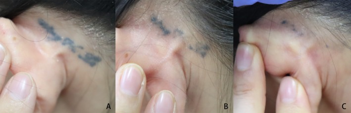
FIGURE 1 A tattoo behind the left ear treated with the PS 1064‐nm laser (fluence: 1.8–4.3 J/cm2, spot size: 3 mm, a repetition rate of 3 Hz). (A) Before treatment, (B) after one treatment, and (C) after two treatments.左耳后纹身经PS 1064 nm激光治疗的临床照片(能量密度1.8–4.3 J/cm²,光斑3 mm,频率3 Hz):(A) 治疗前;(B) 1次治疗后;(C) 2次治疗后。
FIGURE 2 A tattoo on the left cheek treated with the PS 1064‐nm laser (fluence: 2.2–2.4 J/cm2, spot size: 3–4 mm, a repetition rate of 3–4 Hz). (A) Before treatment, (B) after one treatment, (C) after two treatments, and (D) after three treatments.左颊纹身经PS 1064 nm激光治疗的临床照片(能量密度2.2–2.4 J/cm²,光斑3–4 mm,频率3–4 Hz):(A) 治疗前;(B) 1次治疗后;(C) 2次治疗后;(D) 3次治疗后。
FIGURE 3 A tattoo on the left back of the hand treated with the PS 1064‐nm laser (fluence: 3.5–5 J/cm2, spot size: 3 mm, a repetition rate of 2–3 Hz). (A) Before treatment, (B) after one treatment, (C) after two treatments, (D) after three treatments, and (E) after four treatments.左手背纹身经PS 1064 nm激光治疗的临床照片(能量密度3.5–5 J/cm²,光斑3 mm,频率2–3 Hz):(A) 治疗前;(B) 1次治疗后;(C) 2次治疗后;(D) 3次治疗后;(E) 4次治疗后。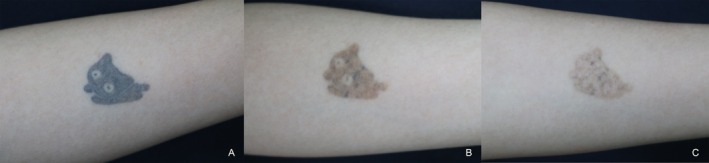
FIGURE 4 A tattoo on the left forearm treated with the QS 1064‐nm laser (fluence: 3.5–4.5 J/cm2, spot size: 3–4 mm, a repetition rate of 2–4 Hz). (A) Before treatment, (B) after one treatment, and (C) after two treatments.左前臂纹身经QS 1064 nm激光治疗的临床照片(能量密度3.5–4.5 J/cm²,光斑3–4 mm,频率2–4 Hz):(A) 治疗前;(B) 1次治疗后;(C) 2次治疗后。
FIGURE 5 A tattoo on the right forearm treated with the QS 1064‐nm laser (fluence: 3.3–3.9 J/cm2, spot size: 3–4 mm, a repetition rate of 3–4 Hz). (A) Before treatment, (B) after one treatment, (C) after two treatments, and (D) after three treatments.右前臂纹身经QS 1064 nm激光治疗的临床照片(能量密度3.3–3.9 J/cm²,光斑3–4 mm,频率3–4 Hz):(A) 治疗前;(B) 1次治疗后;(C) 2次治疗后;(D) 3次治疗后。
FIGURE 6 A tattoo on the right knee treated with the QS 1064‐nm laser (fluence: 2.2–4 J/cm2, spot size: 3 mm, repetition rate of 3–4 Hz). (A) Before treatment, (B) after one treatment, (C) after two treatments, (D) after three treatments, and (E) after four treatments.右膝纹身经QS 1064 nm激光治疗的临床照片(能量密度2.2–4 J/cm²,光斑3 mm,频率3–4 Hz):(A) 治疗前;(B) 1次治疗后;(C) 2次治疗后;(D) 3次治疗后;(E) 4次治疗后。To address the near‐significant baseline imbalance in tattoo color distribution (blue/black: 89.04% in PS group vs. 75.44% in QS group, p = 0.061), we performed a subgroup analysis restricted to patients with blue/black tattoos. Among these 108 patients (65 in PS group, 43 in QS group), the PS 1064‐nm laser achieved an excellent clearance rate of 80% ( n = 52/65) compared to 55.81% ( n = 24/43) for the QS 1064‐nm laser ( p = 0.007). The overall effectiveness rate (> 50% clearance) in the blue/black subgroup was 93.85% ( n = 61/65) for the PS 1064‐nm laser versus 88.37% ( n = 38/43) for the QS 1064‐nm laser ( p = 0.314).
3.3
Both laser modalities maintained excellent safety profiles. Transient adverse effects, such as erythema and edema, were observed in 10.96% ( n = 8/73) of the PS 1064‐nm laser group and 12.3% ( n = 7/57) of the QS 1064‐nm laser group. These reactions resolved spontaneously within 48–72 h. In each group, there were two instances of post‐inflammatory hyperpigmentation (PIH). No severe complications, including blistering, scarring, or permanent dyspigmentation, were observed, and no significant intergroup differences in safety were identified ( p > 0.05).
4
Tattoo removal has evolved into a widely studied and popular cosmetic procedure, primarily driven by an increasing number of individuals who have tattoos and a tendency to remove them for a variety of personal, social, and professional reasons [ 1 ]. Currently, the main techniques used for tattoo removal are based on laser technology, specifically using QS 1064‐nm laser and PS 1064‐nm laser [ 9 ]. Most tattoo pigments consist of particles ranging from 30 to 300 nm with relatively shorter thermal relaxation times (< 10 ns) [ 10 ]. The 5–10 ns pulse duration of QS 1064‐nm laser approximates this TRT, generating shock waves that rupture the targeted ink particles. Previous studies, such as those by Kilmer et al. [ 11 ], reported that 77% ( n = 30/39) of black tattoos had a great response (more than 75% clearance) after approximately four sessions with QS 1064‐nm laser. Moreover, another study revealed that 95.45% (21 out of 22) of patients had an excellent response after one to four treatments [ 12 ]. In contrast, the present cohort achieved an excellent response rate of only 54.39% ( n = 31/57) after one to six sessions, which is slightly lower than findings from previous studies [ 13 ]. This discrepancy might be attributed to the lower fluence used in this research (1–6 J/cm 2 ) compared to earlier studies (5–10 J/cm 2 ) [ 12 , 13 ]. Furthermore, patients with darker skin tones (Fitzpatrick types IV–VI) often require more sessions, longer wavelengths, and face a higher risk of adverse effects, particularly hypopigmentation [ 9 ]. Although repeated QS laser treatments eventually help reduce tattoo visibility, the treatment efficacy is inconsistent, and therapy duration is extended, which induces substantial distress for both practitioners and patients.
With its ultrashort pulse duration and selective photoacoustic effect, the PS 1064‐nm laser has emerged as a benchmark for tattoo removal [ 6 ], offering reduced collateral thermal damage compared to traditional nanosecond lasers. The present investigation revealed that 78.08% (57 out of 73) of patients treated with PS 1064‐nm laser had an excellent response after an average of three sessions. Despite limited comparative data, the present results indicated that the PS laser yields superior outcomes in high‐tier clearance outcomes. This aligns with the findings of Ross et al. [ 7 ], Lorgeou et al. [ 14 ], and Choi et al. [ 15 ], but contradicts the findings of Pinto et al. [ 8 ] and Du et al. [ 16 ], who reported no significant differences between the two laser types. In the study by Du et al. [ 16 ], rats were subjected to a single laser treatment, while in the research by Pinto et al. [ 8 ], the cohort experienced a maximum of two sessions. Participants in studies by Ross et al. [ 7 ], Lorgeou et al. [ 14 ], and our studies received between four and seven treatments. This variation suggests that the PS 1064‐nm laser has more advantages when the number of treatment sessions increases. Moreover, there might be differences in the fluence levels that could explain the inconsistent results. In the study by Du et al. [ 16 ], rats with treatment using PS 1064‐nm laser were given a lower fluence (2.0 J/cm 2 ) compared to QS 1064‐nm laser, which was set at 4.0–5.0 J/cm 2 accordingly, the efficacy between the two was comparable. Conversely, another animal study with guinea pigs demonstrated that the PS 1064‐nm laser (3.0 J/cm 2 ) performed better than the QS 1064‐nm laser (3.4 J/cm 2 ) at comparable fluence levels [ 15 ], which supports these observations. Therefore, the effectiveness of these lasers in tattoo removal depends on both the number of treatment sessions and the energy fluence, with the PS 1064‐nm laser requiring fewer sessions and lower energy input to achieve the best results.
Both modalities exhibited excellent safety for Fitzpatrick skin types (predominantly III–IV) in our cohort. Transient erythema and edema were observed without permanent dyspigmentation. Our findings align with those of Cannarozzo et al. [ 17 ], who reported that Q‐switched 1064 nm picosecond laser treatment for benign hypermelanosis in Asian patients (Fitzpatrick types II–IV) was well tolerated, with only temporary perilesional erythema and edema that resolved within 1–3 days, and no significant adverse events such as persistent dyspigmentation or scarring. The authors emphasized that careful fluence titration is key to minimizing complications in darker skin tones. Together, these observations suggest that historical concerns regarding laser‐induced hypopigmentation in darker skin tones, which may be mitigated by optimized fluence settings and the 1064 nm wavelength's deeper penetration, which minimizes epidermal damage [ 15 , 18 ].
Several key limitations of this study must be addressed. First, the retrospective, non‐randomized design introduces potential selection bias, as treatment allocation was based on device availability rather than strict randomization. This fundamental confounder means that patients in the two groups were inherently different, and unmeasured factors—such as differences in tattoo ink composition, particle size, and depth—could have influenced the outcome [ 9 , 10 ]. The near‐significant baseline imbalance in tattoo color (blue/black: 89.04% in PS group vs. 75.44% in QS group, p = 0.061) is particularly concerning, as color independently drives clearance. While we performed a subgroup analysis restricted to blue/black tattoos, a multivariable analysis would be preferable but is limited by sample size and the retrospective design. Second, the predominance of blue/black tattoos limits the applicability of our conclusions to polychromatic tattoos. Third, the 9‐year study period (2015–2024) introduces the possibility of platform and parameter drift, including potential variations in laser handpiece performance, energy calibration, and operator technique evolution over time. Although all treatments adhered to institutional protocols, we were unable to quantify the magnitude of such drift in this retrospective analysis. This temporal heterogeneity may have introduced noise into our efficacy comparisons and should be considered when interpreting the results. Fourth, the 3‐month follow‐up period may be insufficient to capture long‐term recurrence and side effects. Given these constraints, the difference in the overall effectiveness rate (> 50% clearance) lacking statistical significance (94.52% versus 85.96%, p = 0.094) further underscores that the perceived superiority of the PS 1064‐nm laser in achieving “excellent” clearance should not be overstated. The findings should be interpreted as generating a hypothesis within the constraints of a retrospective comparison, rather than as definitive evidence of superiority.
The PS 1064‐nm laser demonstrated comparable overall effectiveness to the QS 1064‐nm laser for tattoo removal, with potential advantages in achieving excellent clearance and reducing the number of treatment sessions. Given the retrospective design, non‐randomized allocation, and the nonsignificant difference in overall effectiveness, these findings require validation through prospective, randomized controlled trials.
J.C. and Y.L. conceived and designed the study, as well as wrote the manuscript. Y.G., S.M. and B.L. provided study materials and patient data. J.C. and Y.L. collected and assembled the data. J.C. and Y.L. conducted data analysis and wrote the results. B.L. and H.Z. was responsible for reviewing and revising the manuscript.
This work was supported by Guangzhou Municipal Science and Technology Program (Grant no. 2023A03J0470 and 2023A03J0943).
Declaration of Generative AI and AI‐ assisted technologies in the writing process: AI tools (e.g., DeepL) were used to assist with translation and linguistic refinement. The authors reviewed and edited the translated text to ensure accuracy and scientific integrity. All scientific content and analyses were conducted and verified by all authors.
The study protocol was approved by the Ethics Committee of the GuangZhou Dermatology Hospital (gzsp202537). Patients provided written informed consent for the use of their anonymized photographs for research and publication purposes.
Patients signed informed consent regarding the publishing of their data and photographs.
The authors declare no conflicts of interest.
声明:中文精读 · 仅供学术参考。内容来自公开文献检索,不代表本人观点,不构成诊疗建议。 医疗美容需在正规医疗机构由执业医师实施。
基于相同主题推荐 · 继续深挖
前瞻性随机分面对照、评估者盲法试验(泰国 Ramathibodi 医院,2022-11 至 2025-02):12 例活检确诊 LPP(平均 55±11.1 岁,81.8% 女性,
回顾性队列研究:2015年1月至2024年9月广州某医院130例纹身患者,皮秒(PS)1064nm组73例、Q开关(QS)1064nm组57例,分组依设备可用性与医师裁量而非随机;

分脸、双盲、随机、安慰剂对照的单中心临床试验,24名女性(年龄30–48岁,平均38.46±5.17岁)完成;经2周洗脱期后接受单次1064-nm皮秒Nd:YAG激光(Picoca