超声引导中面部三层注射技术:按组织层面匹配流变学特性的透明质酸分层填充概念验证研究
2026年10月13日40 min read
Journal of Cosmetic Dermatology
结论与实践要点
本文发表于 Journal of Cosmetic Dermatology(DOI: 10.1111/jocd.70664),题述为一项「中面部年轻化掌握:新的三层、超声引导技术提案」。作者在 Trévidic 多层方法(按流变学差异分别处理中面部深、浅脂肪室)基础上补充两点:其一,既往多层方法未处理皮肤层,而皮肤治疗是年轻化外观的支柱;其二,超声引导可确保填充剂在不同层次的精准、安全放置。该技术建立在三个基本要素上:全面掌握解剖学、使用高频面部超声引导精准注射、按流变学特性为三个注射平面(深层、浅层、真皮层)选择匹配的 HA 填充剂。需要明确的是,本文为观察性/概念验证研究(proof of concept),在意大利按观察性研究执行,作者称意大利法规对此类研究不要求伦理委员会批准,研究遵循赫尔辛基宣言原则,患者签署知情同意;其证据等级远低于随机对照试验,文中相关表述(如原文称之为现代面部年轻化的重要进展)应理解为作者主张而非已验证的结论。
解剖基础部分强调面部软组织自骨至皮肤的五层结构:深部脂肪室(内侧与外侧 SOOF、深内侧颊脂肪 DMCF)牢固锚定于骨并提供结构支持,随年龄进展性萎缩导致上颊塌陷与假性下垂;浅层脂肪室(眶下、内侧颊、中颊、鼻唇沟脂肪)活动度较大。作者指出,若无超声可视化,深浅脂肪室之间的隔膜无法识别,而 SMAS(高回声、数毫米厚)的识别对确保深层产品正确放置、避免误入浅层或 SMAS 内至关重要。高频超声可客观呈现衰老改变:图 2 对比 26 岁(A)与 57 岁(B)患者,年长者真皮更薄、脂肪层更厚。安全区以两条参考线界定——内侧颊安全线(内眦至下颌角连线)与外侧安全线(颧弓中心垂直下行线)。
血管解剖是安全核心。面部动脉(FA)个体间走行变异大:一项尸体解剖研究显示其主要沿鼻唇沟走行,延续为角动脉者 36.6%、鼻外侧动脉 48.6%、上唇动脉 8.5%,发育不良变异终于下唇动脉者 6.8%;约 30% 个体 FA 分叉为鼻唇干与眶下干。必须强调,这些比例来自离体尸体解剖数据,与在体超声观察到的比例并不一致(原文亦引用一项超声研究报告不同比例),提示血管分布受组织厚度等因素影响变异极大,离体数据不能直接外推为在体临床变异率或结局。眶下动脉分出 2-3 支(睑、鼻、唇支),眶下孔朝向足侧(下颌方向),故该区域应从下方以锐角接近以降低动脉损伤风险;面横动脉(TAF)多数人为单支、起自颞浅动脉,其皮下上行支在侧颊浅层脂肪注射时存在被钝针穿刺的风险;颧面动脉(ZFA)起自 SOOF 内、经颧面孔(多位于外眦外侧)穿出,部分患者 ZFA 与颧面孔可缺如,故该区超声评估不可或缺。
超声贯穿治疗全程:治疗前用于区分年轻与成熟面部(真皮与脂肪层厚度)、绘制包括浅深脂肪室、肌肉位置及主血管神经走行的解剖地图、规划注射路径以避开高危区,从而降低血管内注射与组织损伤风险;治疗中用于识别各沉积层次、需避开的区域与高风险区,实时监控填充剂分布——原文图 7 显示经常用的上外侧入路以钝针无法到达深部脂肪室(钝针停留在浅层颧区),而低位入路方可抵达 SMAS 下深部(图 8),且低位入路同样便利浅层注射(图 9);正确识别 SMAS 与脂肪层可避免填充剂误沉积于 SMAS 内(图 10,无超声引导时可能发生)或同时进入深浅两层(图 11);治疗后用于验证填充剂分布准确性并长期评估其与周围组织的整合情况。
三层注射方案具体为:第一层,深层脂肪——于骨膜上注射高投射能力的「firm」填充剂(高 G′、高强度,文中示例为 Teosyal PureSense Ultra Deep),采用低位钝针入路或上外侧入路锐针,目标是提供骨性支撑、增强颧部投射;第二层,浅层脂肪——将弹性、可塑、延展性更好的填充剂(文中示例为 Teosyal RHA4 或 RHA3)直接注入浅层脂肪室,其组织内整合可减少结节形成与产品迁移,并维持动态表情自然;第三层,真皮皮下层——经同一低位入路、以更锐角度钝针注射非交联 HA(文中示例为 Teosyal PureSense Redensity 1,属 skin booster),含水化、改善皮肤密度与光泽的作用,部分产品复配氨基酸、维生素与矿物质。15 例患者(13 女 2 男,50±8 岁,范围 42-65 岁)以此技术单次疗程治疗,纳入标准为 40-70 岁、既往治疗区无填充史、健康;排除 <40 或 >70 岁、系统性疾病或服用抗血小板/抗凝药物者、妊娠哺乳期者;随访时点为治疗后 1 周与 3 个月。
结果显示:全部治疗患者获益,颊部即时充盈、提升,颧弓更突出;首次对照(1 周)时皮肤更亮、水合、光滑、放松;作者报告无不良反应。两例代表性病例(56 岁与 57 岁女性,正面与 3/4 面观)显示术后颊部即时填充提升、面部椭圆间接提升,皮肤松弛度、光泽与水合改善,病例照片追踪至 7 个月(57 岁例)与 8 个月(56 岁例)时效果仍维持;超声检查(图 15、16)显示填充剂沉积于正确解剖层面,皮下层呈均一高回声模式,提示亲水、可完全吸收的填充剂组织整合良好,无碎裂或包裹征象。作者还提出一个尚待验证的假设:非交联产品以钝针真皮皮下给药的方式,除水化作用外或可延长更深层填充剂的效果,但明确表示需进一步研究证实;另一实用益处是三层治疗共用同一位点入路,可减少多次皮内穿刺造成的不适与短期外观影响。
对整形外科医生的启示在于其可复用的框架而非照搬产品:其一,先超声评估后治疗——术前建立个体化解剖地图(真皮厚度、脂肪室、SMAS、血管走行),术中确认层次,术后验证整合;其二,按层面匹配流变学——深层骨膜上选高 G′、高强度产品以获得支撑与投射,浅层选弹性可塑产品以适应表情,真皮皮下选非交联产品改善肤质;其三,入路设计比产品选择更易被忽视——原文的超声证据表明,钝针经上外侧入路到不了深部脂肪室,低位入路是同时处理深浅层的可行路径;其四,病例选择上,作者认为三层法更适合面部凹陷或皮肤成熟者,而单层深部注射一般更适合容积流失少的年轻患者。最后须注意证据边界:本文为 15 例的概念验证观察性研究,无对照、无盲法、无标准化量表与统计学检验,「无不良反应」仅为作者陈述而无系统的并发症记录;血管比例等关键安全数据来自尸体解剖离体研究;作者自述更大样本队列数据尚在整理、将另行发表。因此该技术应视为一种有超声证据支持的技术提案与概念验证,其安全性、有效性与普适性仍需更大样本、更长随访与对照研究确立,临床采纳须结合个体解剖评估、知情同意与当地法规。
IntroductionAging affects all facial layers: deep fat atrophy reduces midface projection, superficial fat changes alter facial contour, and dermal thinning compromises skin texture. Midface rejuvenation aims to restore malar projection, cheek volume, and youthful contours, thereby improving the nasolabial fold and suborbicular region. Conventional multilayer techniques address both deep and superficial fat compartments using fillers with distinct rheological properties, but often neglect skin quality and the advantages of ultrasound guidance. The approach we propose is grounded on three core principles: the comprehensive anatomical knowledge, the use of high‐frequency ultrasound to ensure precise and safe injections, and the selection of hyaluronic acid fillers specifically tailored to each tissue plane—deep, superficial, and dermal. The aim of our work was to present our technique as a proof of concept for a three‐layer, ultrasound‐assisted technique, which enhances safety, precision, and aesthetic effectiveness, representing a significant advancement in modern facial rejuvenation.
MethodsA three‐layer, ultrasound‐guided midface rejuvenation technique employs tailored hyaluronic acid fillers for each plane. Firm, high G′ fillers target deep compartments through supraperiosteal injections for structural support. Elastic, moldable fillers, directly injected into superficial fat, enhance superficial layers while preserving natural mobility. Non‐cross‐linked formulations, used subdermally, by using a cannula, hydrate and improve skin quality, ensuring precise integration with minimal risk.
ConclusionsThe multilayer technique combines different fillers in deep, superficial, and dermal layers, tailored to the patient's anatomy. This three‐layer method restores volume and preserves natural expression, simultaneously improving skin quality, particularly benefiting hollow or mature faces. Unlike single‐plane injections, it addresses all aging aspects. Although not yet routine, ultrasound integration in multilayer treatments promises higher efficacy and safety. This approach marks a significant advancement in modern facial rejuvenation standards.
Keywords: facial ultrasound、fillers、skin booster、skin layers、three‐layer、three‐layer injection
1
Midface rejuvenation involves enhancing malar projection and restoring full cheek volume to preserve a youthful facial appearance, with beneficial effects on the nasolabial fold and the sub‐orbicular area. Given the multilayer structure of facial soft tissues, a multilayer approach to midface rejuvenation has already been proposed by Trévidic, who suggests treating both the deep and the superficial fat compartments of the midface using fillers with different rheological properties, including variation in elastic (G′) and viscous (G″) moduli, cohesivity, and swelling propensity. However, this approach does not address skin treatment, which remains a fundamental pillar in achieving a youthful appearance. Moreover, we believe that the integration of ultrasound guidance should not be overlooked in this type of treatment, as it offers significant advantages in ensuring the precise and safe placement of fillers across the different layers.
For these reasons, we propose that midface rejuvenation requires a structured approach grounded on three fundamental elements: a comprehensive understanding of anatomy, the use of facial ultrasound to guide precise injection techniques, and the selection of appropriate hyaluronic acid fillers, tailored to the three injection planes according to their rheological properties. The introduction of this three‐layer, eco‐assisted technique may represent a significant turning point in achieving safe and natural aesthetic results.
1.1
To optimize procedural safety and achieve aesthetically consistent and natural results, clinicians must restore volume in the middle third of the face to master its three‐dimensional architecture. A key element of the multilayered treatment approach is understanding the five‐level structure of the face from bone to skin, which includes compartments of static deep fat and movable surface fat pads, separated by the superficial muscular aponeurotic system (SMAS) (Figure 1 ) [ 1 , 2 ].
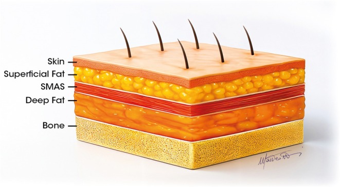
FIGURE 1 Anatomy of the multilayer structure in the midface.中面部多层结构解剖示意图,展示自骨至皮肤的五层结构及被 SMAS 分隔的深部静态脂肪室与浅层可活动脂肪垫。Another important aspect is the anatomy of the arterial vessels, which can vary significantly between individuals, making it essential to master this anatomy to avoid complications. This understanding guides clinical decisions regarding injection technique, depth of intervention, and precautions before and after every treatment.
Ultimately, it is crucial to recognize that the aging process affects each level, leading to interventions aimed at restoring a youthful appearance without compromising its natural integrity [ 3 ]. This section provides a comprehensive overview of the anatomy of the central face area, focusing on the structural and functional aspects for effective volumetric enhancement with fillers. Detailed anatomical studies reported in the scientific literature have revealed complex and variable configurations, often employing different terminologies for these structures.
1.1.1
In the central region of the face, deep fat compartments firmly anchored to the bone provide essential structural support to the overlying soft tissue layers, including the Superficial Musculo Aponeurotic System (SMAS), the superficial fat, and the dermal–subcutaneous layers. The progressive atrophy of these compartments contributes to the collapse of the upper cheeks with age (Figure 2 ) [ 4 , 5 , 6 ]. The identification of the septa that separate the superficial fat compartments and those between the deep compartments is not possible without ultrasound visualization. However, identifying the SMAS is crucial to ensure the correct placement of products intended for the deep compartment, reducing the risk of superficial or intra‐SMAS injections and related complications.
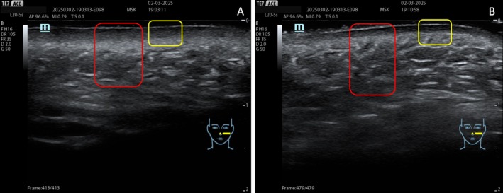
FIGURE 2 The thickness of the dermal and superficial compartments in a 26‐year‐old (A) and a 57‐year‐old patient (B). The dermal layer is highlighted in the yellow box, and the adipose layer is highlighted in the red box. The dermis is thinner, and the adipose layer is thicker in the older patient.高频超声图像对比:26 岁(A)与 57 岁(B)患者的真皮与浅层脂肪室厚度,黄框标示真皮层、红框标示脂肪层;年长者真皮更薄、脂肪层更厚。As people age, volume loss in the deep panniculus adiposus reduces the medial projection of the midface, contributing to pseudoptosis due to excess skin.
Therefore, it is necessary to focus on all fat compartments of the midface: the deep fat [(medial and lateral) suborbicularis oculi fat (medial‐ and lateral‐SOOF), and deep medial cheek fat (DMCF)] (Figure 3 ), and the superficial fat (infraorbital fat, medial cheek fat, middle cheek fat, and nasolabial fat) (Figure 4 ) [ 6 , 7 ], whose treatment is planned according to the multilayer technique [ 8 , 9 ].
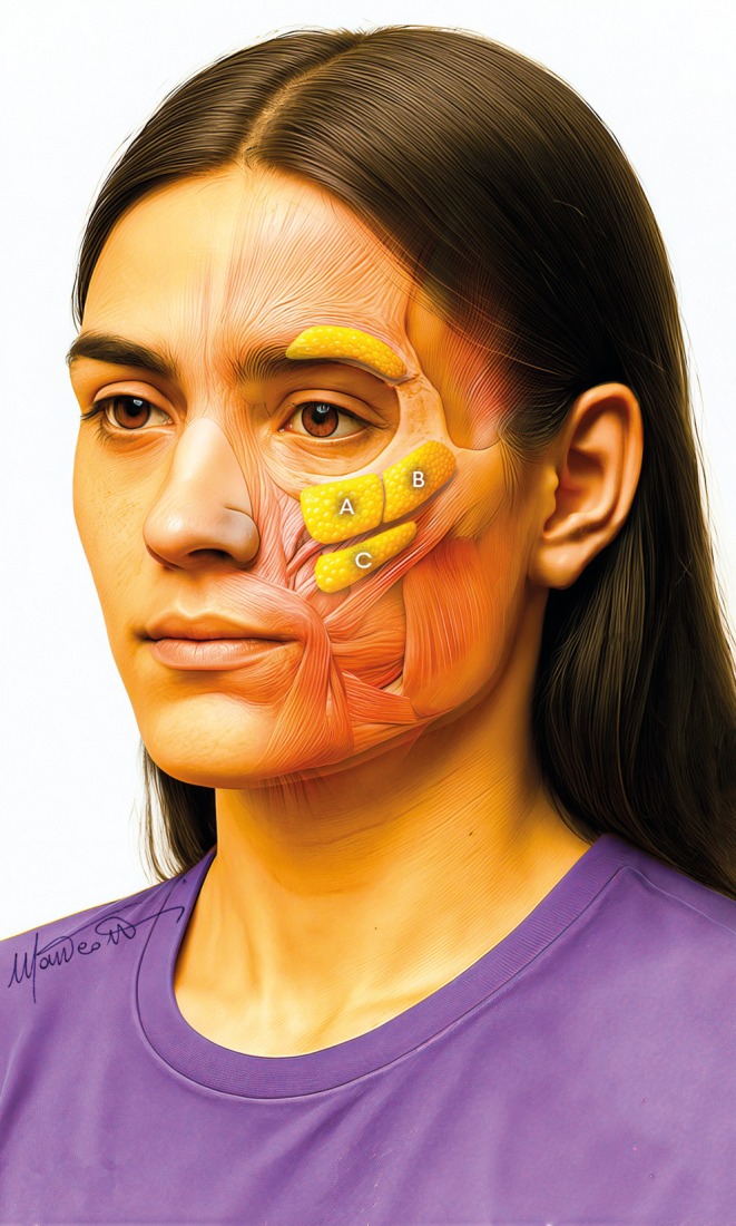
FIGURE 3 Deep compartments in the midface: (A) medial SOOF, (B) lateral SOOF, and (C) deep medial cheek fat.中面部深层脂肪室解剖/超声图像:(A) 内侧 SOOF、(B) 外侧 SOOF、(C) 深内侧颊脂肪(DMCF)。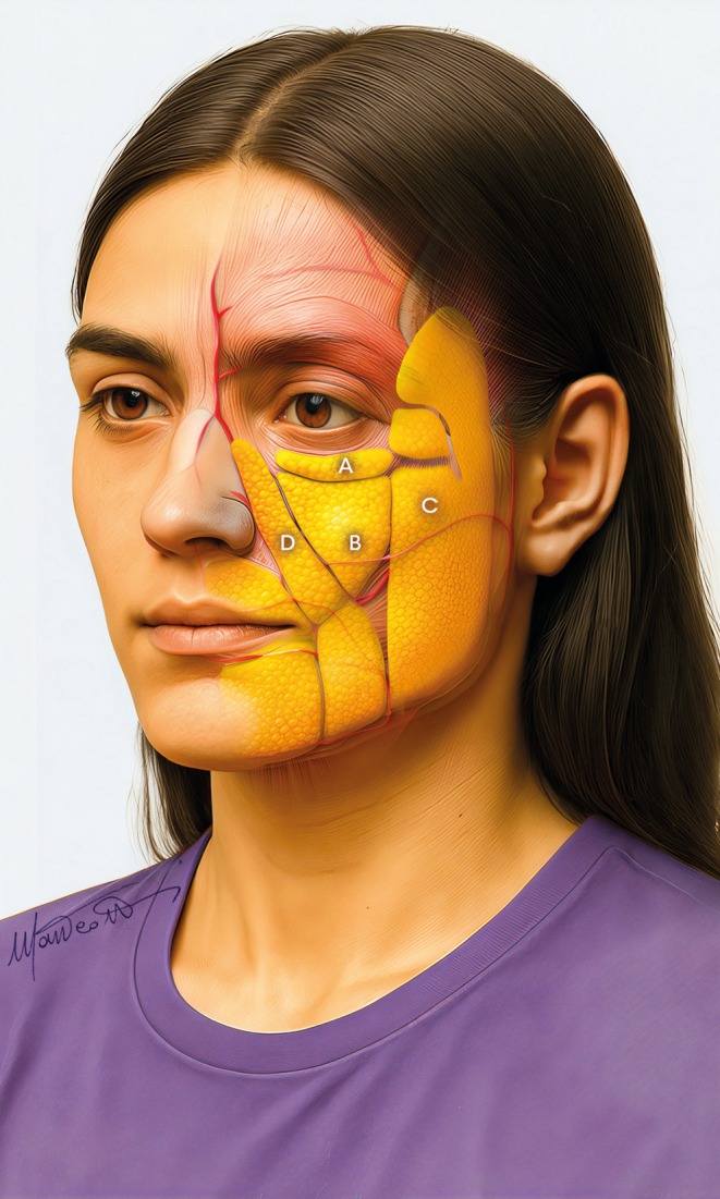
FIGURE 4 Superficial compartments in the midface: (A) Infraorbital fat compartment, (B) Medial cheek fat compartment, (C) Middle cheek fat compartment, and (D) Nasolabial fat compartment.中面部浅层脂肪室解剖/超声图像:(A) 眶下脂肪室、(B) 内侧颊脂肪室、(C) 中颊脂肪室、(D) 鼻唇沟脂肪室。Recent anatomical investigations reveal that the SOOF and DMCF are systematically divided into separate, easily identifiable compartments [ 7 ]. In addition, these structures are permanently adhered to the bone; therefore, their treatment with filling techniques must be targeted and precise.
1.1.2
In clinical practice, anatomical references are used to outline areas known as “safety zones,” which are defined by tracing a straight line from the medial canthus to the mandibular angle (designated as the medial cheek safety line) and a second line that descends vertically from the center of the zygomatic arch (referred to as the lateral safety line) [ 8 ].
The facial artery (FA), a major vessel that supplies blood to the face, originates from the external carotid artery and extends to the facial region through the submandibular area, passing anterior to the masseter muscle. As demonstrated by previous studies, this artery displays considerable variability in its path among individuals (Figure 5 ) [ 10 , 11 , 12 , 13 ].
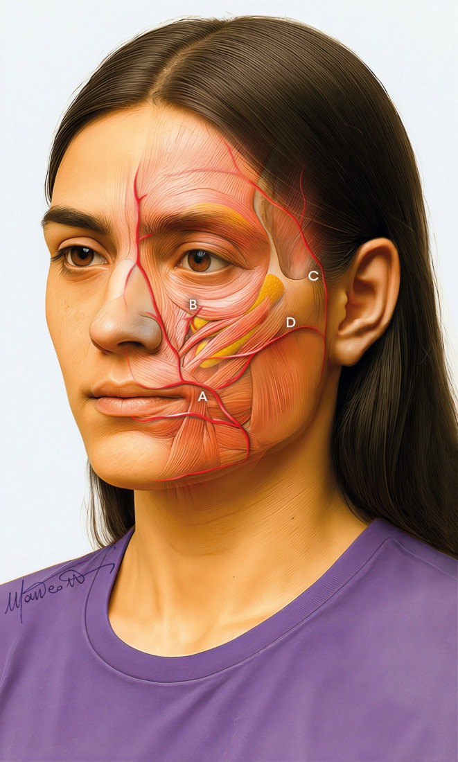
FIGURE 5 Arterial network in the midface: (A) Facial artery, (B) Infraorbital artery, (C) Temporal artery, and (D) Transverse facial artery.中面部动脉网络图像:(A) 面动脉、(B) 眶下动脉、(C) 颞动脉、(D) 面横动脉。A recent study based on cadaveric anatomical analyses showed that FA mainly follows the nasolabial sulcus, entering the angular artery in 36.6% of cases, the lateral nasal artery in 48.6%, and the superior labial artery in 8.5%. Interestingly, a hypoplastic variant of FA ends in the inferior labial artery in 6.8% of individuals. Additionally, about 30% of the population exhibits a bifurcation of the AF into a nasolabial trunk and an infraorbital trunk, which extends vertically toward the nasojugal fold [ 10 , 11 , 12 , 13 ].
At the same time, a recent ultrasound study [ 14 ] shows significantly different percentages concerning the course of the FA tract, identified as the Angular artery. This highlights the variability in vessel distribution related to tissue thickness and lateral‐medial vessel dislocation.
Given this anatomy, it is crucial to take proper precautions during invasive procedures and to monitor any symptoms that may arise.
The infraorbital artery, a vital vessel in the middle third, also needs to be evaluated. It arises from the infraorbital foramen and branches into two or three main branches: the palpebral, nasal, and labial branches. The latter two are located within the DMCF and supply blood, respectively, to the lateral nasal and upper labial areas, mainly below the safety line of the medial cheek [ 15 ]. It is important to note that the infraorbital foramen points downwards toward the mandible; therefore, it is best to approach this area from below at an acute angle to reduce the risk of arterial injury [ 16 ].
In treatments targeting the middle third of the face, evaluating the Transverse Artery of the Face (TAF) is crucial, as it represents a key vascular structure that supplies the cheek region [ 17 ]. Anatomical studies have demonstrated that the majority of individuals have a single TAF, which arises from the superficial temporal artery and splits into an upper and a deeper branch. The two branches supply the parotid gland and its duct as well as the masseter muscle [ 18 ].
During ultrasound‐guided procedures, careful attention to the superior emergent branch is essential. This branch runs into the subcutaneous tissue and poses a risk of cannulation during filler injections into the superficial fat of the lateral cheek [ 19 ].
Moreover, the Zygomatic–Facial artery (ZFA) must also be assessed. This vessel originates within the SOOF, arising from the lacrimal artery and emerging through the zygomatic–facial foramen. Although this foramen has rarely been described, studies have localized it laterally to the lateral canthus [ 20 , 21 , 22 , 23 ]. It has also been observed that both the ZFA and the foramen may be absent in some patients, making careful ultrasound evaluation in this region fundamental.
Lastly, the facial vein, easily recognizable by its origin and course, functions as the main venous drainage vessel for the face. The facial vein, located along the lower edge of the mandible, courses lateral to the FA and proceeds directly toward the medial canthus, passing beneath the zygomaticus major and minor muscles. Since the FA lies beneath the medial cheek slit, it is located below the medial cheek safety line [ 18 ].
1.2
Given these anatomical factors, the integration of ultrasound into clinical practice becomes a critical tool for both assessment and accurate filler placement and distribution.
Ultrasound assessment is pivotal throughout all stages of filler treatment, including pre‐procedural planning, intra‐procedural guidance, and post‐procedural evaluation.
1.2.1
In facial rejuvenation procedures, a pre‐procedure ultrasound examination can facilitate a more accurate distinction between a youthful and a mature face, evaluating the thickness of the dermis and the fat layers. With aging, the dermis becomes progressively thinner, while the fat layer becomes progressively thicker (Figure 2 ) [ 24 , 25 ].
Ultrasound examination can also allow the visualization of anatomical morphology and tissue dynamics, facilitating the accurate identification of both superficial and deep fat compartments, the positioning of muscles, and the pathways of the main blood vessels and nerves. It enables the creation of a precise anatomical map, which would remain theoretical without ultrasound guidance.
Moreover, ultrasound mapping facilitates the planning of injection pathways in targeted areas, thereby avoiding high‐risk zones and reducing the likelihood of intravascular injection or damage to surrounding tissues [ 26 , 27 ]. Ultrasound examination enhances the identification of the SMAS, which appears as a hyperechoic structure measuring several millimeters in thickness, separating the superficial from the deep compartments [ 28 ]. Recognizing the SMAS allows for precise identification of the injection area, whether superficial or deep, preventing filler injections into the SMAS, thereby reducing the risk of complications such as inflammatory nodule formation or unwanted product migration [ 29 , 30 ].
Considering all the above, pretreatment ultrasound evaluation helps delimit safe areas and select the optimal access route to the target areas.
1.2.2
During treatment, ultrasound assists the doctor in identifying (a) the different layers for filler deposition, (b) the areas to avoid for optimal results, and (c) the critical high‐risk zones, especially for preventing complications. Ultrasound‐guided filler injection is crucial for accurately locating the injection sites, monitoring filler distribution during the procedure, and assessing post‐procedural integration to ensure the correct product is administered in the intended location.
For example, only through ultrasound guidance is it possible to realize that, when treating the deep fat compartment of the midface, filler injections into the deep space are not feasible via the commonly used superolateral access (Figure 6 ) due to the compartment's depth and the high flexibility of the cannula (Figure 7 ). Thus, when treating this compartment with a cannula, only a low approach (Figure 8 ) ensures the injection in the target layer. The same result could be achieved with an approach from the zygomatic arch, only with a needle. Simultaneously, a lower approach facilitates injections into the superficial compartments of the middle third (Figure 9 ). Finally, a correct identification of SMAS and fat layers during injection prevents fillers from being inadvertently deposited in the SMAS (Figure 10 ) or, simultaneously, in the superficial and deep compartments (Figure 11 ).
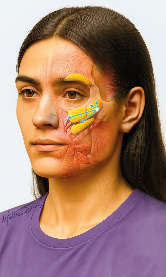
FIGURE 6 Superior–lateral entry point to access the superior deep compartments (medial and lateral SOOF).上外侧入路示意图/照片,用于抵达上方深层脂肪室(内侧与外侧 SOOF)。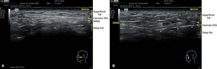
FIGURE 7 (A) Accessing the deep compartments with a cannula from a superior entry point is impossible. The cannula will stay in the superficial zygomatic compartment. (B) Accessing the deep compartments beneath the SMAS is possible by choosing an inferior entry point (B).示意图/超声图:(A) 经上外侧入路以钝针无法进入深层脂肪室,钝针将停留在浅层颧区;(B) 选择低位入路可抵达 SMAS 下方的深层脂肪室。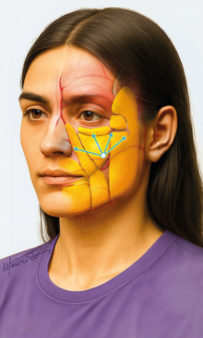
FIGURE 8 Inferior entry point to easily access both superficial and deep compartments of the midface.低位入路示意图/照片,可同时便捷进入中面部的浅层与深层脂肪室。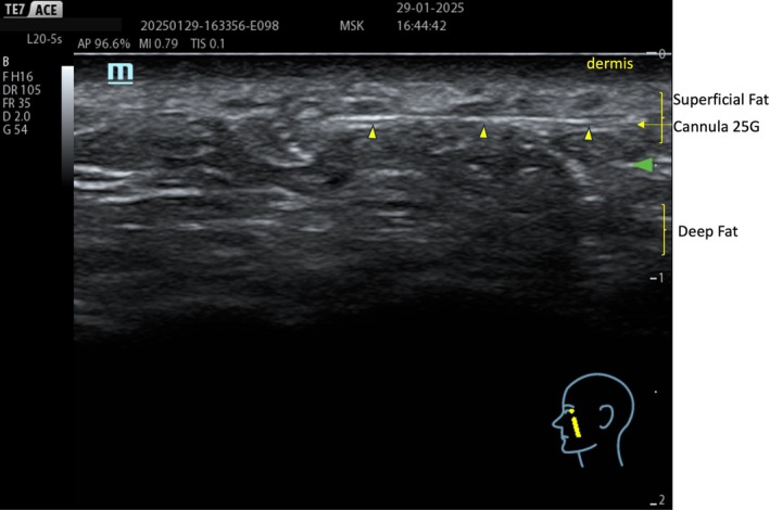
FIGURE 9 An inferior entry point makes accessing superficial planes easier.低位入路示意图/照片,显示该入路更易于进入浅层平面。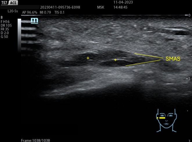
FIGURE 10 HA deposition (+) within the SMAS is possible when the treatment is performed without ultrasound guidance.超声图像,显示无超声引导时 HA 填充剂可能沉积于 SMAS 内的情况。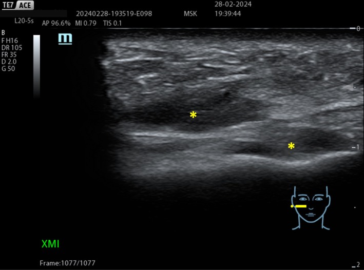
FIGURE 11 Hydrophilic deposits (+) of the same viscoelasticity in both deep and superficial compartments.超声图像,显示相同黏弹性的亲水填充剂同时沉积于深层与浅层脂肪室的情况。1.2.3
After treatment, ultrasound support enables real‐time verification of the accurate distribution of the filler and, over time, completes the evaluation of outcomes. Indeed, ultrasound analysis provides visual evidence of optimal product integration and permanence, highlighting the significance of facial ultrasound in monitoring procedure execution as well as in the long‐term assessment of outcomes.
1.3
Careful selection of fillers represents a crucial aspect of the therapeutic approach to midface rejuvenation. The choice of the correct hyaluronic acid (HA) filler must rely on its specific rheological properties, which are designed to integrate within various tissue compartments, including both superficial and deep layers. This helps achieve long‐lasting aesthetic results while maintaining a natural look during dynamic expressions, such as smiling [ 9 , 30 ]. Although the effectiveness of HA fillers for midface rejuvenation is well known, few have outlined a systematic multilayer approach that matches fillers with specific rheological properties to targeted tissue compartments [ 30 ].
Since deep fat compartments are anatomically distinct from septa and ligaments, while superficial compartments exhibit less segmentation, selecting the most suitable filler for specific compartments is a crucial key to preventing undesirable effects, such as filler migration and visible nodules.
In modern filler assessment, viscoelastic properties are measured using rheological parameters such as elastic modulus (G′) and viscous modulus (G″), which determine the filler's hardness and resistance to deformation. These parameters are important, but not the only factors to consider. Other mechanical parameters that have been recently defined [ 30 ], such as strength and stretch scores, must also be considered to address the limitations of measuring G′ under quasi‐static conditions. More specifically, the strength of a gel indicates its ability to maintain its characteristics under a wide range of mechanical stresses, while the stretch describes its tendency to deform and adapt to mechanical constraints, such as those in response to the endless facial movements [ 30 ]. The application of these fillers, called “resilient”, is essential for the harmonious remodeling of both deep and superficial compartments, particularly in patients with variable skin thickness, whether thin or thick. These fillers, characterized by a softer and more moldable consistency, exhibit slightly lower strength but greater stretchability. These properties allow better adaptation to facial movements and help minimize the risk of visible irregularities [ 9 ]. Fillers considered “firm”, namely those characterized by high G′ and significant strength, are ideal for injections in the deep static adipose compartment: the filler should be placed at bone contact, where significant structural support and pronounced zygomatic projection are required. In individuals with thinner skin, softer and more moldable fillers with slightly lower strength but greater stretch are preferable to avoid the appearance of visible irregularities [ 9 ].
Unlike cross‐linked fillers, which incorporate cross‐linking agents to create a more stable, gel‐like matrix for volumizing effects, non‐cross‐linked hyaluronic acids are specifically designed for application within the dermal layer. They are classified as skin boosters and are more fluid to spread in the target layer, providing hydration, thus improving skin quality and luminosity. These products are often combined with essential nutrients, such as amino acids, vitamins, and minerals, which help stimulate collagen and elastin synthesis to boost dermal density.
2
The new three‐layer ultrasound‐guided approach we propose for midface rejuvenation, as its name suggests, targets the reported three facial compartments (Figure 12 ) to improve both procedural accuracy and treatment outcomes.
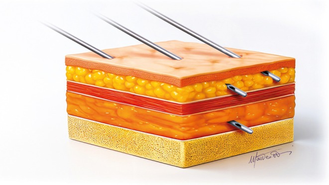
FIGURE 12 The cannulas showing the correct position in the three compartments to be treated.照片,显示钝针在拟治疗的三个脂肪室中的正确位置。2.1
Treatment begins with supraperiosteal injections targeting the deep fat folds, primarily within the fat compartments beneath the inferior orbital rim, such as medial and lateral SOOF, SOOF, and DMCF. In this area, “firm” fillers with high projection capacity are preferred, such as Teosyal PureSense Ultra Deep (Teoxane, Rue de Lyon 105, 1203 Geneva, Switzerland). Firm fillers in deep fat compartments aim to provide structural support for a solid foundation and enhance malar projection. A low entry point, using a cannula, or a superolateral entry point with a needle, is used for targeting this plane (Figures 6 and 8 ).
2.2
The second phase involves injections into the superficial fat compartments using more elastic, moldable fillers, such as Teosyal RHA4 or Teosyal RHA3 (Teoxane, Rue de Lyon 105, 1203 Geneva, Switzerland). These fillers, directly injected into superficial compartments, help maintain a natural look and allow for dynamic facial expressions. Their specific integration in the tissue prevents nodule formation and product migration.
2.3
The treatment concludes with subdermal infiltrations of non‐cross‐linked products such as Teosyal PureSense Redensity 1 (Teoxane, Rue de Lyon 105, 1203 Geneva, Switzerland). This type of filler, when applied in the subdermal layer, effectively restructures the deep dermis, improving skin density and appearance [ 31 ]. These layers are approached by the same low approach (Figure 8 ), but the cannula must be directed in the appropriate plane at a more acute angle.
Fifteen patients (13 women, two men, age 50 ± 8 years old) (range 42–65) were treated with the reported technique in one shot. They were controlled after 1 week and at 3 months distance to assess the midterm outcome. Inclusion criteria: patients in the age range 40–70, both sexes, without previous filler treatment in the treated area, in a healthy status, without systemic disease. Exclusion criteria: patients younger than 40 years and older than 70 years, with systemic diseases or taking antiplatelet or anticoagulant medications, pregnant, or breastfeeding. The treated patients were informed about the given treatment and signed an informed consent to be treated.
3
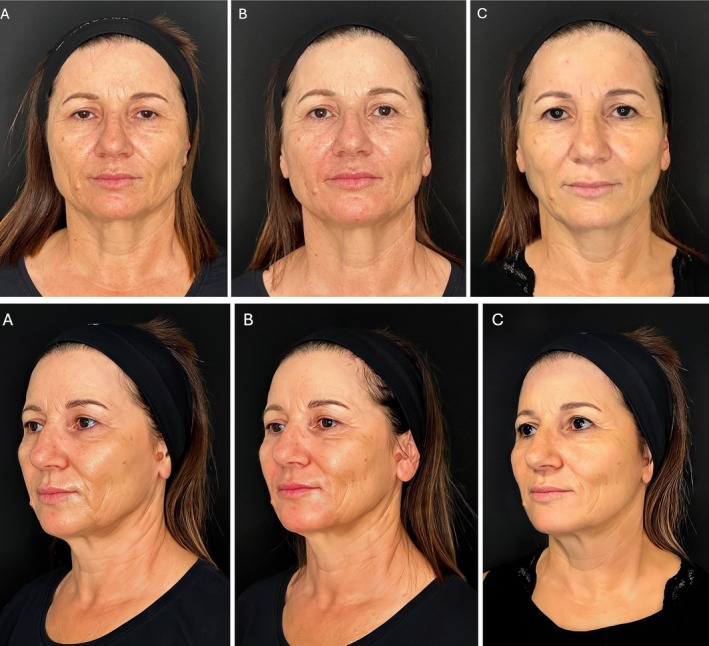
The treatment resulted in benefits for all the treated patients. In particular, the cheeks resulted in immediate filled and lifting with a more prominent zygomatic arch. At the first control, the skin appeared brighter, hydrated, smoother, and relaxed. No side effects were reported (Figures 13 and 14 ).
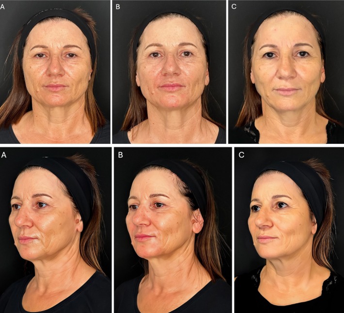
FIGURE 13 Front and three‐quarter views of a 56‐year‐old female. (A) Pretreatment: Patient with an aged face, showing tissue ptosis and skin laxity. The cheeks are hollowed out, and age furrows, visible in all projections, mark the skin, which is dull, devitalized, dehydrated, and uneven. (B) Post‐3‐layer midface treatment: The cheeks are immediately filled, lifted, and elevated, leading to noticeable improvements in all projections. Both frontal and three‐quarter views show the effect of indirect lifting on the face's oval, caused by the traction from the volumetric increase of the malar area. The skin also appears visibly better—more relaxed, brighter, and hydrated, with a noticeable improvement in surface smoothness and furrow depth. (C) Eight months after treatment: The filler is still completely integrated into the tissues, and the result remains evident: The cheeks are filled with a homogeneous volume, the cheekbones are well‐projected, and the lifting effect is still visible. The face remains overall rejuvenated, with harmonious proportions, and the skin appears glowing, relaxed, and more compact.56 岁女性病例临床照片(正面与 3/4 面观):(A) 治疗前,面部老化、组织下垂、皮肤松弛、颊部凹陷、皮肤暗哑脱水;(B) 三层中面部治疗后即刻,颊部充盈提升、颧部突出、各角度均改善、皮肤更放松水润;(C) 治疗后 8 个月,填充剂仍与组织完全整合,效果维持。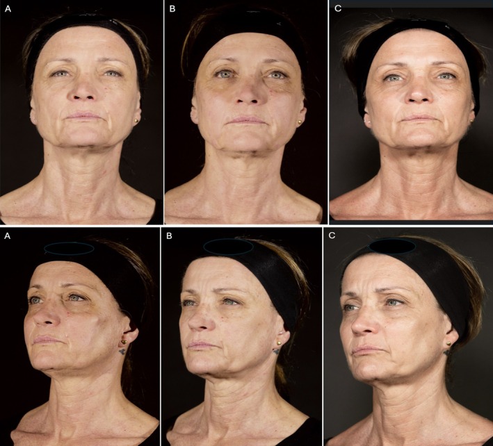
FIGURE 14 Front and three‐quarter views of a 57‐year‐old female. (A) Pretreatment: Patient with a tired face, with skin showing aging signs, in which tissue ptosis and skin laxity are evident (especially at three‐quarters and laterally). The cheeks are emptied, the tissues have slipped down, and the skin is moderately marked, dehydrated, dull, and devitalized, which is evident in all projections. In addition, especially in lateral viewing, there is a flat zygomatic bone and a lack of zygomatic prominence. (B) Post‐3‐layer midface treatment: The cheeks are immediately filled and lifted with a fresh and rested appearance. In the three‐quarter view, a moderate improvement in the zygomatic prominence can be appreciated. The skin is also improved, bright, more hydrated, and relaxed. (C) Seven months after treatment: The filler is integrated into the face tissues, and the aesthetic result is even more evident, especially concerning the lifting and projection effect. The skin is also visibly improved, more relaxed, glowing, compact, and hydrated. The face is overall rejuvenated, fresh, and rested.57 岁女性病例临床照片(正面与 3/4 面观):(A) 治疗前,疲态面容、组织下垂松弛、颊部空虚、颧骨平坦;(B) 三层治疗后即刻,颊部填充提升、皮肤改善更水润;(C) 治疗后 7 个月,填充剂与面部组织整合,提升与投射效果更明显,皮肤更紧致发光。The ultrasound examination revealed the filler deposition in the correct anatomical plane, its perfect filler integration in the patient tissue without any fragmentation (Figures 15 and 16 ).
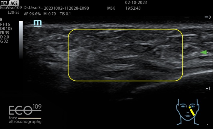
FIGURE 15 Before: Normal dermal and subcutaneous ultrasound stratification is observed.超声图像(治疗前):显示正常的真皮与皮下层分层结构。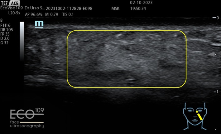
FIGURE 16 After: A homogeneous and uniform echogenic pattern is visible within the subcutaneous layer, indicating a recent injection of a hydrophilic, fully absorbable filler with high tissue integration, with no evidence of fragmentation or encapsulation.超声图像(治疗后):皮下层呈均一、高回声模式,提示近期注射的亲水、可完全吸收填充剂组织整合良好,无碎裂或包裹征象。4
The multilayer technique we propose as a proof‐of‐concept involves injecting different fillers into specific facial compartments. Selecting the proper filler is essential and must be customized to the patient's unique features, including bone structure, skin thickness, and the degree of volumetric loss.
This method can rebalance the volume in the mid‐third of the face, restoring a natural appearance (Figures 13 and 14 ) while simultaneously enhancing skin quality. Questions may arise regarding the optimal method for treating the most superficial layer, as it is typically addressed with intradermal injections using very thin needles, following the nappage technique.
In our method, the non‐cross‐linked filler is injected into the subdermal layer via a cannula. We hypothesize that when administered in this manner, this product may prolong the effects of deeper fillers beyond its hydrating effect; however, further studies are required to confirm this hypothesis.
Another practical benefit of this technique, even if it lacks scientific significance, is that the injection is made at the same entry point as the previous treatment. This helps avoid multiple intradermal injections, which can cause discomfort to the patient and an unpleasant appearance for several hours.
Compared to single‐plane injections, the three‐layer technique offers enhanced effectiveness, addressing all aspects of facial aging while maintaining a natural look. Therefore, this approach is particularly well‐suited for patients with a hollow face or mature skin, as it provides the necessary projection, preserves natural mobility and expressiveness, and improves skin quality. In contrast, single‐plane filler injections, which mainly target deep fat compartments, are generally indicated for younger patients with minimal volumetric loss.
The introduction of ultrasound as a supportive tool in treatments with HA fillers marks a significant advancement: in the pretreatment phase, enabling real‐time visualization of anatomical structures; during treatment, enhancing accuracy by injecting the proper filler into the target space while reducing associated risks and complications; in the posttreatment phase, allowing real‐time monitoring of the interaction between the filler and surrounding tissues.
Integrating ultrasound into daily practice requires adequate operator training; however, the benefits in treatment safety and efficacy are undeniable.
Though ultrasound assessment is not yet integrated into clinical practice, particularly within the multilayer technique, current evidence demonstrates its potential to elevate the standards of modern aesthetic medicine.
A limitation of the present work lies in the absence of well‐structured data derived from a large case series. As the ultrasound‐guided three‐layer technique represents a novel approach, such comprehensive datasets are not yet available. However, the really encouraging results coming from this preliminary study convinced us to treat a substantial number of patients consistently demonstrating favorable outcomes, including concurrent improvements in skin quality and midface volumetric restoration. With the wider clinical adoption of this technique, it is anticipated that a larger patient cohort will be treated, thereby enabling more rigorous validation of our preliminary observations. The data coming from this case series are under close examination and will be the object of a new publication.
5
Treating the three layers of the midface simultaneously (including the deep and superficial fat pads alongside the dermal layers), combined with the careful selection of fillers tailored to their specific rheological properties for each tissue compartment, represents a significant advancement in facial rejuvenation. Performed under ultrasound guidance, this approach enhances procedural precision and efficacy while maximizing safety, thereby setting new standards in aesthetic medicine.
The authors have nothing to report.
This is an observational study. For this kind of study, Italian Authorities do not request an Ethical Committee approval. The study was performed according to the Helsinki Declaration principles.
The patients who received the new three‐layer treatment were fully informed about the treatment and gave their informed consent.
Paola Molinari, Simone Ugo Urso, and Giovanni Mosti wrote the manuscript and checked the references; Chiara Faso controlled the manuscript, especially regarding the English form, and suggested some changes to make its reading more fluent; Ilenia Iacovone helped us with the reference research and verified that the details regarding the used products are correct. All authors declare no conflicts of interest regarding their author contributions.
声明:中文精读 · 仅供学术参考。内容来自公开文献检索,不代表本人观点,不构成诊疗建议。 医疗美容需在正规医疗机构由执业医师实施。
基于相同主题推荐 · 继续深挖
研究设计:意大利团队提出的三层(深层脂肪、浅层脂肪、真皮皮下层)超声引导中面部年轻化技术,属观察性/概念验证研究;15 例患者(13 女 2 男,50±8 岁,范围 42-65 岁
研究类型:系统综述联合单臂荟萃分析(遵循PRISMA,PROSPERO前瞻性注册),纳入11项研究共967例受试者,评估VYC-17.5L(Volift/Vollure,17.5
:整合解剖与美学的唇部评估框架/13565382_fig1.webp)
提出「唇部复杂度分类(LCC)」:按 5 个维度(相对唇容量、形态与对称、口周老化征象、组织质量、既往治疗史)将唇部分为 I–III 级,而非仅按容量评估。