多聚核苷酸HPT治疗眶周黑眼圈的真实世界研究
2026年10月20日27 min read
Clinical, Cosmetic and Investigational Dermatology
结论与实践要点
黑眼圈(眶周色素沉着)成因多因素,包括解剖/结构(泪槽、韧带牵拉、容积缺失)、色素、血管及混合型。PN HPT为源自鳟鱼性腺的高纯化多聚核苷酸,具即刻容积填充与长期组织重塑(促Ⅰ型胶原、弹性纤维、细胞外基质)双重作用。其通过水合形成生物相容性支架,补充真皮/皮下间隙、改善 Hollow 与肤质。
本文为真实世界观察性队列研究(非RCT、无对照组),证据等级低于随机对照试验,结论支持“关联/观察性改善“而非因果确证。作者自述主要弱点为缺乏正式对照组;计划中的前瞻性RCT尚未完成,相关结论待更高等级研究验证。原文未提供统一的最佳剂量/疗程标准,当前3次、每侧约1 mL方案为实际临床实践描述,具体参数应依术者判断。
Introduction and PurposeAnatomic factors and age‑related changes in the midface, along with sun exposure, prolonged television viewing, anemia, and hormonal derangements, contribute to the development of periorbital hyperpigmentation, also known as dark circles. Intradermally injected PN HPT (Polynucleotides High Purification Technology) from trout gonads has an immediate volume-enhancing effect and a longer-term indirect restructuring action on dermal and subdermal tissues.
MethodsOpen-label multinational observational cohort of 61 real-life multinational subjects of both genders and unskewed age distribution (mean 41.5 ± 10.5 years, range 20 to 67), with subjectively disturbing dark circles across all Huang subtypes. PN HPT hydrogel (7.5 mg/mL) in phosphate buffer was administered with needle or cannula at baseline (T0), after two to three weeks (second injection, T1), and two to three weeks after T1 (third injection, T2); usually 1 mL on the right periorbicular area and 1 mL on the left (lower doses allowed at the specialist’s discretion). Post-treatment follow-up visits: Snapshot photographs were taken at 1 month (T3) and three months after the final T2 injection (T4) and digitally analyzed for color, pigmentation, and outcomes compared with T0 (primary endpoint). Every participating specialist and every treated subject also assessed hyperpigmentation and skin quality categories with GAIS-like scoring scales.
ResultsRegarding the primary endpoint, the overall picture showed a generalized, highly significant shift toward less severe periorbital hyperpigmentation (p <0.001), especially among needle-treated subjects with the structural subtype. The progressive improvements at T3 and T4 in the evaluated perceptual skin quality categories were also remarkable for both participating specialists and treated subjects.
ConclusionThe double-edged rationale of low-concentration PN HPT — immediate correction of hollows and tissue loss, and longer-term restructuring of periorbital connective tissues — may be a sound option for improving the appearance of dark circles and the lives of subjects with this aesthetic nuisance.
Keywords: dark circles、huang classification、periorbital hyperpigmentation、PN HPT、polynucleotides high purification technology
Dark circles have a multifactorial etiology, with several contributing factors; moreover, there are no established guidelines. A cosmetic approach can be effective for blending skin tone and concealing darker areas, possibly by using mineral makeup concealers of hues opposite each other on the color wheel. For example, concealing an erythematous area may benefit from adding green pigment to the makeup base. 1 Another cosmetic approach is to exploit the different refractive indexes of the micro-ionized mineral particles of boron nitride, mica, polymethylmethacrylate, silica, and zinc oxide, designed to reflect light away from the skin, contained in optical diffusers. After the application of a mineral optical diffuser, a combination of transmittance, spectral, and diffuse reflection creates the impression of optically smooth skin. 2 Even tattooing may have a role, with the pigment granules endocytosed in the fibroblasts of orbicularis connective tissue helping smooth color transition in hyperpigmented areas (“blepharopigmentation”), although with risks of adverse events. 3 , 4
Several therapeutic options have been tested to improve skin pigmentation indices and reduce capillary density. 5 Topical agents have been the treatment mainstay of hyperpigmentation, supporting conversion to hypopigmentation by primarily targeting tyrosinase, with adjuncts to improve local microcirculation. 6 Locally slowed microcirculation and hypoxia in vascular dark circles provide a rationale for carboxytherapy-based microinvasive strategies. 7 Energy-based procedures — radiofrequency techniques to increase the transdermal absorption rate of drugs and Q‑switched lasers with nanosecond pulse technology, especially the 1064-nm neodymium-doped yttrium aluminum garnet laser — have recently become popular for treating dark circles primarily due to hyperpigmentation changes. 8–10 Retinoids block inflammatory mediators, improve skin quality and smoothness by inducing neocollagenesis, and promote a correct reorganization of collagen bundles. They also attenuate skin dyschromia by reducing the skin’s melatonin content. 11 Injectables, mainly collagen or sodium hyaluronate solutions with added antioxidants and tranexamic acid to block interactions between keratinocytes and melanocytes, or fat grafting, are other non-topical, microinvasive strategies that address dark shadings due to periorbital volume deficiencies and under‑eye contour irregularities. 12–15
Polynucleotides High Purification Technology (PN HPT), DNA fragments extracted from the gonads of salmon trout ( Oncorhynchus mykiss) fish-farmed for human consumption through an original high-purification technology (HPT) to ensure high safety and tolerability, are a well-known ingredient in CE-marked medical devices, with efficacy independent of ethnicity, for several aesthetic issues. 16–23 In formulation for intradermal administration, such as the PLINEST eye medical device evaluated in this real-world post-marketing clinical data collection (under another brand name in some non-EU countries), the PN HPT ingredient develops an immediate hollow-filling effect followed by long-lasting hydration of the dermal microenvironment. Over the longer term, the PN HPT ingredient replenishes the tissue pools of nitrogen bases and nucleotide precursors by passive hydrolysis. 16–23
PN HPT recently demonstrated promising efficacy in attenuating melasma hyperpigmentation in East Asian subjects when combined with gold-standard laser treatment. 24 This real-world, observational study illustrates the outcomes of three-month, low-concentration treatment with PN HPT in a multinational cohort of subjects with dark cycles of all subtypes and mixed variants.
The study was conducted in accordance with the ethical principles of the 1975 Declaration of Helsinki, as revised in 2000 and 2008, and Good Clinical Practice. Sixty-one real-life multinational subjects aged ≥18 years, without gender preference, with subjectively disturbing dark circles across the full range of the Huang subtypes, composed the open-label observational cohort. 25 Nineteen subjects were from the United Kingdom, 15 from Italy, 11 from Malaysia, 9 from Turkey, and 7 from Indonesia. All subjects, 55 women (90.2%) and 6 men (9.8%), had sought specialist advice to help them with what they invariably described as the emotionally heavy burden they experienced, although well aware that dark circles are an aesthetic issue and not a medical concern. They should not have undergone filler treatment in the affected areas during the previous year, nor undergone other aesthetic procedures in the treated areas, such as laser, ultrasound, surgery, or peeling, during the previous six months. There were no specific exclusion criteria besides those reported in the product information sheet.
After being informed of the available options best suited to their overall situation, all subjects and their practitioners agreed that micro-invasive treatment with low-concentration PN HPT was the best ambulatory option for them and signed the informed consent forms required in their national regulatory legislation for ambulatory practice.
The age distribution of cohort subjects was unskewed, with a mean ± standard error of the mean (SEM) of 41.7 ± 10.8 years (range 20 to 67), and a median age of 42.0 years. The small subgroup of male subjects was somewhat younger than the predominant subgroup of female subjects (37.6 vs. 41.9 years old, respectively). Most subjects were non-smokers (53, 86.9%) vs. 8 smokers; most subjects reported no alcohol consumption (48, 78.7%). The reported average sleep was 6–7 hours for 31.7% of the cohort, 7–8 hours for 27.7%, 8–9 hours for 24.4%, and 10 hours for 3.1%. Table 1 illustrates the distribution of periorbital dark circle subtypes; Table 2 illustrates the distribution of Fitzpatrick skin phenotypes.
Table 1 Distribution of the Periorbital Dark Circle Subtypes in the Observational Cohort
| Periorbital Dark Circle Subtypes in the Study Cohort |
|---|
| Periorbital Dark Circle Subtypes |
| Structural |
| Pigmented |
| Vascular |
| Mixed |
| Unknown |
Table 2 Distribution of the Fitzpatrick Skin Phenotypes in the Observational Cohort
| Fitzpatrick Skin Phenotypes in the Study Cohort |
|---|
| Fitzpatrick Skin Phenotypes |
| I |
| II |
| III |
| IV |
| V |
| VI |
| Unknown |
PN HPT hydrogel (7.5 mg/mL) in phosphate buffer in sterile, single-use 2-mL pre-filled glass syringes with two fine-gauge 30G/13 mm needles (PLINEST eye, Mastelli, Sanremo, Italy). Needle and cannula administration were both feasible and allowed.
Three deep intradermal administrations at baseline (T0), after two to three weeks (T1), and after another two to three weeks after T1 (T2); usually 1 mL on the right periorbicular area and 1 mL on the left (lower doses allowed at the specialist’s discretion). A point-injection technique was used to distribute the hydrogel evenly across the target areas. Injecting into the deep ligamentous structures and in a pre-periosteal plane helped to immediately expand volumes in the areas of dense attachments between the skin and deeper tissue and bone created by the orbitomalar ligament, the orbitozygomatic ligament, and the confluence of the lower eyelid retractors, orbital septum, and tarsal plate.
A first one (T3) one month and a second one (T4) three months after the final T2 injection, respectively.
A snapshot photograph, taken at baseline for each patient before the first injection, without makeup and with the dark-circle area highlighted, the usual procedure for all involved practitioners, served as the individual reference benchmark for further assessments. All participating specialists received a brief, preliminary online training on photographic techniques to obtain high-quality diagnostic images. A neutral background, same perspective and distance, and artificial light only were standard procedures for all snapshots, ideally with the same camera (usually, a 5-megapixel digital camera). Digital, computerized analysis for color and pigmentation followed. Snapshot photographs were taken with identical techniques and digitally analyzed at the T3 and T4 monitoring visits.
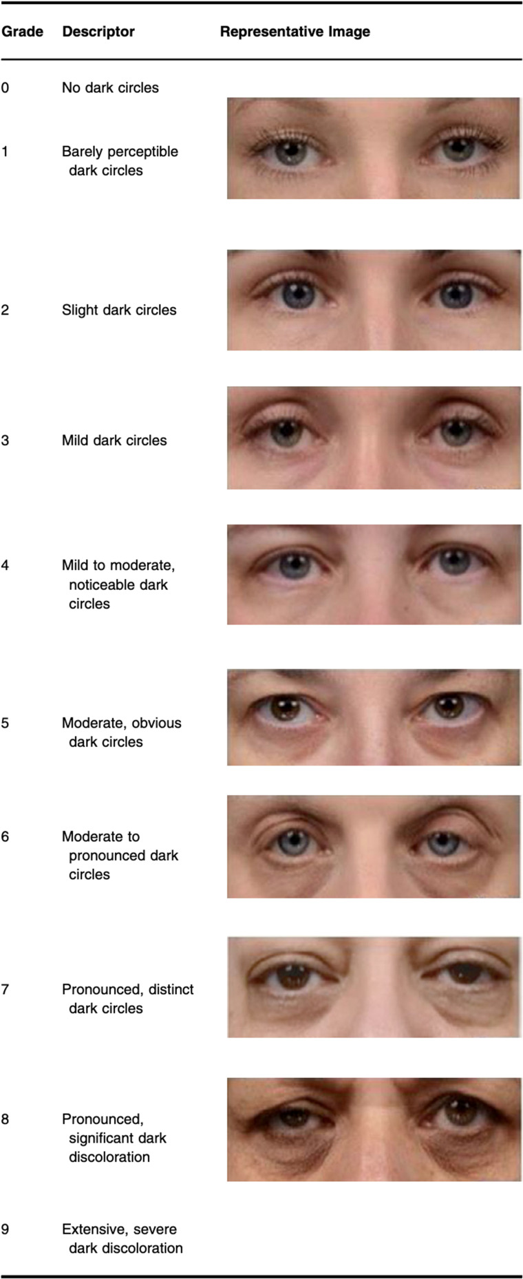
To monitor the aesthetic evolution of dark circles at follow-up visits, investigators used a validated 10-point photonumeric scale specific to dark circles, which has already demonstrated discriminative value in a split-face randomized clinical trial ( Figure 1 ). 26 The photonumeric scale is robust, with statistically significant correlations between color data and plots of corresponding grades, is sensitive to changes in grade that are visibly perceptible to the human eye, and demonstrates more than 90% agreement between experienced evaluators and consistent within-grader repeatability. 26
To the extent possible, independent local evaluators performed the photonumeric dark-circle grading.
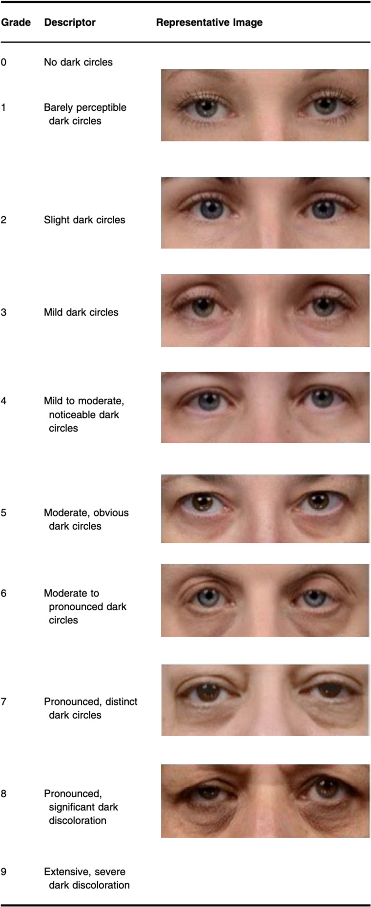
Figure 1 Descriptors of the scores 2 to 8 of the validated 10-score photonumeric scale and corresponding images used in the reported real-world, single-cohort observational study. Reproduced without changes from Ref. 26 under the terms of the Creative Commons Attribution License 3.0, permitting use, distribution, and reproduction in any medium, provided the original work is properly cited.
An advisory board of international dermatologists and aesthetic physicians recently characterized skin quality across all ethnicities into four perceptual categories—skin tone evenness, skin surface evenness, skin firmness, and skin glow. 27 Their considerations were applied to the reported real-world study on dark circles. After a preliminary online training, every participating specialist and every treated subject expressed a subjective opinion on the three skin quality perceptual categories most relevant for dark circles in relatively young subjects at T3 and T4 with a semi-quantitative GAIS-like, five-score scale (GAIS = Global Aesthetic Improvement Scale; 1=worse, 2=no change, 3=improved, 4=much improved, 5=very much improved) ( Table 3 ). 27 An analogous five-point scale was used by treated subjects to evaluate possible changes in skin pigmentation at T3 and T4.
Table 3 The Skin Quality Perceptual Categories and Their Descriptors Most Relevant to the Dark Circle Issue, as Assessed by Participating Specialists Using a GAIS-Like Five-Score Scale
| Skin Quality Perceptual Categories |
|---|
| Skin tone evenness |
| Skin firmness |
| Skin glow |
Moreover, analysis using commonly used commercial three-dimensional optical skin analysis systems could complement the primary evaluation at baseline and the two follow-up visits. The evaluation was optional, in line with the real-world clinical routine of participating specialists. The outcome was a simple qualitative statement that confirmed or added information about pigment content, texture, roughness, and capillary density to the photonumeric scale outcomes. Evaluation of the treated subject’s satisfaction was based on a five-point impromptu Likert-like scale ( Figure 2 ).
Adverse events based on spontaneous patient reporting and objective evaluations, with special attention devoted to those distinct from those commonly observed with infiltrative treatments (bruising and minor local irritation with transitory wheals, redness, and edema), and the need for treatment was reported as a percentage of the cohort patients.
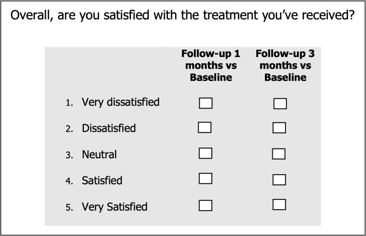
Figure 2 The Likert-like score scale for evaluating the treated subject’s satisfaction at the end of the treatment cycle.
Based on the assumptions of being able to triple at T4 the number of subjects classified at baseline as Grade 1 (“Barely perceptible dark circles”) to 3 (“Mild dark circles”) and a 90% power of avoiding false-negative type II errors (ß=0.10), the estimate was for a minimum of 55 subjects as an adequate sample size. 28
Primary endpoint: chi-square tests applied to 10×3 contingency tables (ten dark circle grades, three observational times, unstratified subjects, and subjects stratified according to Huang subtypes). Secondary endpoints: chi-square tests applied to 5×2 contingency tables (skin perceptual categories and hyperpigmentation: five scores, T3 and T2 observational times). Statistical program: GPower version 3.14. 28
Ten specialists accepted to participate in this observational study (“The Periorbital Hyperpigmentation and PN HPT Research Group”); aesthetic surgeons treated 24 subjects, aesthetic physicians 13, dentists 11, dermatologists 9, with this information unknown for 4 subjects. The PN HPT was administered intradermally with a needle in 30 subjects, with a cannula in 25, with both needle and cannula in 6; 49 subjects (80.3%) underwent the optional three-dimensional optical skin quality analysis.
Figure 3 illustrates the evolution of the overall, unstratified photonumeric grades from baseline until the end of the PN HPT treatment cycle. The overall picture shows a highly significant shift toward less severe periorbital hyperpigmentation (contingency table 10x3, chi-square= 78.79, p<0.001), but with only a relatively modest association between the overall evolution of photonumeric grades and PN HPT treatment (Cramer’s V= 0.67). At baseline (T0), none of the subjects lamented a hyperpigmentation less severe than Grade 3 (“Mild dark circles”); in contrast, most subjects of the observational cohort were Grade 4 (20.2% with “Mild to moderate, noticeable dark circles”) Grade 5 (33.2% with “Moderate, obvious dark circles”), and 6 (18.1% with “Moderate to pronounced dark circles”).
The progressive shift toward lower photonumeric grades and less aesthetic severity of periorbital dark circles appeared to differ in subjects undergoing the cannula and needle procedures. Scores at baseline (mean ± SEM) were similar (5.2 ± 1.19 vs. 5.4 ± 1.46, respectively), and appeared to improve rapidly at T3 in subjects treated with cannulas (3.4 ± 1.14 vs. 4.3 ± 1.66, p <0.05, respectively). The trend in favor of the cannula procedure was confirmed at the final T4 monitoring visit (2.2 ± 1.92 vs. 3.2 ± 1.93; p<0.01).
Stratifying the analysis by dark circle subtypes reveals that the shift toward progressively less severe periorbital hyperpigmentation after PN HPT treatment was especially noteworthy in the small subgroup of 11 subjects with the structural subtype ( Figure 4 ). However, the efficacy of PN HPT was also statistically significant in the 20 subjects with the pigmented subtype (p<0.05). The subjects classified as troubled by vascular periorbital hyperpigmentation are too few for any meaningful conclusion, while the efficacy of PN HPT, although not significant, was somewhat less than borderline in the subjects with the various mixed subtypes.
Regarding the secondary outcomes, Figures 5 and 6 compare the GAIS-like scores between the T3 and T4 follow-up visits; the difference across the three skin quality categories most relevant for dark circles in relatively young subjects is highly significant for both the participating specialists and the treated subjects who answered the query, indicating a prolonged and cumulative effect. Figure 7 shows how the treated subjects who expressed their GAIS-like judgments evaluate the skin pigmentation in their dark circles at T3 and T4, once again reporting a highly significant difference and a prolonged, cumulative effect. Figure 8 highlights the overall judgment on the PN HPT treatment at the two monitoring follow-up visits by the 60 subjects who answered the query.
Figures 9 and 10 show five exemplary demonstrations of improvements in periorbital dark circles from baseline, before beginning PN HPT treatment, and T4. Notwithstanding the delicate nature of periorbital skin, safety was excellent, with only 21 subjects (34.4%) reporting mild adverse events, such as edema or erythema, which are expected with microinvasive procedures and resolved within a few hours or on the same day. In only two subjects, the edema and local induration persisted for less than three days.
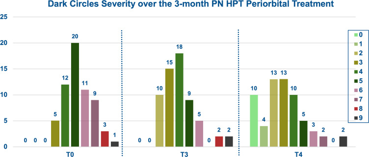
Figure 3 Highly significant overall progressive shift to the left of the distribution of periorbital dark circles toward lower photonumeric grades (less aesthetic severity) from baseline (T0) to the final monitoring visit (T4), three months after the final injection session (p <0.001 vs. T0).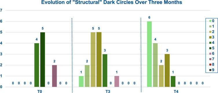
Figure 4 Highly significant progressive shift to the left (less aesthetic severity) of the distribution of “structural” periorbital dark circles from baseline (T0) to the T4 follow-up visit (p <0.001 vs. T0).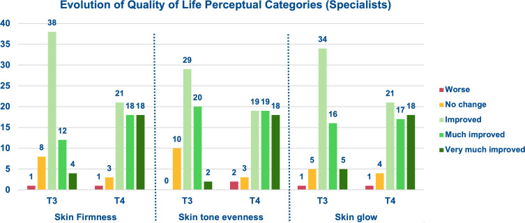
Figure 5 Progressive improvements of GAIS-like scores for the evaluated perceptual skin quality categories at the T3 and T4 monitoring visits, one and three months after the final injection session, respectively (p <0.01, T3 vs. T4).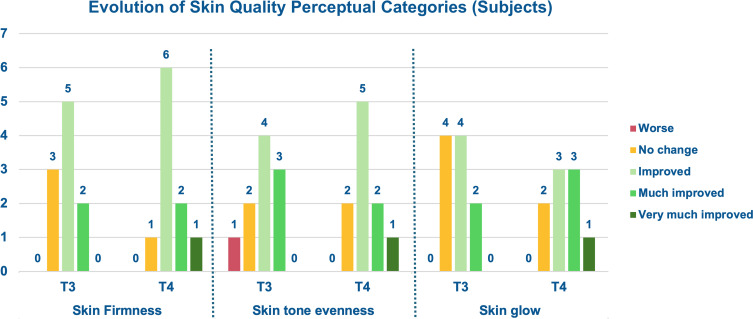
Figure 6 Progressive improvements of GAIS-like scores for the evaluated perceptual skin quality categories between the T3 and T4 follow-up visits, one and three months after the final injection session, respectively (p <0.01, T3 vs. T4), according to the 10 subjects who understood and answered the query.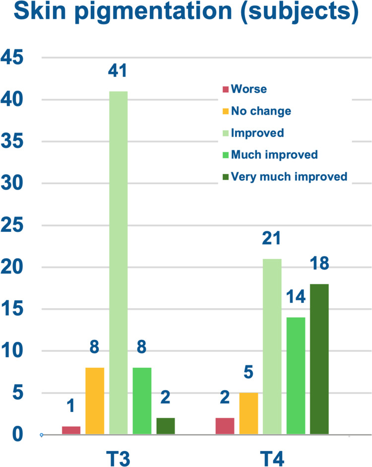
Figure 7 Progressive improvements of GAIS-like scores for the evaluated perceptual skin quality categories between the T3 and T4 follow-up visits, one and three months after the final injection session, respectively (p <0.01, T3 vs. T4), according to the 60 subjects who answered the query.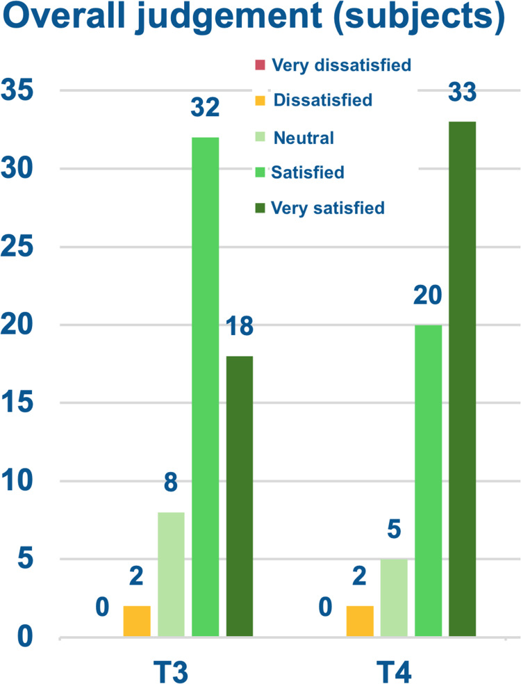
Figure 8 Overall judgment on the PN HPT treatment by the 60 subjects who answered the query.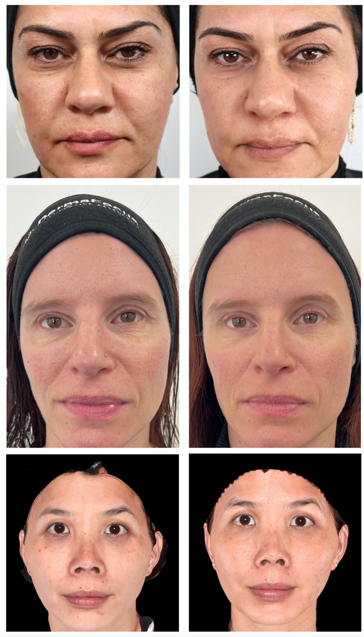
Figure 9 Improvements in periorbital hyperpigmentation with microinvasive PN HPT between T0 (photographs on the left) and T4 (photographs on the right) in three women of different ethnicities: Turkish (upper couple, courtesy of Dr. Gonka Gökdemir, Istanbul, Turkey), European (middle couple, courtesy of Dr. Raquel Amado, West Malling, Kent, UK), Malaysian Chinese (lower couple, courtesy of Dr. Tristan Tan, Kuala Lumpur, Malaysia). All subjects provided written informed consent for the publication of the photographic documentation of their clinical history for scientific purposes.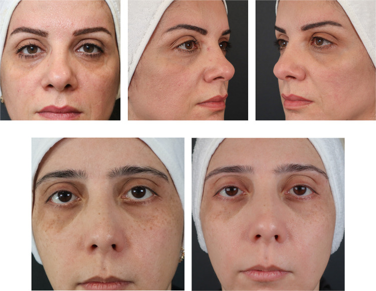
Figure 10 Further examples of improvement in periorbital hyperpigmentation with PN HPT between T0 (photographs on the left) and T4 (central and right photographs) in two Turkish women. Courtesy of Dr. Ayse Serap Karadag, Istanbul, Turkey). All subjects provided written informed consent for the publication of the photographic documentation of their clinical history for scientific purposes.
Contrary to what many have always thought, there appears to be no relationship between sleep quality and the emotionally troubling aesthetic nuisance known as dark circles. 29 In the literature, a vascular etiology underlies most dark circle episodes, with dilation of blood vessels and slowing down of the circulation of inadequately oxygenated blood. 7 , 30 Accumulated blood pigment from blood extravasation, hemoglobin and its degradation products, most often as the result of chronic inflammation, co-determines the hyperpigmentation dark circles variant. 7 , 30 However, contact-free hyperspectral imaging usually identifies melanin as the dominant chromophore. 31 , 32 The variant that mixes vascular and pigmented features is usually the second most common subtype in the Huang classification, but subtypes mixing structural and hyperpigmentation features, or structural and vascular features, are not uncommon. 25 , 31
Although primarily affecting the lower eyelids bilaterally, significantly darker skin color and accentuated color deviations may extend to the upper eyelids, eyebrows, temporal region, and malar region across all age groups, both genders, and all human types. 29 Throughout cultures, periorbital dark circles translate into a less youthful, exhausted, and melancholy appearance. 1
Anatomic features play a relevant role in dark circle development, especially in the lower eyelid area ( Figure 11 ). 1
Examples of anatomic predisposing features include prominence of the orbicularis oculi muscle and a high density of capillaries within the thin eyelid skin in intimate relationship with the orbicularis muscle, which helps explain the frequent family history of dark circles. 1 , 25 , 32 Age‑related midfacial changes, such as facial fat atrophy, fat descent downwards, and malar bone volume loss, lead to orbital rim recession, tightening and tethering of the orbital and facial retaining ligaments, orbital rim and facial hollowing and shadowing. The tear trough area in the inferomedial orbit is particularly prone to shadowing due to the thinness of eyelid skin. 1 , 25 , 32
Other acknowledged contributors and risk factors include sun exposure, prolonged television viewing, facial pallor due to anemia, and estro-progestin contraception. The common, recurrent worsening of dark circles during pregnancy further supports the likely contribution of hormonal factors. 29
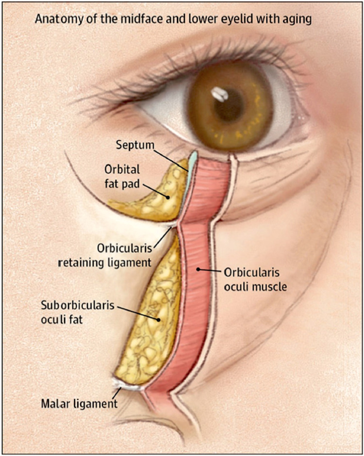
Figure 11 Soft tissues and osseous and ligamentous structures that are involved in eyelid aging and the development of dark circles, primarily in the infraorbital areas. Reproduced from Ref. 1 under the terms of the Creative Commons Attribution License 3.0, permitting use, distribution, and reproduction in any medium, provided the original work is properly cited.
PN HPT bind water molecules in aqueous solutions and evolve into a hydrogel, providing a biocompatible scaffold for cells. 16 , 17 , 23 The immediate volume-replacing effect of PN HPT is similar to that of hyaluronic acid fillers. However, their longer-term effects—such as promoting neocollagenesis and replenishing fibroblasts in the extracellular matrix—distinguish PN HPT from hyaluronic acid-based medical devices. 22 , 33 Besides their short-term hollow-filling effect through rapid hydration, PN HPT also facilitates dermal fibroblasts to produce Type I collagen, elastin fibers, and extracellular matrix by passively restoring nitrogenous bases and nucleotide precursors in the dermal and submucosal pools. These two properties of PN HPT work in synergy, as hydration of the extracellular matrix is essential for fibroblast vitality. 16 , 17 , 22 , 23 Currently, understanding how PN HPT help reduce periorbital hyperpigmentation is not straightforward. It is likely that restoring fibroblast vitality and the dermal microenvironment also contributes to the improvement of dark circles. A previous pilot study assessed the effectiveness of low-concentration PN HPT in subjects with poor periocular skin quality and reported positive outcomes. This larger multinational study confirmed the value and rationale of low-concentration PN HPT as a minimally invasive alternative to reduce severity and, indirectly, the psychological impact of periorbital dark circles. The notable shift towards lower scores, less burdensome Huang dark-circle grades, and improved skin quality parameters was evident with both injection techniques, according to investigators, as shown in Figures 3–6 . Although the aesthetic outcomes with both techniques appear satisfactory, the outcomes were more rapid and the final aesthetic results better in subjects undergoing the cannula procedure. Differences in aesthetic outcomes at the two follow-up visits underscore the need for prospective studies that are more discriminating than what retrospective data collection can provide. The improved hyperpigmentation scores reflect an enhanced dermal and subdermal environment; the optical skin analysis could only offer additional qualitative insights.
For example, frequent observations by participating specialists about the overall periocular condition at T3 and even more so at T4 included brighter skin, the subject looks more awake, the picture very much improved, improved hollows, reduced pigmentation with mild hollows, improved skin texture, improved skin thinning, lines, wrinkles, improved puffiness and palpebral bags, improved elastin and collagen deterioration, decreased dermal thickness.
The especially favorable results in the dark-circle structural subtype are not surprising, considering the immediate filling of hollows and the powerful long-term restructuring of dermal and subdermal tissues, passively triggered by low-concentration PN HPT. As expected for a medical device, PN HPT act non-pharmacologically as a supportive, hydrating extracellular matrix, replenishing intercellular hollows and restoring and maintaining volume and spatial integrity over time. Additionally, functioning as a hydrated scaffold, PN HPT promotes cell adhesion, migration, and alignment, as well as intercellular communication and tissue resilience. 23
The data collection has several notable weaknesses that warrant acknowledgment. The primary issue is the lack of a formal control group. Developing methodologically sound controls would have been challenging given the real-world observational approach, which involved actual ambulatory patients seeking advice and help for their psychologically burdensome periorbital hyperpigmentation. However, even without controls, the statistically significant reduction in dark circle severity, improvement in quality of life, and greater satisfaction among treated subjects—progressively improving at T3 and T4—are undeniable. Unlike this study, which remains a pilot, the upcoming investigation, currently in the planning stage, will not be observational but will be a prospective, randomized controlled study.
Using a validated photonumeric scale to grade periorbital hyperpigmentation, the primary endpoint, is a strong point of the observational study; 26 using impromptu GAIS-like scoring scales to evaluate skin quality and hyperpigmentation (secondary endpoints) is another weak point, especially since many subjects had difficulty answering the GAIS query, but the authors believe these may be only minor biases. The issue is that there are still no universally accepted criteria for evaluating treatment strategies for this aesthetic concern; compromises are inevitable. The photonumeric scale based on the Huang classification seemed to be the best compromise in designing their observational pilot study. In conclusion, until more methodologically robust studies are available, the dual rationale that inspired this study to investigate the value of low-concentration PN HPT in real-world conditions—immediate correction of hollows and tissue loss, as well as longer-term restructuring and reactivation of periorbital skin connectives—seems to be a solid option for improving the appearance of dark circles and enhancing the life for individuals who complain about this aesthetic concern.
声明:中文精读 · 仅供学术参考。内容来自公开文献检索,不代表本人观点,不构成诊疗建议。 医疗美容需在正规医疗机构由执业医师实施。
基于相同主题推荐 · 继续深挖
本开放标签、多国观察性队列纳入61例受黑眼圈困扰者(女性90.2%,平均41.5±10.5岁,范围20–67),覆盖Huang各亚型,于眶周真皮内注射PN HPT(7.5 mg/m
回顾性大样本病例系列(2016年11月至2025年1月),共1999例连续手术,中位年龄36岁(IQR 31至42),中位BMI 29(IQR 26至32),最短随访6个月。
研究类型:前瞻性自身对照split-face研究,40例(37例完成疗效评估,平均31岁、92.5%女性)接受3次、间隔3周的聚核苷酸(Rejuran,多聚核苷酸)注射,左颊皮内、