胰岛素抵抗作为CO₂激光换肤术后炎症后色素沉着(PIH)的潜在预测因子:一项双步法方案真实世界研究(《Clinical, Cosmetic and Investigational Dermatology》)
2026年10月22日27 min read
Clinical, Cosmetic and Investigational Dermatology
Clinical, Cosmetic and Investigational Dermatology
结论与实践要点
CO₂激光换肤是皮肤再生与年轻化的重要手段,但炎症后色素沉着(PIH)是其常见且棘手的并发症。传统上PIH风险被认为与较深Fitzpatrick肤色类型及高紫外线暴露环境相关,导致深肤色患者常被谨慎对待甚至回避治疗。本研究在马格里布地区(高紫外线、以中高肤色为主)开展,探索胰岛素抵抗(以HOMA-IR评估)是否可作为CO₂激光术后PIH的附加预测因素。
本研究为前瞻性、非随机真实世界研究,纳入45例接受双步法CO₂激光(Dual Technique)方案治疗的患者:第一步SuperPulse(SP)模式1.5 W、5 Hz进行微剥脱汽化,第二步Deep Pulse(DP)分段模式10 W、脉宽700 µs、间距400 µm,于同一次治疗内连续完成。结果显示,PIH在Fitzpatrick I–III型患者中发生率为75%(15/20),反而高于IV–VI型的36%(9/25)(χ²=6.790,p=0.0092)。PIH严重程度与HOMA-IR呈强正相关(Spearman ρ=0.8624,p<0.0001,n=37),且胰岛素抵抗(HOMA-IR>2.5)在浅色组更普遍(60% vs 20%,χ²=7.563,p=0.0060);PCOS患病率组间无显著差异(p=0.8)。研究未提供比值比(OR)或多变量校正,关联以相关分析与组间比较呈现。
临床意义上,该发现提示除肤色外,代谢状态(尤其是胰岛素抵抗)可能影响个体对激光后PIH的易感性,支持从单纯按肤色分层转向更个体化的风险评估,并为术前代谢筛查(如HOMA-IR)提供了探索方向。但本研究存在样本量小、非随机、适应症异质、缺乏对照与多变量校正等局限,无法确立HOMA-IR为独立预测因子,结论属假说生成性质,尚不足以常规推荐代谢评估用于激光风险分层,需更大规模、控制良好的研究加以验证。
PurposeLaser-assisted skin regeneration is a cornerstone of modern dermatologic practice; however, post-inflammatory hyperpigmentation (PIH) remains a major limiting complication of CO 2 laser treatments. Risk has traditionally been attributed to darker phototypes, particularly in high ultraviolet (UV) exposure settings. Emerging evidence suggests that metabolic factors, especially insulin resistance measured by HOMA-IR, may influence pigmentary responses. This study evaluated a Dual Technique CO 2 laser protocol designed to enhance skin regeneration while reducing the number of treatment sessions and explored the association between PIH and metabolic status.
Patients and MethodsA prospective, non-randomized, real-world study included 45 patients treated in a private practice in the Maghreb region, characterized by high UV exposure and predominantly medium-to-high phototypes. The Dual Technique combined ablative fractional vaporization with subablative thermal stimulation. Skin quality was assessed using the Vancouver Scar Scale (VSS), and PIH severity using the Dermal Pigmentation Area and Severity Index (DPASI). Outcomes were analyzed in relation to phototype, HOMA-IR, and polycystic ovary syndrome (PCOS).
ResultsSignificant clinical improvement was observed across indications following treatment, typically within two sessions. PIH occurred in 15/20 (75%) patients with Fitzpatrick phototypes I–III compared with 9/25 (36%) patients with phototypes IV–VI (p = 0.0092). PIH severity showed a strong positive correlation with HOMA-IR (Spearman ρ = 0.8624, p < 0.0001). PCOS prevalence did not differ significantly between phototype groups (p = 0.8). PIH improved significantly following standardized management.
ConclusionThe Dual Technique CO 2 laser protocol was associated with favorable clinical outcomes using relatively few treatment sessions. In this cohort, higher HOMA-IR was strongly associated with greater PIH severity, suggesting that insulin resistance may represent an additional factor associated with individual PIH susceptibility. These findings are hypothesis-generating and require confirmation in larger, controlled studies before metabolic assessment can be recommended for routine laser-treatment risk stratification.
Keywords: CO2 laser、fractional laser resurfacing、insulin resistance、polycystic ovary syndrome、post-inflammatory hyperpigmentation、skin phototype、ultraviolet radiation
Laser-assisted skin regeneration has become a fundamental component of modern dermatologic and reconstructive practice. Since the introduction of the carbon dioxide (CO 2 ) laser in the 1960s and its clinical adoption in dermatology in the 1980s, this technology has evolved significantly, particularly with the development of fractional photothermolysis. 1 , 2 CO 2 lasers operate at a wavelength of 10,600 nm, allowing precise ablation of water-containing tissues and inducing controlled thermal injury. 3 This process stimulates a cascade of wound healing responses, including fibroblast activation, neocollagenesis, elastogenesis, and extracellular matrix remodeling, ultimately improving skin texture, elasticity, and overall quality. 4 As a result, CO 2 laser resurfacing has become a gold standard for the treatment of scars, photoaging, and various dermatologic conditions. 5
Expert consensus on clinical recommendations for fractional ablative CO 2 laser in facial skin rejuvenation generally suggests performing 2–4 sessions for rejuvenation and up to 6–8 sessions for scar management. While these multi-session protocols are associated with improved clinical outcomes, they may also increase the cumulative risk of adverse effects, It is important to note that variations in laser devices, energy settings, and treatment parameters can significantly influence both efficacy and safety profiles, contributing to heterogeneity in clinical protocols and a lack of standardization across practices. 6
Despite its well-documented efficacy, CO 2 laser treatment is not without limitations. Among the most frequently reported adverse effects are erythema, edema, infection, and delayed healing; however, post-inflammatory hyperpigmentation (PIH) remains one of the most clinically significant and challenging complications. 7 , 8 PIH results from an increased melanocytic response following cutaneous inflammation or injury, leading to increased melanin production and deposition in the epidermis and/or dermis. 9 This complication can significantly affect patient satisfaction and may persist for prolonged periods, particularly in susceptible individuals.
The risk of PIH has traditionally been associated with higher Fitzpatrick skin phototypes (IV–VI), where melanocytes exhibit greater reactivity to inflammatory stimuli. 10 , 11 Consequently, the use of ablative laser technologies in darker skin types has often been approached with caution or even avoided altogether. This concern is further amplified in geographical regions characterized by high ultraviolet (UV) radiation exposure, where UV-induced melanogenesis may potentiate pigmentary complications. 12 In such environments, clinicians frequently limit the use of CO 2 lasers due to the perceived increased risk of adverse pigmentary outcomes, thereby restricting access to highly effective regenerative treatments.
Beyond phototype and environmental factors, emerging evidence suggests that PIH may also be influenced by systemic and metabolic conditions. 13 Pigmentary disorders have been increasingly linked to metabolic alterations, particularly insulin resistance and endocrine dysregulation. 14 Conditions such as polycystic ovary syndrome (PCOS), which is frequently associated with insulin resistance, have been implicated in altered melanocyte activity and dysregulated inflammatory responses. 15 Insulin and insulin-like growth factors (IGFs) may directly or indirectly stimulate melanogenesis through complex signaling pathways, including activation of tyrosinase and other melanogenic enzymes. 16 Hyperinsulinemia enhances insulin/IGF-1 receptor signaling, activating pathways such as PI3K/Akt and MAPK, which upregulate microphthalmia-associated transcription factor (MITF), the key regulator of melanocyte activity. This results in increased melanin synthesis and transfer to keratinocytes. Additionally, insulin resistance is associated with a pro-inflammatory and oxidative environment that may further amplify pigmentary responses following skin injury. 17
Clinically, this mechanism may contribute to PIH susceptibility in addition to conventional phototype-related risk. The management of PIH is often challenging and prolonged, requiring strict photoprotection and topical therapies, with variable outcomes. This can significantly affect patient satisfaction, highlighting the importance of identifying metabolic risk factors such as insulin resistance. 18 Importantly, direct evidence linking HOMA-IR specifically to laser-induced PIH remains limited, and the observed relationship should therefore be considered exploratory. HOMA-IR was selected as a practical surrogate of insulin resistance that could be readily incorporated into clinical assessment. Nevertheless, PIH is multifactorial, and phototype, UV exposure, baseline inflammation, treatment characteristics, and adherence to post-procedure photoprotection may influence the observed association.
The pathophysiological mechanisms underlying this relationship are multifactorial. Chronic low-grade inflammation, oxidative stress, and hormonal imbalances contribute to melanocyte hyperactivity and increased susceptibility to pigmentary dysregulation following cutaneous injury. 19 Additionally, insulin resistance may impair normal wound healing processes, leading to prolonged inflammation and an increased likelihood of PIH development. 20 These findings suggest that PIH should not be considered solely a local cutaneous phenomenon but rather a multifaceted condition influenced by both intrinsic and extrinsic factors.
To address these challenges, optimized CO 2 laser protocols have been developed to balance tissue ablation and thermal stimulation, aiming to achieve effective remodeling while minimizing excessive inflammation and pigmentary complications. 21 The Dual Technique CO 2 laser protocol combines sequential ablative fractional vaporization with subablative thermal stimulation to promote controlled skin regeneration while maintaining treatment safety across different phototypes. However, whether metabolic status influences susceptibility to PIH following CO 2 laser treatment remains insufficiently characterized. Therefore, we investigated the association between insulin resistance, assessed by HOMA-IR, and PIH occurrence and severity, hypothesizing that higher HOMA-IR would be associated with greater PIH susceptibility in addition to conventional phototype-based risk. Its potential predictive value remains exploratory, and the observational design does not allow causal inference.
This prospective, non-randomized, real-world study was conducted over two years (January 2024–January 2026) in a private dermatology center in Oran, Algeria (Maghreb region), an area characterized by high UV exposure and predominantly medium-to-high skin phototypes.
A total of 45 patients (42 females, 3 males; mean age 37 ± 15 years) were treated using the dual CO 2 laser technique for various indications, including scars (burn, surgical, traumatic, acne), skin rejuvenation, benign lesions, and striae distensae. Patients were stratified by Fitzpatrick phototype into light (I–III) and dark (IV–VI) groups to assess differences in pigmentary risk.
Exclusion criteria included autoimmune or coagulation disorders, active inflammatory or infectious conditions, untreated dermatologic diseases, pregnancy or breastfeeding, hypersensitivity to anesthetics, smoking, and use of medications affecting healing or inflammation (eg, isotretinoin, antibiotics, statins, anti-inflammatory drugs). Eligible patients were prospectively recruited during the study period among individuals presenting for CO 2 laser treatment who fulfilled the study eligibility criteria and provided written informed consent. Recruitment was therefore prospective among eligible patients rather than based on consecutive sampling.
All treatments were performed using a SmartXide 2 CO 2 laser system (DEKA, Italy; maximum power: 40 W). A standardized “one-shot” dual technique protocol was applied in all patients, consisting of two sequential steps within the same session.
First Step (Tissue Approximation): The SuperPulse (SP) mode was delivered at 1.5 W and 5 Hz, enabling controlled ablative action with minimal thermal diffusion. This step facilitated precise tissue vaporization and mechanical approximation of lesion edges.
Second Step (Fractional Remodeling): Subsequently, the Deep Pulse (DP) fractional mode was applied at 10 W, with a pulse duration of 700 µs and a spacing of 400 µm. This phase aimed to induce controlled dermal injury, promote collagen remodeling, neocollagenesis, and overall improvement in dermal architecture. The two-step protocol was performed sequentially within the same treatment session.
All procedures were performed by the same operator. Treatment covered the clinically affected area, with the clinical endpoint based on adequate and uniform treatment of the target tissue while avoiding excessive thermal injury. Additional treatment sessions were determined according to clinical response and residual findings at follow-up, rather than a predefined number of sessions, with a minimum interval of one month between sessions.
Topical anesthesia was achieved using lidocaine 10.56% cream (J-Cain Cream, Inist Bio Pharmaceutical Co., Ltd., South Korea), applied for 30 minutes prior to treatment. No external cooling devices were used before or during the procedure.
Clinical tissue characteristics and treatment-related changes were assessed using the Vancouver Scar Scale (VSS), which evaluates pigmentation, vascularity, pliability, and height. 22 In the present heterogeneous cohort, the VSS was used as a pragmatic longitudinal assessment tool, principally applicable to scar-related tissue changes rather than as a validated global skin-quality instrument for every indication. Standardized clinical photographs obtained before treatment initiation and at the final follow-up visit regardless of the number of sessions performed, were evaluated. The VSS scoring system allowed standardized comparison of pre- and post-treatment clinical tissue characteristics, particularly for scar-related outcomes.
PIH was diagnosed as newly developed or increased pigmentation occurring within the laser-treated area during post-treatment follow-up and was subsequently quantified using DPASI. PIH assessment was incorporated into the scheduled follow-up visits at Day 15, Day 30, and monthly thereafter when clinically indicated. HOMA-IR was calculated from fasting glucose and fasting insulin measurements obtained during metabolic assessment. Clinical response was defined by improvement from baseline based on standardized photography and VSS assessment, whereas persistence or worsening of the treated condition was considered an insufficient response.
PIH was diagnosed as newly developed or increased pigmentation occurring within the laser-treated area during post-treatment follow-up and was quantitatively assessed using the Dermal Pigmentation Area and Severity Index (DPASI), 23 a scoring system originally validated for acquired dermal macular hyperpigmentation. In the present study, DPASI was used as a pragmatic quantitative measure of the extent and severity of laser-associated PIH and not as an instrument specifically validated for this indication. In this study, DPASI scoring was applied specifically in patients who developed PIH, both at the time of diagnosis and following the implementation of therapeutic management strategies. DPASI measurements were closely monitored throughout the follow-up period, allowing for dynamic evaluation of PIH progression and response to treatment interventions. The number of participants included in individual analyses varied according to the availability of the corresponding paired clinical and metabolic measurements.
Standardized high-resolution digital photographs were obtained using a portable digital camera, maintaining consistent angle, distance, background, and lighting conditions across all time points. All images were analyzed independently by blinded observers to minimize bias and participants provided written informed consent, including authorization for clinical photography and publication. The study was conducted in accordance with the principles outlined in the Declaration of Helsinki.
The predefined primary follow-up endpoint was 1 month after treatment. Patients were additionally evaluated at Day 15 for early treatment response and adverse events. Extended follow-up was performed for up to 3 months, particularly for monitoring PIH. In patients who developed PIH, follow-up continued until complete clinical resolution or the 3-month endpoint, whichever occurred first; most cases resolved before 3 months.
Post-procedure care was standardized and included strict photoprotection with daily broad-spectrum sunscreen use, along with emollients and barrier-repair formulations to support healing. Patient education was emphasized to ensure adherence and minimize complications.
HOMA-IR was determined from fasting glucose and insulin measurements obtained after a 12-hour fast, with a value >2.5 considered indicative of insulin resistance. Female patients were screened for PCOS using established criteria. Patients with elevated HOMA-IR and/or PCOS were classified as a high-risk subgroup to assess the impact of metabolic status on outcomes, particularly PIH.
Patients with HOMA-IR >2.5 were referred for endocrinological evaluation; those between 2.5–3.0 received lifestyle and dietary recommendations, while values >3.0 were managed with metformin under specialist supervision.
The main adverse effects were erythema, edema, and PIH. Erythema was typically transient; persistent cases were managed with topical azelaic acid from Day 15. Edema was mild to moderate and resolved spontaneously. PIH was the most relevant complication and was managed with early use of topical depigmenting agents (eg, hydroquinone), strict photoprotection, and, in selected cases, topical anti-inflammatory therapy.
Data were analyzed using Prism version 11.0.1 (GraphPad Software, USA). Categorical variables were compared using Pearson’s chi-square (χ 2 ) test without continuity correction, with exact p-values reported for the corresponding 2×2 comparisons. Welch’s unpaired t -test was used for comparisons of continuous variables between independent groups, whereas paired t-tests were used for within-subject pre- versus post-treatment comparisons. Normality was assessed using the Shapiro–Wilk test. The association between HOMA-IR and DPASI was assessed using Spearman’s rank correlation coefficient (ρ), given the non-normal distribution of these variables and the bounded nature of the DPASI score. A p-value <0.05 was considered statistically significant.
The number of participants included in individual analyses varied according to the availability of the corresponding paired clinical and metabolic measurements and is reported for each analysis.
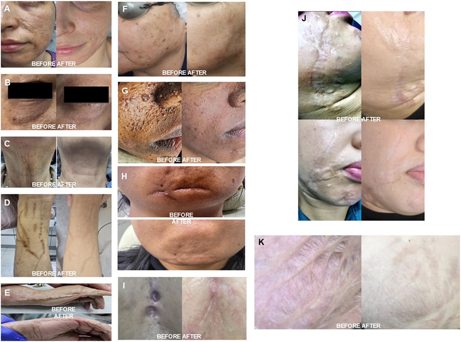
As illustrated in Figure 1 , patients demonstrated substantial clinical improvement in skin quality, scar appearance, and overall tissue regeneration following treatment with the Dual Technique across a wide range of dermatologic and aesthetic indications.
A long-standing burn scar of more than 25 years’ evolution showed marked improvement in texture, pliability, and integration with surrounding tissue after three sessions (a). Periorbital rejuvenation was achieved with a single session, demonstrating visible tightening and reduction of fine wrinkles (b), while cervical skin laxity improved after two sessions (c).
Self-inflicted scars on the arms exhibited significant structural and aesthetic improvement after two sessions, with better blending into adjacent skin (d). A case of retractable adhesions of the hand showed both functional and structural improvement after a single session (e). Full-face rejuvenation combined with the removal of seborrheic keratoses and benign solar lentigines was successfully achieved in one session, with clear enhancement in both texture and pigmentation (f).
Facial lesions associated with tuberous sclerosis complex demonstrated visible improvement in lesion prominence and overall skin homogeneity after one session (g). A traumatic facial scar improved in contour and visibility following two sessions (h), while a post-surgical thoracic scar showed reduced thickness and improved color uniformity after two sessions (i). Post-traumatic scars secondary to a dog bite also demonstrated significant improvement after a single session (j). Additionally, striae distensae on the breasts following surgical augmentation showed marked improvement in texture and coloration after two sessions (k).
Acne scar management, presented in Figure 2 , also demonstrated consistent clinical benefit. Patients with atrophic boxcar and ice-pick scars showed clear structural improvement after two sessions, although mild PIH was observed in some cases (a). A particularly challenging case required six sessions due to severe atrophic scarring in the context of repeated acute inflammatory episodes on a chronically inflamed tissue background; this was managed using a vertical elevation approach and showed progressive improvement, albeit with increased PIH correlating with the higher number of sessions (b). Predominantly boxcar scars improved after two sessions, with an additional secondary skin-lightening effect (c). Rolling scars demonstrated improvement in both structure and skin tone after a single session (d), while other cases showed enhanced texture, tone, and reduction in inflammatory signs after two sessions (e).
Overall, these clinical improvements were supported by a statistically significant reduction in VSS scores following completion of the treatment protocol, as shown in Figure 3 , supporting improvement in the clinical tissue characteristics captured by the VSS following treatment.
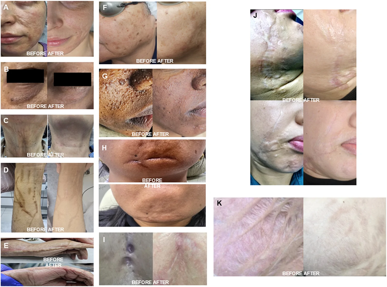
Figure 1 Clinical outcomes across diverse indications following the Dual Technique CO2 laser protocol (A) Long-standing facial burn scar of >25 years’ duration before treatment and after three sessions. (B) Periorbital rejuvenation before treatment and after one session. (C) Cervical skin laxity before treatment and after two sessions. (D) Self-inflicted scars of the forearm before treatment and after two sessions. (E) Retractable hand adhesions before treatment and after one session. (F) Full-face rejuvenation with treatment of seborrheic keratoses and solar lentigines before treatment and after one session. (G) Facial lesions associated with tuberous sclerosis complex before treatment and after one session. (H) Traumatic facial scar before treatment and after two sessions. (I) Post-surgical thoracic scar before treatment and after two sessions. (J) Post-traumatic facial scars secondary to dog bite before treatment and after one session. (K) Striae distensae of the breast following surgical augmentation before treatment and after two sessions.展示双步法CO₂激光在多种适应症(如长期烧伤瘢痕、眶周年轻化、颈部皮肤松弛等)治疗前后的临床对比照片,体现皮肤质地与组织再生的改善。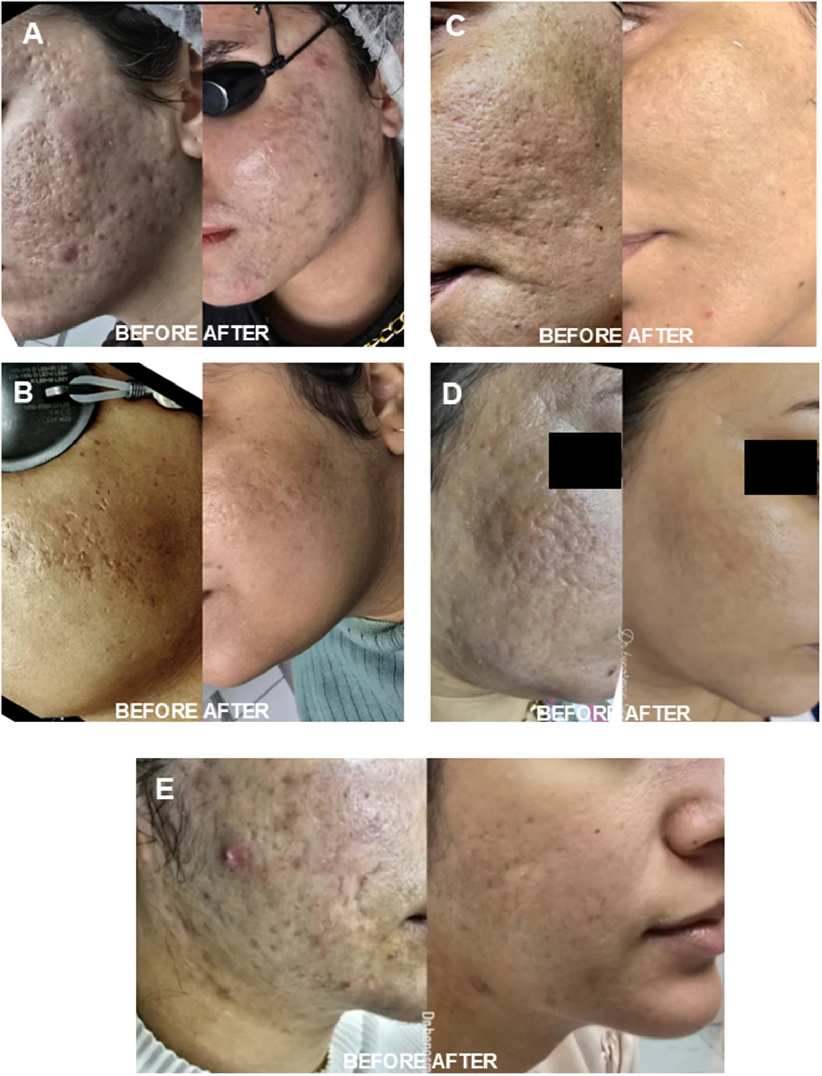
Figure 2 Clinical outcomes in acne scar management following the Dual Technique CO2 laser protocol (A) Atrophic boxcar and ice-pick scars after two sessions, with mild post-inflammatory hyperpigmentation; (B) severe atrophic acne scars treated with six sessions using a vertical elevation approach, showing progressive improvement with associated PIH; (C) predominantly boxcar scars after two sessions, with secondary skin-lightening effect; (D) rolling scars after one session, with improvement in skin texture and tone; (E) mixed atrophic scars after two sessions, demonstrating enhanced skin quality and reduction of inflammatory signs.展示痤疮瘢痕患者经双步法治疗后的临床改善,涵盖厢车型、冰锥型、滚动型等不同瘢痕亚型,部分病例伴轻度炎症后色素沉着(PIH)。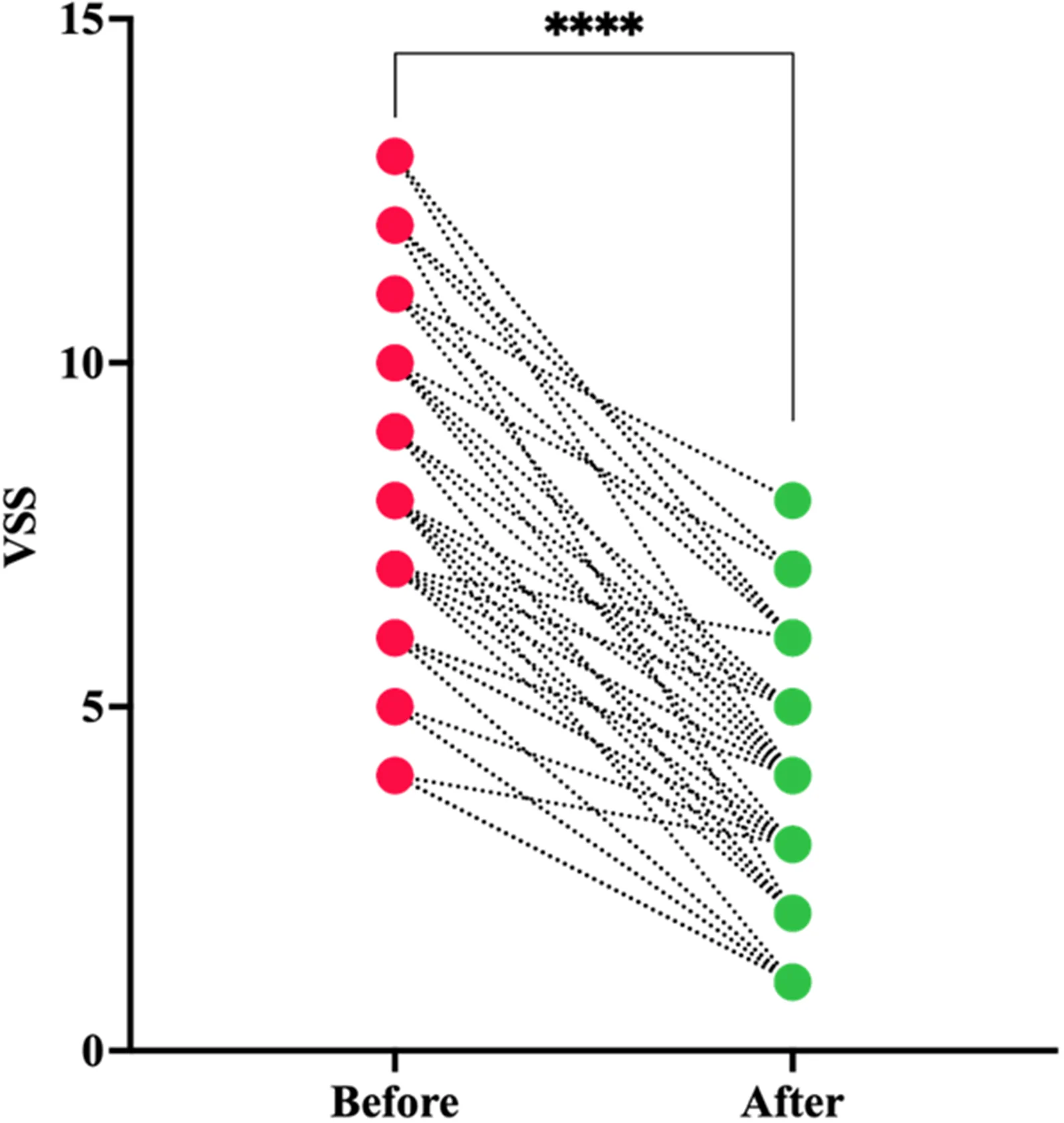
Figure 3 Changes in VSS-assessed clinical tissue characteristics following Dual Technique CO2 laser treatment Comparison of Vancouver Scar Scale (VSS) scores before treatment and after completion of the Dual Technique CO2 laser protocol. A significant reduction in VSS scores was observed following treatment, indicating improvement in the evaluated scar characteristics. Statistical analysis was performed using a paired t-test (****p ≤ 0.0001; n = 45).以配对点图展示温哥华瘢痕量表(VSS)评分治疗前后变化,治疗后评分显著下降(配对t检验,p≤0.0001,n=45),提示瘢痕特征改善。
As summarized in Table 1 , PIH occurred in 15/20 (75%) patients with Fitzpatrick phototypes I–III compared with 9/25 (36%) patients with phototypes IV–VI (χ 2 = 6.790, p = 0.0092).
Table 1 Post Inflammatory Hyperpigmentation Prevalence in Skin Phototypes
| PIH | Present | Absent | Total n = 45 | |
|---|---|---|---|---|
| Group 1 | 15 (75%) | 5 (25%) | 20 | p = 0.0092 |
| Group 2 | 9 (36%) | 16 (64%) | 25 |
Elevated HOMA-IR was observed in 12/20 (60%) patients with Fitzpatrick phototypes I–III and 5/25 (20%) patients with phototypes IV–VI (χ 2 = 7.563, p = 0.0060) as presented in Table 2 . Metabolic analysis also demonstrated a significant difference in the prevalence of elevated HOMA-IR between phototype groups, with higher prevalence in Fitzpatrick I–III than in Fitzpatrick IV–VI (60% vs 20%; χ 2 = 7.563, p = 0.0060).
Table 2 HOMA-IR in Light and Dark Skin Phototypes
| HOMA-IR | High (≥2.5) | Normal (<2.5) | Total n = 45 | |
|---|---|---|---|---|
| Group 1 | 12 (60%) | 8 (40%) | 20 | p = 0.0060 |
| Group 2 | 5 (20%) | 20 (80%) | 25 |
In contrast, the prevalence of PCOS did not differ significantly between phototype groups ( Table 3 ). A strong positive correlation was observed between PIH severity, assessed by DPASI, and HOMA-IR (Spearman ρ = 0.8624, p < 0.0001; n = 37), indicating that higher insulin resistance was associated with greater pigmentation severity ( Figure 4A ).
Table 3 PCOS in Light and Dark Skin Phototypes
| PCOS | PCOS + | PCOS- | Total n = 43 | |
|---|---|---|---|---|
| Group 1 | 10 (50%) | 10 (50%) | 20 | p = 0.8 |
| Group 2 | 11 (47.8%) | 12 (52.2%) | 23 |
Importantly, appropriate and timely management of PIH resulted in successful clinical resolution, even in patients presenting higher baseline HOMA-IR values and more severe pigmentation. This therapeutic response is illustrated in Figure 4B and was quantitatively confirmed by a significant reduction in DPASI scores across all affected patients ( Figure 4C ).
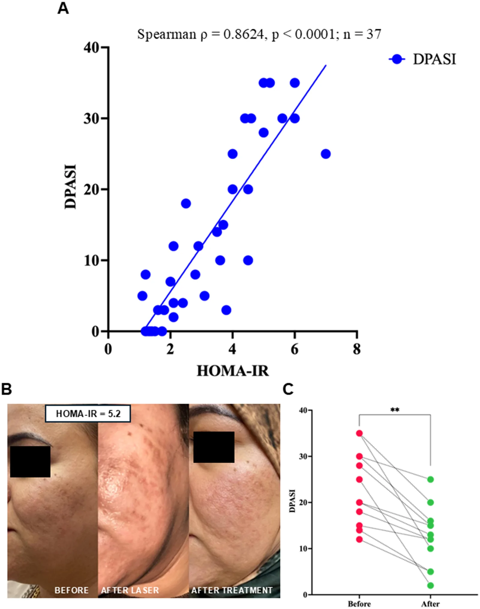
Figure 4 Association between post-inflammatory hyperpigmentation severity, metabolic status, and treatment response (A) Association between HOMA-IR and DPASI among participants with paired HOMA-IR and DPASI data available for the correlation analysis (n=37). (B) Representative patient with elevated HOMA-IR (5.2), showing the clinical appearance before CO2 laser treatment, development of PIH after laser treatment, and improvement following PIH management. (C) Paired comparison of DPASI scores among patients with available pre- and post-treatment DPASI measurements (n=12). Statistical analysis: (A) Spearman rank correlation coefficient (ρ = 0.8624, p < 0.0001; n = 37); (C) paired t-test, **p ≤ 0.01; n = 12.展示PIH严重程度与代谢状态关系:HOMA-IR与DPASI评分呈强正相关(Spearman ρ=0.8624,p<0.0001,n=37),并附典型高HOMA-IR(5.2)患者治疗前、激光后PIH发生及管理后改善的对照照片。
The present study demonstrates that the Dual-Technique CO 2 laser protocol achieves significant clinical improvement across a broad spectrum of dermatologic indications with a reduced number of treatment sessions, while maintaining a favorable safety profile. Importantly, the incidence of PIH was lower than expected when considering only patients treated with this protocol. Furthermore, our findings reveal a novel and clinically relevant association between PIH severity and insulin resistance, suggesting that metabolic factors may contribute to individual PIH susceptibility alongside established clinical risk factors.
A key finding of this study is that effective clinical outcomes were achieved with a limited number of sessions, most commonly two, across various conditions including scars and skin rejuvenation. This contrasts with traditional protocols, which often recommend multiple sessions, typically up to 6–8 for scar management, to achieve optimal results. Reducing the number of sessions may not only improve patient adherence and satisfaction but also decrease cumulative tissue injury and the risk of complications such as PIH. 24 , 25 The observed efficacy with fewer sessions likely reflects the optimized balance between ablative and thermal effects achieved with the Dual Technique.
Interestingly, PIH was more frequently observed in patients with lighter phototypes, which contrasts with the traditional paradigm associating higher risk with darker skin types. While melanin activity is known to vary across phototypes, emerging evidence suggests that factors beyond baseline pigmentation, such as inflammatory response variability and individual susceptibility may influence pigmentary outcomes. 26 In this context, our findings support a shift from a purely phototype-based risk model toward a more individualized approach.
The most relevant and novel observation in this study is the positive correlation between PIH severity and insulin resistance. Although PCOS has been associated with pigmentary disorders, these findings suggest that metabolic status, particularly insulin resistance, may contribute to individual susceptibility to PIH and could partly explain pigmentary risk not accounted for by phototype alone. Hyperinsulinemia has been shown to stimulate melanocyte activity through insulin-like growth factor pathways and may exacerbate inflammatory responses, thereby promoting melanogenesis following cutaneous injury. 27 , 28 Additionally, insulin resistance is associated with impaired wound healing and prolonged inflammatory states, both of which may contribute to the development and persistence of PIH. 29
From a clinical perspective, these findings align with real-world observations. Initially, PIH was presumed to be primarily related to phototype; however, the occurrence of PIH in fair-skinned patients treated with identical parameters suggested the involvement of additional factors. Subsequent analysis identified an association with insulin resistance, prompting greater clinical consideration of metabolic status during patient assessment. Whether correction of insulin resistance may influence the subsequent course or resolution of PIH remains to be determined in prospective studies incorporating serial metabolic and pigmentation assessments.
Importantly, PIH was effectively managed in all cases, regardless of baseline HOMA-IR, with complete resolution following appropriate treatment. This highlights that, while metabolic factors may increase susceptibility, they do not preclude successful management when early and targeted interventions are implemented.
This study has several limitations, including its relatively small sample size, non-randomized design, heterogeneous treatment indications, absence of a comparator group, lack of multivariable adjustment, and the possibility of residual confounding. Recruitment from a single real-world clinical setting may also introduce selection bias, while the predominantly female composition of the cohort limits generalizability across sexes. These factors limit causal inference and preclude establishing HOMA-IR as an independent predictor of PIH. The observed association should therefore be interpreted as exploratory and hypothesis-generating. Nevertheless, the prospective real-world design, standardized treatment protocol performed by a single operator, and blinded outcome assessment provide a clinically relevant basis for future controlled studies designed to validate metabolic risk stratification in CO 2 laser treatment. Additionally, most cases were related to scar treatment rather than purely aesthetic rejuvenation, which should be considered when interpreting the results.
Taken together, these findings support a hypothesis in which metabolic status may contribute to individual susceptibility to pigmentary complications following CO 2 laser treatment. The unexpected distribution of PIH across phototypes and the strong unadjusted association between HOMA-IR and DPASI suggest that phototype alone may not fully capture PIH risk. However, these observations should be considered hypothesis-generating and require confirmation in larger, more homogeneous prospective cohorts with appropriate multivariable adjustment.
Future research should focus on larger, multicenter, and blinded studies to validate the predictive role of HOMA-IR in PIH development. If validated prospectively, metabolic assessment could ultimately contribute to more individualized pre-laser risk stratification.
The Dual Technique CO 2 laser protocol was associated with favorable clinical outcomes using relatively few treatment sessions. In this cohort, higher HOMA-IR was strongly associated with greater PIH severity, suggesting that metabolic status may represent an additional factor relevant to individual PIH susceptibility. However, these findings are exploratory and require confirmation in larger, controlled studies to determine whether HOMA-IR represents an independent predictor or a clinically useful screening marker. Routine metabolic assessment for laser-treatment risk stratification cannot be recommended on the basis of the present findings alone.
声明:中文精读 · 仅供学术参考。内容来自公开文献检索,不代表本人观点,不构成诊疗建议。 医疗美容需在正规医疗机构由执业医师实施。
基于相同主题推荐 · 继续深挖
研究类型与对象:前瞻性、非随机真实世界研究,纳入马格里布地区高紫外线暴露环境下的45例患者,采用CO₂激光双步法(SuperPulse 1.5 W/5 Hz + Deep Puls
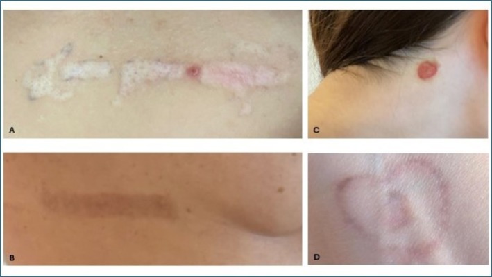
多中心回顾性队列纳入111名成人(均龄33.3±7.8,88.3%为女性),接受1064/532 nm调Q Nd:YAG文身清除治疗,平均2.5±1.55次(范围1–8次)。
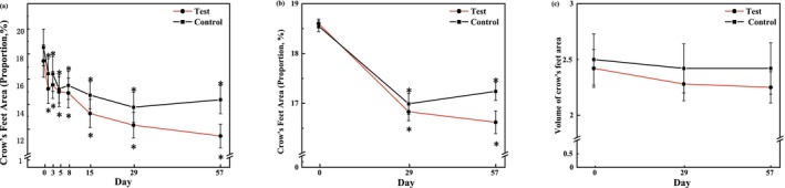
本研究为分脸、双盲、随机、安慰剂对照的单中心临床试验,共纳入24名30–48岁女性(均值38.46±5.17岁),均完成试验。