增强吸脂联合环形去上皮与腺体切除治疗Ⅲ度男性乳腺发育(82 例回顾)
2026年10月24日32 min read
BMC Surgery
结论与实践要点
本文报告一种针对Ⅲ度(以腺体增生为主、常伴皮肤冗余)男性乳腺发育的综合手术路径:先以吸脂处理脂肪成分,再经环乳晕圆形去上皮(round-block de-epithelialization)保留乳头乳晕血供的前提下去除表皮,随后皮下锐性分离并完整切除纤维腺体组织,最后缝合重建。该思路兼顾脂肪与腺体两类成分,并尽量隐藏瘢痕于乳晕缘。
结果方面,78% 的总体满意度与多数维度 65%–75% 的满意率显示该术式可接受;但水肿(31.7%)与血清肿(12.2%)发生率偏高,提示术后引流与加压包扎管理很关键。乳头乳晕复合体坏死虽仅 3.7%,仍是该区域手术须严防的严重并发症。值得注意的是,研究未报告长期(>1 年)随访,皮肤回缩与轮廓的长期稳定性尚待观察。
临床启示:对Ⅲ度 GYN,单纯吸脂不足以去除腺体,环乳晕入路联合腺体切除仍是可靠选择;圆形去上皮有助于处理皮肤冗余而不显著增加瘢痕。须与患者充分沟通术后肿胀/血清肿的预期与瘢痕位置,并严格保护乳头血供。
BackgroundGynecomastia is associated with significant psychological and cosmetic concerns.
ObjectiveSurgical approaches for gynecomastia that are minimally invasive, associated with few complications, allow for rapid recovery, and result in high patient satisfaction are highly valuable. This study aims to evaluate the aesthetic efficacy and complications of accumulated techniques for treating patients with grade III gynecomastia.
MethodsThe preferred strategy includes a combination of enhanced liposuction, round-block de-epithelialization, complete subcutaneous dissection of fibroglandular tissue, and reconstruction methods. Demographic characteristics, complications, satisfaction questionnaires with a 5-point Likert scale, and postoperative quality of life with WHO-QOL-BREF questionnaire were evaluated during follow-up.
ResultsThis study included 82 male patients with gynecomastia grade III (including 3 transgender patients) with a mean age of 30.18 ± 7.60 and the mean BMI of 30.97 ± 3.96. The overall postoperative satisfaction of the patients was 78%. Most patients reported being satisfied or very satisfied with the breast appearance (65.8%), symmetry (75.6%), skin tightness (73.2%), excessive skin resection (64.6%), healing of the incision (62.2%), and contour irregularity (75.6%). Seroma (12.2%), and edema (31.7%) were the most reported postoperative complications which gradually resolved, whereas other complications were insignificant such as bleeding (2.4%), subcutaneous hematoma (3.7%), NAC necrosis (3.7%), and infection (2.4%). No evidence of hemo/pneumothorax, intrathoracic organ damage, and fat embolism, were observed. Regarding scar characteristics, 1.2% had prominent scars and 86.6% had small or hidden scars. Regarding incision characteristics, wound disruption occurred in 8.5% of the cases, whereas 89% of the patients had minimal and good length incisions.
ConclusionEnhanced liposuction, round-block periareolar de-epithelialization, and complete subcutaneous breast tissue resection approach in patients with grade III gynecomastia and severe ptosis is a feasible, and safe approach with rapid recovery, hidden scars and acceptable patient satisfaction.
Trial registrationNot applicable. This study is a retrospective observational case series and does not require trial registration.
Supplementary InformationThe online version contains supplementary material available at 10.1186/s12893-026-03823-1.
Keywords: Gynecomastia、Grade III、Ptosis、Round-block de-epithelialization、Liposuction、Subcutaneous breast tissue resection
Gynecomastia (GYN) is a benign enlargement of one or both breasts in men resembling female breast tissue, with a prevalence of approximately 32–65% [ 1 , 2 ]. Patients usually suffer from psychological and cosmetic concerns [ 3 ]. Therefore, timely and aesthetically focused management can improve patient quality of life and satisfaction. The use of medical therapy is limited due to poor patient compliance, side effects, long process, delayed effectiveness, and limitations compared to surgical treatment. The breast’s gland and adipose tissue can be resected in surgical treatment combined with liposuction and significant appearance improvement occurs quickly resulting in high patient acceptability and less recurrence [ 4 – 6 ]. Traditional techniques result in complications or discontent such as contour asymmetry and irregularity, subtotal glandular resection, collapse of nipple areola complex (NAC), NAC distortion, necrosis, ischemia, and significant scarring [ 7 – 9 ]. Novel minimally invasive surgical methods in managing gynecomastia have been improved to meet better aesthetic outcomes.
Simon grading is an appropriate one for gynecomastia staging including four stages which various surgical approaches are used for each: Grade I refers to mild hypertrophy in glands without excess skin; Grade IIa demonstrates moderate glands hypertrophy without excess skin; Grade IIb is a moderate glands hypertrophy with a small excess skin; and Grade III consists severe glands hypertrophy with redundant skin [ 10 ].
A wide variety of surgical methods are used for gynecomastia, but performing a smooth contour to surrounding subcutaneous tissue, without redundant skin, besides providing proper NAC shape and position resulting in minimal unpleasant and hidden scarring are the most significant requirements for improving the outcome of surgery. To our knowledge, this is the first study to evaluate a combined approach of enhanced liposuction, round-block periareolar de-epithelialization, and complete subcutaneous glandular excision specifically in patients with Grade III gynecomastia and severe ptosis, which provides optimal aesthetic outcomes with minimal retraction and ptosis, hidden scars, a low complication rate, and a high patient satisfaction. Although each component of this technique has been previously described, the present study focuses on their structured combination and systematic application in patients with Grade III gynecomastia, a subgroup that remains surgically challenging.
This study was designed as a retrospective single-center case series of 82 males with grade III gynecomastia treated at the Department of Plastic Surgery of the Niyavaran Limited Surgical Center in Tehran, in 2023 and examined in the 1-year follow-up period. The study was conducted following the recommendations of the Declaration of Helsinki, reported in accordance with the STROBE guidelines for observational studies, and obtaining the approval of the Research Ethics Committee of Alborz University of Medical Sciences, and the relevant documents of written informed consent regarding the risks and complications of surgery were obtained from all patients.
82 patients diagnosed with grade III gynecomastia were recruited as study subjects from the Department of Plastic Surgery in Tehran, in 2023 and examined in a 1-year follow-up period. The mean age of the patients was 30.18 ± 7.60 years, and female patients accounted for 3.70% and referred for gender reassignment surgery with judicial order. All of the patients had bilateral gynecomastia.
All patients included in the study met the following criteria: (1) clinical and confirmed diagnosis of grade III gynecomastia in male patients based on Simon grading for breast sign who have wrinkle and excessive skin following liposuction and removal of excess skin tissue result in dissatisfaction; (2) females referred for gender reassignment surgery; (3) surgery requirement due to the effect of psychological and cosmetic problems (4) absence of contraindications to surgery; and (5) completed monitoring up to 12 months after surgery. Simon grading used and the current approach was assigned to grade III [ 10 ]. Patients with contraindications of surgery were excluded.
Auxiliary tests were examined in included patients including complete blood cells (CBC), electrolytes (K, Na), blood glucose, blood lipids, anticoagulation tests (PT, PTT, INR), liver and kidney function tests, electrocardiogram (EKG), ultrasound of both breasts, and other required assessments regarding past medical history and existing risk factors such as chest radiography and immune function tests. Patients underwent preoperative evaluations including clinical, ultrasonography and laboratory assessments to help differentiate glandular gynecomastia from pseudogynecomastia. The volume and texture of the breast glandular tissue, and axillary, supraclavicular and inferior lymph nodes were evaluated by the specialist, and also the flaccidity degree of breast skin, and breast diameter were assessed.
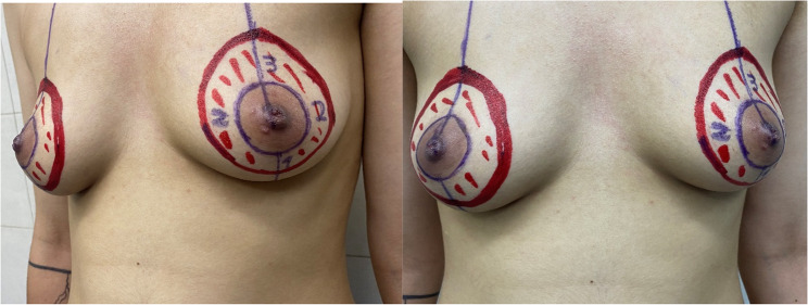
The surgery was performed by an established surgical team with a complete understanding of techniques. (1) In order to determine liposuction tunnels and glandular tissue access along the peri-areolar region markers were conducted in standing position before anesthesia with both hands located on the anterior superior iliac spine, chest out, chin up and staring eyes straight ahead, and feet apart in shoulder-width. A round region at a distance of 3 cm from the superior and lateral, 2–3 cm from the medial and 1–2 cm from the inferior of the NAC was marked as the intended area for de-epithelialization (Fig. 1 ). Meanwhile, the standardization of the location of the NAC has been determined according to the distance of the nipple on the meridian line from the supra-sternal notch and the equal distance of the edges of the areola from the midline. (2) All cases underwent general anesthesia in the supine position with the arms along the body. No additional local anesthetic infiltration was required. (3) The surgical region was draped and disinfected.
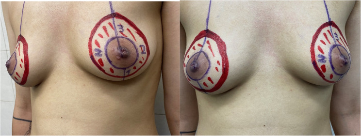
Fig. 1 Round marked region for determining the round block epithelialization area确定环形去上皮区域的标记范围。
Enhanced liposuction is used for removing the intra glandular fat, make the operation area identical and only the glandular tissue is remained so that the dissection becomes facilitate. A stab incision (3 mm) located at the intersection of inframammary fold and the anterior axillary line is performed slightly with Bisturi No 15, and cannulas inserted in this incision. Tumescent solution accompanied by Klein–Nelligan standards contains 20 ml of 2% lidocaine and 1 ml of epinephrine in 1000 ml of normal saline is injected through the cannula to the breast and the surrounding fatty tissue in the breast contour area (between parasternal to anterior axillary line and subclavian muscle to infra mammillary fold). The volume of solution is based on breast size, approximately about 150% to 200% of the breast volume between 500 mL and 1,000 mL per patient. After 20 minutes’ in order to evenly distribution the solution, manual liposuction is conducted as wet technique. As described in the literature, modern liposuction techniques incorporate refined tumescent infiltration, improved cannula control, and systematic fat removal to enhance contouring and reduce complications [ 11 ]. The use of tumescent anesthesia in gynecomastia surgery has been well described in recent literature, with established safety profiles and dosing strategies supporting its use in liposuction-assisted procedures [ 12 ].
In this step, the surface epithelium of the round marked region is removed with Metzenbaum scissors, without damaging the dermis, which is the place of blood supply (Fig. 2 ). In this technique it is possible to make the areolar smaller.
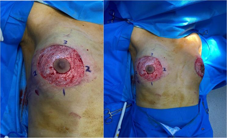
Fig. 2 Round block de-epithelialization环形去上皮(round-block de-epithelialization)操作步骤。
Through the arc-shaped incision of approximately 1 cm was performed at supro-lateral (10-clock position margin of round de-epithelialization area (out of NAC margins to prevent skin bumping), sharp dissection with blunt scissors under direct vision was performed and complete breast tissue was pulled out slowly to pectoralis muscle fascia and held with an Allis clamp (Fig. 3 ).
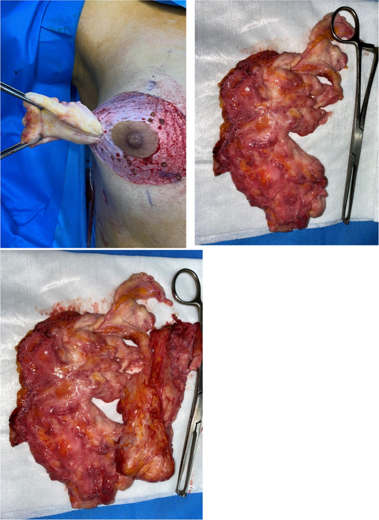
Fig. 3 Subcutaneous dissection of fibro glandular tissue皮下纤维腺体组织的锐性解剖分离。
There was no remained fibroglandular breast tissue under skin whereas by preserving the dermis and subdermis tissues containing vessels, along with careful surgical technique, allowed maintenance of skin vascularity and contour, thereby preventing complications such as crater deformity, contour depression, or vascular compromise of the NAC. Careful hemostasis of subcutaneous bleeding was thoroughly achieved and The defect for tissue dissection was sutured in layers with Monocryl 3 − 0 suture. Then for NAC pexy, round block de epithelialization was closed in two layer sutures with Nylon 3 − 0 in clockwise pattern means superior, lateral, inferior, and medial respectively (Fig. 4 ). Then, sutures are completed with simple ones in the middle of the empty spaces with Monocryl 4 − 0. A drainage tube was placed and fixed through the liposuction incision at the intersection of inframammary fold and the anterior axillary line. As a routine, the resected tissues were sent for histopathologic assessments.
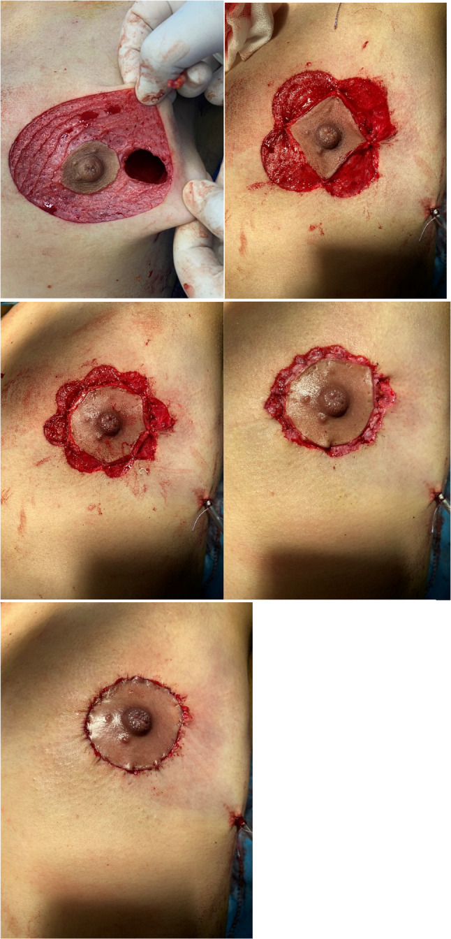
Fig. 4 Reconstruction重建步骤示意。
Follow up visits are performed a day, a week, 1, 3, and 12 months after surgery (Fig. 5 ). Daily activities are allowed immediately after surgery. A day after surgery patients are completely educated about the care of drainage tube and incision. Patients received pressurized bandaging till 3 days and regular pressurized dressing for about 7–10 days. Most of the patients can be discharged on the day of surgery or stay for 1 day. The sutures and drainage tube can be removed 7 days after surgery when the drainage volume was less than 10 ml at 24 h, and also daily use of the silicone gel on incision line is recommended for 3 months after complete healing of wounds. At the first month following surgery visit, (1) Complete training of physical and sports activities after surgery (2) Postoperative conditions such as bleeding, edema, incision features (length, opening), and scar features were assessed. (3) Complications including infection, NAC necrosis, hematoma, seroma, hemo/pneumothorax, fat emboli, intrathoracic organs damage, healing of incision were also monitored during 12-month follow-up period, with most resolving spontaneously or with conservative management within the first 3 months. (4) Cosmetic outcomes such as breast appearance, symmetry, NAC shape, position and sensation, contour irregularity, excessive skin, skin tightness, and overall satisfaction using the 5-point Likert scale were evaluated in which 1 was very dissatisfied, and 5 was very satisfied. Patient satisfaction was assessed using an author-constructed questionnaire designed based on previously published studies and developed under the supervision of three experienced plastic surgeons to ensure clinical relevance. However, it has not undergone formal psychometric validation. The post-operative quality of life was also evaluated using WHOQOL-BREF questionnaire [ 13 ]. Recurrence rate and revisionary surgery requirement are also examined at the 1-year follow-up period. Complications and recurrence were assessed using a multimodal approach, including patient-reported perception, through clinical examination during routine postoperative visits, and when clinically indicated based on standard surgical principles, ultrasonography was requested.
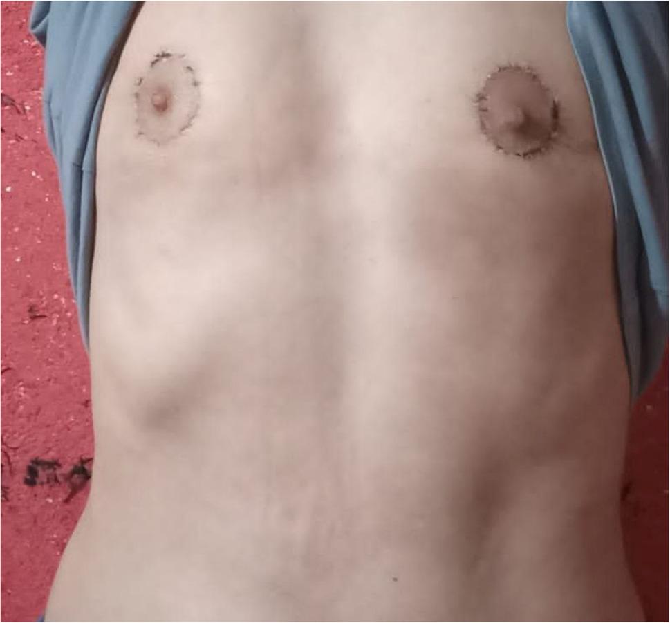
Fig. 5 3 months after surgery术后 3 个月外观。
The statistical Analysis was conducted using SPSS 16.0 software. Quantitative variables (e.g., age, BMI, surgery duration, recovery time) were summarized using mean ± standard deviation, minimum, and maximum values. Categorical variables (e.g., sex, post-operative satisfaction, complications, scar and incision features, WHOQOL-BREF domains) were reported as frequencies and percentages. Although SPSS version 16.0 is an older version, it was sufficient for the descriptive analyses conducted in this study.
This study was conducted in 82 male patients with grade III gynecomastia operated at the Department of Plastic Surgery in Tehran, in 2023 and examined in the 1-year follow-up period. More than 90% of the patients were male (96.3%) with a mean age of 30.04 ± 7.65 and 3.7% were female with a mean age of 24.3 ± 1.53. The overall mean age of the patients was 30.18 ± 7.60 with a range of 17 to 51 years, and the mean height and weight were reported as 181.71 and 102.54, respectively. The mean body mass index (BMI) was 30.97 ± 3.96 (Kg/M 2 ) with minimum and maximum values of 19.70 and 39.9 (Table 1 ). Histopathological examination of all resected specimens demonstrated benign findings, with no evidence of malignancy or unexpected pathological abnormalities.
Table 1 Demographic information of the patients (n = 82)
| Characteristics | n (%) | Mean ± SD (minimum–maximum) |
|---|---|---|
| Age | 82 (100) | 30.18 ± 7.60 (17–51) |
| Height | 82 (100) | 181.71 ± 8.47 (160–199) |
| Weight | 82 (100) | 102.54 ± 15.77 (59–137) |
| BMI | 82 (100) | 30.97 ± 3.96 (19.7–39.9) |
| Sex | ||
| Male | 79 (96.30) | |
| Female | 3 (3.70) |
Based on analysis of the results of post-operative satisfactory questionnaire (Table 2 ), the majority of the patients were satisfied (40.2%) and very satisfied (25.6%) with their surgery outcomes. More than 65% of the subjects were satisfied and very satisfied with the breast symmetry, skin tightness, and excessive skin (75.6%, 73.2% and 64.6%). In terms of healing of the incision, only 1 of the operated cases was completely dissatisfied with the operation, and 35.4% and 26.8% of the people were satisfied and very satisfied with the result of their operation, respectively. In terms of the characteristics of NAC, 2.4% were very dissatisfied with its shape and sense and 1.2% with its position, and more than 50% were satisfied with the result of the operation. In terms of recurrence, 2 people (2.4%) were dissatisfied, and 45.1 and 42.7% were satisfied and very satisfied, respectively. However, these complaints were not confirmed by clinical examination or objective clinical assessment and therefore were not considered true recurrence. None of the patients had reported contour irregularity at a very dissatisfied level, and 7.3% of patients were dissatisfied in this regard, but the majority of them were satisfied and very satisfied (75.6%). In terms of the overall satisfaction of the patients after the surgery, about 5% of the patients (4 people) were very dissatisfied and dissatisfied with the result of the operation, and about 80% of the people were satisfied and very satisfied with the result of the operation.
Table 2 Post-operative satisfactory questionnaires with 5-point likert scale
| Variable | 5-point Likert scale |
|---|---|
| Very satisfied n (%) | Satisfied n (%) |
| Breast appearance | 21 (25.6) |
| Symmetry | 22 (26.8) |
| Skin tightness | 16 (19.5) |
| Excessive skin | 21 (25.6) |
| Healing of incision | 22 (26.8) |
| NAC shape | 18 (22.0) |
| NAC position | 19 (23.2) |
| NAC sensation | 17 (20.7) |
| Recurrence | 35 (42.7) |
| Contour irregularity | 26 (31.7) |
| Overall satisfaction | 33 (40.2) |
Post-operative complications were also evaluated in 82 patients who participated in this study, according to which, 2.4% bleeding, 3.7% subcutaneous hematoma, 3.7% partial NAC necrosis, and 2.4% infection had been reported. The most reported complications were seroma (12.2%), and edema (defined as persistent swelling beyond the expected postoperative course [ 14 ], more than 72 h, or swelling associated with symptoms requiring prolonged compression or intervention) (31.7%). 14.6% of the cases also needed revisionary surgery. No evidence of hemo/pneumothorax, intrathoracic organs damage, and fat emboli, were observed after surgery (Table 3 ).
Table 3 Post-operative complications
| Complications | Yes n (%) | No n (%) |
|---|---|---|
| Bleeding | 2 (2.4) | 80 (97.6) |
| Seroma | 10 (12.2) | 72 (87.8) |
| Subcutaneous hematoma | 3 (3.7) | 79 (96.3) |
| NAC necrosis | 3 (3.7) | 79 (96.3) |
| Infection | 2 (2.4) | 80 (97.6) |
| Edema | 26 (31.7) | 56 (68.3) |
| Revisionary surgery | 12 (14.6) | 70 (85.4) |
| Hemo/pneumothorax | 0 (0) | 82 (100) |
| Intrathoracic organs damage | 0 (0) | 82 (100) |
| Fat emboli | 0 (0) | 82 (100) |
About scar features 1.2% had prominent scars. Patients required secondary scar revision due to not responding to conservative scar treatments such as silicone-based therapies. These revisions were performed under local anesthesia, with excision of the affected scar, and the outcomes were satisfactory. Importantly, none of the revision procedures were performed due to recurrence. Open incision occurred in 8.5% of the cases, whereas 89% of the patients had minimal and better length incisions (Fig. 6 ).
Surgical data were assessed that revealed the recovery and drainage tube extraction time was less than or equal to 7 days in 98.8% of the patients. Only one person had a longer recovery (two months) and drainage period (15 days). Also, the mean duration of the surgery was 123.84 ± 28.82 min (Table 4 ).
Table 4 Surgical data
| Variable | Time | n (%) |
|---|---|---|
| Recovery time | ≤ 7 days | 81 (98.8) |
| > 7 days | 1 (1.2) | |
| Drainage tube extraction time | ≤ 7 days | 81 (98.8) |
| > 7 days | 1 (1.2) | |
| Duration of the surgery (Mean ± SD) | 123.84 ± 28.82 min |
Among the patients who underwent surgery, 38 cases completed the WHOQOL-BREF questionnaire and their post-operative quality of life was examined. Based on this, the mean scores of the physical, mental and social domains of their quality of life were reported as 58.21, 58.63 and 66.43 respectively, which revealed that these people were at an acceptable level. The mean score of the environmental domain of their quality of life was reported as 58.71 ± 16.45. The mean overall quality of life score of the patients was 71.95 ± 16.35 with a median of 75 (Table 5 ).
Table 5 Evaluation of the quality of life of the patients (n = 38)
| Variable | Mean ± SD | Median | Minimum | Maximum |
|---|---|---|---|---|
| Physical domain (0 -100) | 58.21 ± 10.20 | 59.00 | 39 | 86 |
| Mental domain (0 -100) | 58.63 ± 13.79 | 54.00 | 33 | 92 |
| Social relationship domain (0 -100) | 66.43 ± 17.21 | 67.00 | 16 | 100 |
| Environmental domain (0 -100) | 58.71 ± 16.45 | 63.00 | 16 | 91 |
| Overall quality of life (0 -100) | 71.95 ± 16.35 | 75.00 | 25 | 100 |
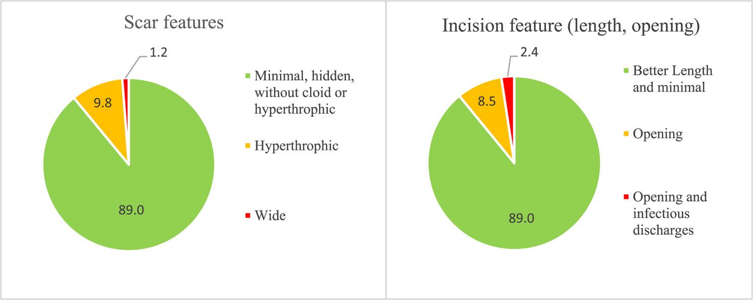
Fig. 6 Incision and scar features切口与瘢痕特征。
The current study provides the preliminary evidence regarding the feasibility and safety of a minimally invasive and fast recovery surgical technique that is associated with reduction in breast volume while preserving breast shape, symmetry, contour, and NAC shape, position, and sensation in treating grade III gynecomastia. The present study does not propose a new surgical technique, but rather evaluates the outcomes of a specific combination of established procedures when applied in a standardized manner to advanced gynecomastia. Furthermore, due to the descriptive, retrospective, and uncontrolled modality of the study, no comparative conclusions can be drawn from these results.
Gynecomastia mostly occurs as a result of imbalanced levels of estrogen and androgen which leads to abnormal fibro-glandular or fatty tissue hyperplasia [ 15 ]. Simon grading is the most used grading method which includes four grades consist of Grade I with mild hypertrophy in glands without excess skin, Grade IIa with moderate glands hypertrophy without excess skin, Grade IIb with moderate glands hypertrophy with a small excess skin; and Grade III with severe glands hypertrophy plus excessive skin [ 10 ].
Surgical methods in managing gynecomastia have been considered as the most effective with high satisfaction due to better aesthetic outcomes and emotional improvements. For the first time, in 1538, surgical methods were used, initially through predominantly transverse thoracic incisions, causing marked scar, and then switched to periareolar incisions [ 10 ]. In 1980, liposuction was used alone for treating gynecomastia by Lewis, which can be effective for fat hyperplasia and cannot resect the fibro glandular tissue, so that can cause residual fat hyperplasia, and postsurgical recurrence rate has been reported as high as 35%. Liposuction has progressed through the years to reduce the operative area complications such as fat embolism, subcutaneous effusion, and paralysis [ 16 , 17 ]. A study conducted by Zhenzhen et al. reported that the combined use of liposuction with small-incision glandular excision had fewer complications, and patient satisfaction than the open resection surgical method [ 18 ].
Grade III gynecomastia, which is a significant breast enlargement and remarkable excess skin, is managed through a wide range of surgical techniques, and the choice of method depends on individual patient characteristics [ 8 ]. In these patients, mastectomy with skin resection (MSR) is considered one of the main techniques, in which both glandular tissue and excess skin are removed simultaneously, with the correction of ptosis [ 19 ]. For protecting the neurovascular supply of the NAC, various subcutaneous pedicled techniques can be used, whereas in more severe cases with a large nipple-to-inframammary fold distance, simple mastectomy via the inframammary fold (IMF) with free nipple grafting is preferred. Less invasive techniques such as subcutaneous simple mastectomy (SSPM), alone or in combination with liposuction, are mainly used for glandular excision through small periareolar incisions; however, in Grade III cases they are usually insufficient as standalone procedures except in selected situations [ 20 , 21 ]. Suction-assisted techniques such as vacuum-assisted mastectomy (VAM) and microdebrider excision and liposuction technique (MELT) also allow tissue removal through small incisions, but their limitation are the correction of redundant skin [ 22 , 23 ]. In addition, endoscopic subcutaneous mastectomy (ESCM), using endoscopic assistance and instruments such as ultrasonic scalpels, enables tissue removal with minimal cutaneous scarring; however, similar to other minimally invasive methods, it remains limited in addressing skin laxity [ 24 , 25 ]. Overall, in Grade III gynecomastia, techniques that include skin excision (such as MSR or mastectomy with free nipple grafting) are preferred, whereas minimally invasive methods are more suitable for patients who prioritize minimal scarring and can tolerate residual skin redundancy.
In the current study we used a comprehensive technique including manually liposuction, round block periareolar de-epithelialization, complete subcutaneous breast tissue resection through periareolar incisions, NAC pexy, and minimally invasive reconstruction methods (in comparison to conventional simple mastectomy techniques for Grade III gynecomastia, which typically require larger incisions and more extensive tissue dissection). This technique is associated with several practical and potential advantages, including relative ease to performance, limited specialized equipment requirement, short time of surgery, fast post-operative recovery, and the use of small peri-areolar incisions resulting in hidden scars, low risk of nipple injury, and subcutaneous hematoma formation. Furthermore, the neurovascular function of the NAC has been preserved and no recurrence was observed during the 1-year follow-up period. There was no remained fibroglandular breast tissue and irregularity. Lifting was performed without excessive flattening, retraction, wrinkles and ptosis. It should be noted that favorable aesthetic outcomes, including skin re-draping and natural wrinkle formation, may not be fully apparent until 12 to 18 months postoperatively, following complete wound healing and tissue remodeling. During the follow-up period, 14.6% of patients required revision surgery. These procedures were not performed to address recurrence or residual tissue, infection, or functional impairments, but rather to improve the appearance of prominent or hypertrophic scars in the superficial cutaneous layers. The relatively high rate of scar revision may be related to several factors, including the anatomic location of the incision, which may cause mechanical tension, variability in scar biology, and wound-healing features. Moreover, revision procedures were performed due to patient concerns, with a relatively low threshold for secondary intervention in the context of high aesthetic expectations [ 26 ]. No keloid scars were reported. Open incision occurred in 8.5% of the cases, whereas 89% of the patients had minimal and good length incisions.
In a retrospective study of 98 patients, the causes of secondary surgery after gynecomastia correction were evaluated [ 27 ]. Of these, most of the patients required revision due to undercorrection rather than overcorrection. The main cause of undercorrection was incomplete removal of fibroglandular tissue, whereas overcorrection was mainly due to excessive resection of retro-glandular fat. After revision surgery, patient satisfaction significantly improved (from approximately 1.5–2.8 to 4.3–4.7), with sustained high satisfaction and no further need for reoperation during follow-up. Overall, these findings suggest that complete excision of fibroglandular tissue while preserving adequate retro-glandular fat is essential to reduce revision rates and achieve optimal aesthetic outcomes.
The overall post-operative satisfaction of the patients was about 80%. The majority of them were satisfied and very satisfied with the breast appearance (65.8%), symmetry (75.6%), skin tightness (73.2%), excessive skin (64.6%), healing of the incision (62.2%), and contour shape (75.6%). In terms of the characteristics of NAC, 2.4% were very dissatisfied with its shape and sense and 1.2% with its position, and more than 50% were satisfied with the result of the operation. Seroma (12.2%), and edema (31.7%) were the most reported post-operative complications whereas other complications such as bleeding, subcutaneous hematoma, NAC necrosis, and infection were insignificant. No evidence of hemo/pneumothorax, intrathoracic organs damage, and fat emboli, were observed. Early complications, such as hematoma, bleeding, and seroma, typically occurred within the first 7 days postoperatively and were readily detected through drainage output. In the subsequent follow-up phase, complications were initially assessed through clinical examination, and when necessary, confirmed with ultrasonography. Management of seroma and hematoma was performed via percutaneous aspiration using a 20 mL syringe, which was sufficient for complete resolution. Regarding infections, no abscesses were observed; reported cases corresponded to mild cellulitis, which were effectively treated with topical mupirocin ointment and, if required, oral ciprofloxacin.
Gynecomastia can affect the quality of life of the patients due to its impact on their daily activities, causing various psychological problems, which can lead to the worsening of a person’s mental condition. A longitudinal study evaluated the impact of gynecomastia surgery on adolescents’ quality of life [ 28 ]. Compared to healthy controls, patients initially had significantly lower self-esteem and poorer physical, social, and mental health. After surgery, substantial improvements were observed in both physical and psychological domains, with outcomes becoming comparable to healthy peers over follow-up (up to 5 years). These findings suggest that surgical treatment not only improves physical appearance but also significantly enhances psychological and social well-being. Therefore, effective, timely, and minimally invasive intervention with good cosmetic outcomes which restore normal male breast contour can prevent these problems and improve patient satisfaction. In the present study we evaluate patient’s post-operative quality of life using WHOQOL-BREF with 5 domains that each can be reported from 0 to 100 scores [ 13 ]. The results revealed relatively good life satisfaction after surgery and moderate scores for each physical, mental, social, and environmental domains as about 58.21, 58.63, 66.43, and 58.71 respectively. The average overall quality of life score was 71.95 ± 16.35. These findings should be interpreted with caution, as the assessed subgroup may not fully represent the entire study. The WHOQOL-BREF questionnaire was completed by 38 of 82 patients (46.3%) and the remaining patients did not complete this assessment. In addition, the absence of preoperative QoL assessment limits the ability to evaluate postoperative improvement.
Although the present technique does not represent a completely new procedure, its novelty lies in the deliberate, simultaneous, and integrated use of previously established, safe, and well-validated surgical methods into a single standardized approach tailored for the management of Grade III gynecomastia. The inclusion of transgender patients was intended solely to demonstrate the applicability and versatility of the proposed surgical technique in scenarios requiring removal of a large volume of fibroglandular and subcutaneous breast tissue (i.e., subcutaneous mastectomy), along with chest contouring and prevention of postoperative ptosis through lifting and NAC repositioning. Due to the very small number of transgender patients ( n = 3), a formal statistical comparison was not meaningful. However, no clinically differences were observed between transgender and cisgender patients in terms of postoperative complications, satisfaction, or need for revision surgery.
The potential limitation of our study is the small sample size, heterogeneity of the study population (including both transgender and cisgender patients), and retrospective design which can limit the generalizability of the results and availability of preoperative baseline data and validated patient-reported outcome measures (PROM), which restricted the ability to perform pre–post comparative analyses. Furthermore, the lack of randomization, control groups, and subgroup analysis may be considered confounding factors and limit achievement of accurate, definitive, efficient, and practical conclusions about the efficiency of the method. The main limitations of this study is the absence of a comparative control group. Therefore, the outcomes observed cannot be directly compared with other established surgical techniques for Grade III gynecomastia, such as concentric mastopexy or free nipple grafting. Furthermore, a standardized etiologic work-up was not systematically performed as part of the study design. Clinical examination and preoperative breast ultrasound were used as part of routine evaluation to confirm the presence of fibroglandular tissue; however, imaging findings were not used as formal inclusion stratification criteria. Additionally, the heterogeneity of the cohort, particularly regarding the distinction between true gynecomastia and pseudogynecomastia in patients with a relatively high mean BMI, may limit the generalizability of the findings. An additional limitation of this study is the use of a non-validated, author-constructed satisfaction questionnaire, which may limit the reliability and comparability of the patient-reported outcomes. Furthermore, quality-of-life data were available only for a subset of patients, which may introduce selection bias. In addition, Outcome assessment was performed by the operating surgeons during routine follow-up visits, which may introduce observer bias in the evaluation of postoperative outcomes. The lack of preoperative quality-of-life data limits the ability to assess the magnitude of improvement attributable to surgery, and only postoperative QoL status can be reported. For confirmation, interpreting, and generalization of the results, resolving the limitations, providing further studies with larger sample sizes, and consideration of randomized designs, control groups, variety of subgroups, and transgender as a distinct group are required.
Comprehensive technique including enhanced liposuction, round block periareolar de-epithelialization, complete subcutaneous breast tissue resection, and NAC pexy approach in patients with grade III gynecomastia and severe ptosis appears to be feasible, and associated with low complications, hidden scars and acceptable patient satisfaction.
声明:中文精读 · 仅供学术参考。内容来自公开文献检索,不代表本人观点,不构成诊疗建议。 医疗美容需在正规医疗机构由执业医师实施。
基于相同主题推荐 · 继续深挖
男性乳腺发育(尤其Ⅲ度)影响心理与外观;本研究评估「增强吸脂+环乳晕圆形去上皮+皮下腺体完整切除+重建」综合术式。
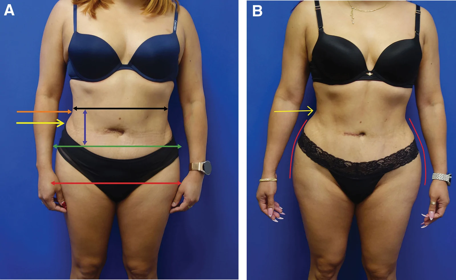
回顾性大样本病例系列(2016年11月至2025年1月),共1999例连续手术,中位年龄36岁(IQR 31至42),中位BMI 29(IQR 26至32),最短随访6个月。
回顾性无对照研究(深圳市宝安区人民医院,2023-05 至 2025-05):32 例(17 女 15 男,平均 28.9 岁)下睑成形(LLB)后黑眼圈加重,接受自体微脂肪移植矫