点阵激光与微针治疗非痤疮性瘢痕及瘢痕样真皮纤维化的系统综述与荟萃分析
2026年10月24日36 min read
Lasers in Medical Science
结论与实践要点
本文聚焦「非痤疮性」瘢痕(外伤、烧伤、术后等所致萎缩或增生性瘢痕及瘢痕样真皮纤维化),这是一类与痤疮瘢痕病理相近但病因不同的纤维化病变。作者纳入 9 项 RCT 进行荟萃分析,发现点阵激光在改善瘢痕评分与促进胶原纤维再生上显著优于微针,但二者在弹性纤维、表皮增厚与不良事件上无差异。
需客观解读:① 点阵激光的「优势」幅度有限(SMD 接近 -1,且置信区间较宽),且多项结局异质性高,说明研究间差异大、结论不宜绝对化;② 亚组分析未显示 CO₂ 与非 CO₂、机械与射频微针间的一致差异,提示设备选择对结果影响有限;③ 本研究未纳入停工期、疼痛与色沉风险等临床决策关键维度。
临床落地:对增生性或萎缩性非痤疮瘢痕,点阵激光证据略占优,尤其适合需较强重塑者;但微针停工期更短、炎症后色沉风险更低,对深肤色患者更友好。实际选择应结合瘢痕性质、皮肤类型与治疗可及性,并以综合治疗思维制定方案。
BackgroundScarring caused by trauma, burns, surgery, and other dermal fibrotic conditions can lead to functional limitation and cosmetic distress. The comparative effectiveness of fractional laser therapy and microneedling for non-acne scars remains uncertain.
MethodsWe conducted a systematic review and meta-analysis of randomized controlled trials comparing fractional laser therapy with microneedling for non-acne scars and scar-like dermal fibrotic lesions. Following a PROSPERO-registered protocol and PRISMA guidelines, we searched PubMed, EMBASE, Web of Science, Cochrane Library, and CNKI from inception to May 2026 without language restrictions. Parallel-group and split-body randomized trials were eligible. Random-effects models were used to calculate standardized mean differences (SMDs) for continuous outcomes and odds ratios (ORs) for dichotomous outcomes.
ResultsNine randomized controlled trials were included. Fractional laser therapy showed a statistically significant advantage over microneedling in scar scores (SMD = -0.99, 95% CI [-1.83, -0.15], P = 0.02) and collagen fiber regeneration (SMD = -2.14, 95% CI [-3.57, -0.72], P = 0.03). No statistically significant differences were found between the two interventions for elastic fiber improvement, epidermal thickness, or adverse events. Subgroup analyses did not show clear or consistent significant differences according to laser type, including comparisons between traditional and non-traditional fractional lasers and between CO₂ and non-CO₂ fractional laser systems. Substantial heterogeneity was observed across several outcomes, indicating considerable between-study variability.
ConclusionBased on currently available randomized evidence, fractional laser therapy may provide superior improvement in overall scar severity and collagen fiber regeneration compared with microneedling for non-acne scars and scar-like dermal fibrotic lesions. However, no clear differences were observed for elastic fiber improvement, epidermal thickness, or adverse-event incidence. Given the substantial heterogeneity, limited sample sizes, and possible reporting bias, these findings should be interpreted cautiously. Further large, standardized trials with longer follow-up are needed.
Supplementary InformationThe online version contains supplementary material available at https://doi.org/10.1007/s10103-026-05017-w.
Keywords: Cicatrix、Laser Therapy、Needles、Burns、Collagen
Scarring, whether resulting from trauma, surgery, or burns, represents a significant clinical problem, often leading to functional impairment, aesthetic disfigurement, and substantial psychological morbidity [ 1 – 4 ]. The burden of pathological scarring is immense, with hypertrophic scars and keloids affecting millions worldwide and presenting a persistent challenge in clinical practice due to their disfiguring nature and tendency for recurrence [ 5 ]. While numerous therapeutic modalities exist, including silicone sheeting, intralesional corticosteroids, and pressure therapy, their efficacy is often variable and limited, leaving clinicians and patients in search of more effective and less invasive options [ 6 ].
In recent years, two minimally invasive techniques have emerged as promising interventions for scar revision: fractional laser therapy and microneedling. Fractional photothermolysis, introduced in the early 2000s, revolutionized laser resurfacing by delivering microscopic columns of thermal injury to the dermis while leaving surrounding tissue intact, thereby stimulating neocollagenesis with a favorable safety profile compared to traditional ablative lasers [ 7 – 9 ]. Concurrently, microneedling, also known as percutaneous collagen induction therapy, has gained popularity for its ability to create controlled micro-injuries that initiate a wound-healing cascade, leading to collagen remodeling and scar improvement without significant epidermal damage [ 10 ]. Both modalities have demonstrated efficacy for a range of scar types, including non-acne scars, burn scars, and other scar-like dermal fibrotic lesions.
Despite their widespread use, a critical gap exists in the current literature. Direct comparative studies between fractional laser therapy and microneedling for the treatment of non-acne scars and scar-like dermal fibrotic lesions are scarce and often limited by small sample sizes or heterogeneous methodologies. The absence of a comprehensive, high-quality synthesis of available evidence, such as a systematic review and meta-analysis, prevents clinicians from making evidence-based decisions regarding the relative efficacy and safety of these two interventions. Individual studies may be underpowered to detect clinically meaningful differences, and conflicting results across trials can lead to uncertainty in clinical practice.
To address this critical knowledge gap, we conducted a systematic review and meta-analysis of randomized controlled trials (RCTs) to compare the effectiveness of fractional laser therapy versus microneedling for the treatment of non-acne scars and scar-like dermal fibrotic lesions, including burn scars, hypertrophic scars, traumatic scars, surgical scars, striae distensae, and other atypical dermal fibrotic lesions. In accordance with PRISMA guidelines and adhering to PRISMA guidelines, we performed a comprehensive, language-unrestricted search of major databases up to May 2026. Eligible studies were randomized controlled trials with either parallel-group or split-face/split-body randomized designs. Our analysis will employ rigorous methods, including risk of bias assessment using the Cochrane RoB 2 tool, evaluation of heterogeneity, and planned subgroup analyses by laser type (CO₂ vs. non-CO₂) and microneedling type (mechanical microneedling vs. radiofrequency microneedling). By pooling data from eligible RCTs, this meta-analysis aims to provide a more comprehensive evidence-based comparison that can guide clinical decision-making and inform future research priorities in scar management.
This systematic review and meta-analysis was conducted in accordance with the Preferred Reporting Items for Systematic Reviews and Meta-Analyses (PRISMA) guidelines.
Participants: Patients with non-acne scars and scar-like dermal fibrotic lesions, including but not limited to burn scars, surgical scars, traumatic scars, hypertrophic scars, contracture scars, striae distensae, and other dermal fibrotic lesions with scar-like characteristics, regardless of age, sex, ethnicity, or lesion duration.Interventions: Treatment with fractional laser therapy as the primary intervention, including ablative fractional lasers (e.g., fractional CO₂ laser) and non-ablative fractional lasers (e.g., fractional Nd: YAP laser, fractional erbium: YAG laser, or other clearly specified fractional laser systems), using any reported treatment parameters (such as wavelength, energy, density, or pulse duration).Comparator: Treatment with microneedle-based therapies clearly specified as either mechanical microneedling or radiofrequency microneedling, including manual or automated mechanical microneedling devices and microneedling radiofrequency systems.Outcomes: Studies were required to report at least one of the following outcomes: scar/lesion severity scores (e.g., Vancouver Scar Scale, POSAS, or other validated scar assessment systems); histopathological changes, including collagen fibers and elastic fibers; epidermal thickness; or adverse reactions (e.g., erythema, edema, post-inflammatory hyperpigmentation, or infection).Study Design: Randomized controlled trials (RCTs), including parallel-group or split-body designs, directly comparing fractional laser therapy with microneedling for the treatment of non-acne scars or scar-like dermal fibrotic lesions. Non-randomized studies, case reports, reviews, animal studies, and studies without a direct comparator were excluded.
Studies in which the full text was unavailable despite comprehensive retrieval efforts.Studies with insufficient data for quantitative synthesis, and for which the corresponding authors could not be contacted or failed to provide the necessary information.Studies published only as abstracts, conference proceedings, or other forms without a full-text article containing complete methods and results.Studies in which the outcome assessment methods were fundamentally non-comparable or non-standardized between groups, and no valid method of data conversion or harmonization was possible.Studies focusing on acne scars only or on lesions not consistent with scar-like dermal fibrotic lesions.
A systematic literature search was conducted to identify relevant studies published up to May 2026. The following electronic databases were searched without language restrictions: PubMed, EMBASE, Web of Science, Cochrane Library, and CNKI (China National Knowledge Infrastructure). The search strategy combined controlled vocabulary terms (e.g., MeSH and Emtree, where applicable) and free-text keywords related to fractional laser therapy, microneedling, and scar/fibrotic lesions. The search syntax was adapted for each database. The detailed database-specific search strategies are provided in Supplementary Table S1 .
In brief, the search terms included the following concepts: laser therapy (“laser therapy”, “fractional laser”, “fractionated laser”, “carbon dioxide laser”, “erbium laser”, etc.), microneedling (“microneedling”, “micro needling”, “percutaneous collagen induction”, “dermaroller”, etc.), and scar/fibrotic lesions (“scar”, “cicatrix”, “fibrosis”, “dermal fibrosis”, “hypertrophic scar”, “atrophic scar”, etc.). In addition, the reference lists of included studies and relevant reviews were manually screened to identify any potentially missed articles.
All identified records were imported into EndNote X21 (Clarivate, PA, USA) for duplicate removal. Two independent reviewers (Author 1 and Author 2) screened the titles and abstracts of the remaining records. Full texts of potentially eligible studies were retrieved and assessed against the PICOS criteria. Any discrepancies during the selection process were resolved through discussion or consultation with a third senior reviewer (Author 3).
A standardized data extraction form was used to collect relevant information from the included studies, including basic study characteristics (first author, publication year, country, and sample size), participant characteristics (scar type, lesion duration, and baseline characteristics), intervention characteristics (fractional laser modality and microneedling type), study design characteristics (parallel-group RCT or split-face/split-body randomized design, follow-up duration, and outcome assessment methods), and outcome data (means and standard deviations for continuous outcomes, as well as event counts for dichotomous outcomes). Data extraction was independently performed by two reviewers, and any disagreements were resolved through consensus or adjudication by a third reviewer.
The methodological quality of the included RCTs was independently assessed by two reviewers using the Cochrane Risk of Bias 2 (RoB 2) tool. This tool evaluates bias across five domains: (1) randomization process; (2) deviations from intended interventions; (3) missing outcome data; (4) measurement of the outcome; and (5) selection of the reported result. Each domain was judged as “Low risk,” “Some concerns,” or “High risk” of bias. The overall risk of bias judgment was determined according to the RoB 2 guidance as follows: studies were rated as having an overall “Low risk” of bias when all domains were judged as low risk; as having “Some concerns” when at least one domain was judged as some concerns but no domain was judged as high risk; and as having an overall “High risk” of bias when at least one domain was judged as high risk. Any disagreements between the two reviewers were resolved through discussion or consultation with a third reviewer.
All statistical analyses were performed using Review Manager (RevMan) version 5.4 (The Cochrane Collaboration, Copenhagen, Denmark) and Stata version 18.0 (StataCorp LLC, College Station, TX, USA). For continuous outcomes, including scar scores, collagen fibers, elastic fibers, and epidermal thickness, pooled effect sizes were calculated as standardized mean differences (SMDs) with 95% confidence intervals (CIs), because different measurement scales were used across studies. For dichotomous outcomes, such as adverse reactions, odds ratios (ORs) with 95% CIs were calculated. Statistical heterogeneity among studies was assessed using the Cochran Q test and quantified using the I² statistic, with a P value < 0.10 for the Q test or an I² value > 50% considered indicative of substantial heterogeneity. When substantial heterogeneity was present, a random-effects model was applied; otherwise, a fixed-effects model was used. Prespecified subgroup analyses were conducted, where feasible, to explore potential sources of heterogeneity according to laser type (CO₂ fractional laser vs. non-CO₂ fractional laser) and microneedling type (mechanical microneedling vs. radiofrequency microneedling), and these analyses were performed only when sufficient studies were available for meaningful comparison. To further investigate potential sources of between-study heterogeneity, meta-regression analyses were conducted using Stata 18.0 for the main outcomes with sufficient data, namely scar score and collagen fiber outcomes. The study-level covariates evaluated included publication year and follow-up duration, and separate univariable meta-regression models were constructed for each covariate and each outcome. Given the limited number of included studies, the findings from meta-regression were considered exploratory and interpreted cautiously. Sensitivity analyses were performed using the leave-one-out method by sequentially omitting each study and recalculating the pooled effect estimate to evaluate the robustness and stability of the pooled results for the major outcomes. Publication bias for the primary outcomes was assessed by visual inspection of funnel plot asymmetry, with funnel plots additionally examined after stratification by laser subtype where applicable. Because the number of included studies for several outcomes was small and between-study heterogeneity was substantial, the assessment of publication bias was considered preliminary and interpreted with caution. A two-sided P value < 0.05 was considered statistically significant, except for the heterogeneity test, for which P < 0.10 was used.
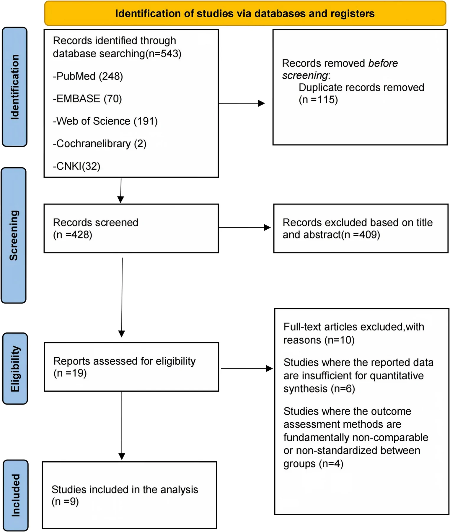
The initial database search identified 543 potentially relevant records, including PubMed ( n = 248), EMBASE ( n = 70), Web of Science ( n = 191), Cochrane Library ( n = 2), and CNKI ( n = 32). After removing 115 duplicate records, 428 unique records remained for title and abstract screening. During this stage, 409 records were excluded based on title and abstract. Subsequently, the full texts of the remaining 19 articles were retrieved and assessed for eligibility. Of these, 10 articles were excluded, including 6 studies with insufficient data for quantitative synthesis and 4 studies in which the outcome assessment methods were fundamentally non-comparable or non-standardized between groups. Ultimately, 9 studies were included in the analysis. The detailed study selection process and reasons for exclusion are presented in Fig. 1 .
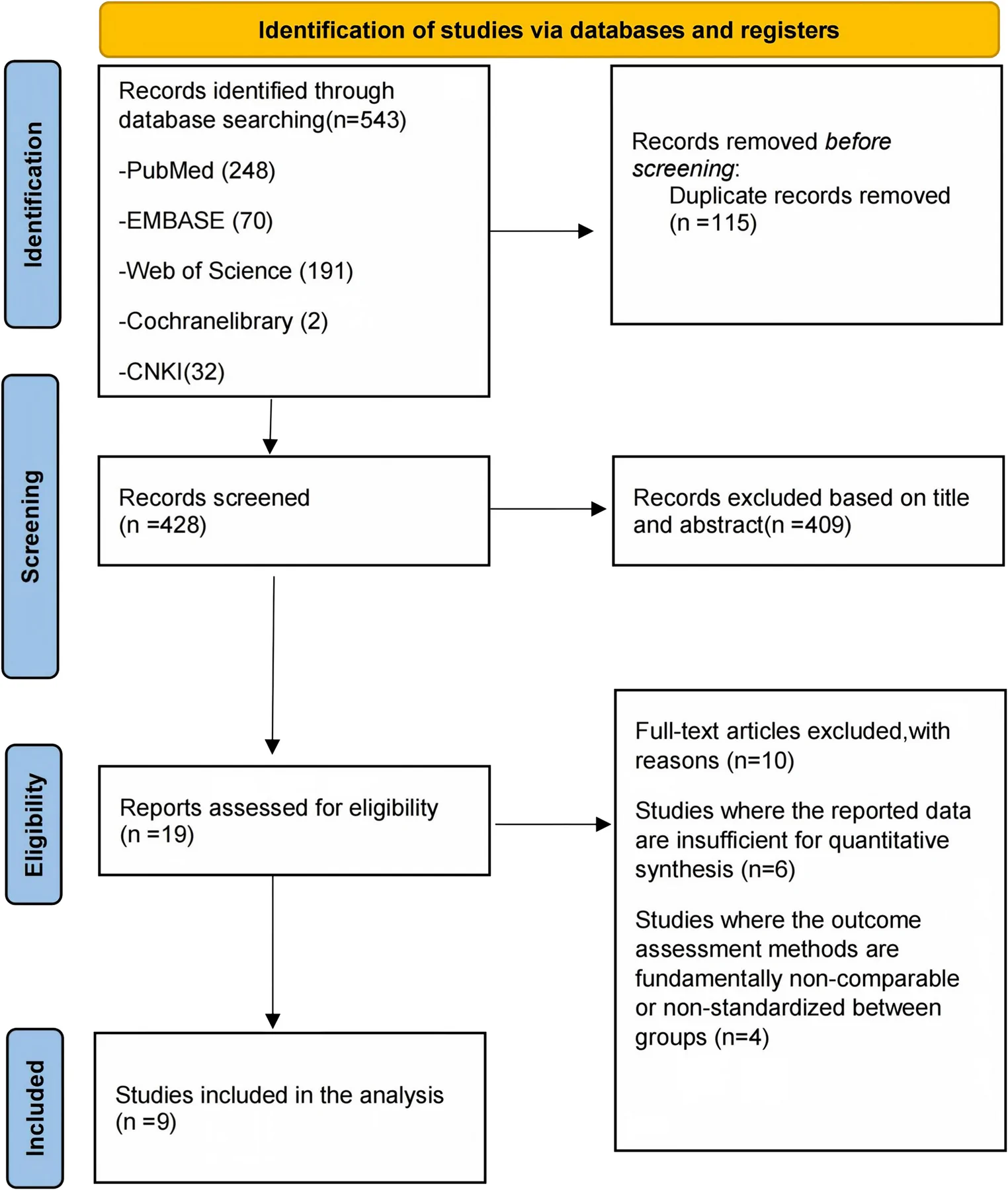
Fig. 1 PRISMA flow diagram of the literature search and selection processPRISMA 文献检索与筛选流程图。
A total of 9 RCTs were included in the meta-analysis [ 11 – 19 ]. The baseline characteristics of these studies are summarized in Table 1 .
Table 1 Characteristics of included studies for systematic review and meta-analysis
| Year | Number of Enrolled Participants | Scar Type | Laser Type | Microneedling Type | Study Type | Outcome Measures | Follow-up Duration(months) | |
|---|---|---|---|---|---|---|---|---|
| Amira A. Zayed et al. [ 10 ] | 2019 | 50 | Burn scar | Dot matrix CO₂ laser | Mechanical microneedling | Split-face randomized study | ①②③ | 4 |
| Ana Paula Naspolini et al. [ 11 ] | 2019 | 40 | Expansion lines | Non-ablative fractional Nd: YAP laser | Mechanical microneedling | Split-face randomized study | ①②③④ | 6 |
| Hesham A. Nada et al. [ 12 ] | 2020 | 40 | Expansion lines | Dot matrix CO₂ laser | Radiofrequency microneedling | Split-face randomized study | ①④⑤ | 5 |
| M. Yulianto Listiawan et al. [ 13 ] | 2021 | 44 | Expansion lines | Dot matrix CO₂ laser | Radiofrequency microneedling | Randomized controlled trial | ② | 6 |
| Mai M. El-Shahawy et al. [ 14 ] | 2025 | 60 | Morphea | Dot matrix CO₂ laser | Mechanical microneedling | Randomized controlled trial | ①⑤ | 3 |
| Mona Soliman et al. [ 15 ] | 2018 | 60 | Expansion lines | Dot matrix CO₂ laser | Mechanical microneedling | Split-face randomized study | ⑤ | 4 |
| Shereen O. Tawfic et al. [ 16 ] | 2022 | 40 | Burn scar | Dot matrix CO₂ laser | Radiofrequency microneedling | Split-face randomized study | ①②③ | 3 |
| Samia Esmat et al. [ 17 ] | 2021 | 60 | Traumatic facial scar | Dot matrix CO₂ laser | Mechanical microneedling | Randomized controlled trial | ①②③⑤ | 1 |
| Mahshid Sabetghadam et al. [ 18 ] | 2025 | 48 | Hypertrophic scar | Photodynamics Therapy | Mechanical microneedling | Split-face randomized study | ①④ | 3 |
The studies were published between 2018 and 2025. The sample sizes ranged from 40 to 60 participants. In terms of scar types, 4 studies focused on expansion lines, 2 on burn scars, 1 on hypertrophic scar, 1 on morphea, and 1 on traumatic facial scar. Regarding laser intervention modalities, 7 studies adopted dot matrix CO₂ laser, 1 used non-ablative fractional Nd: YAP laser, and 1 applied photodynamic therapy. For microneedling types, mechanical microneedling was used in 6 trials, and radiofrequency microneedling was adopted in 3 trials. The majority of the studies (7 out of 9) employed a split-face randomized design, while the remaining 2 were standard randomized controlled trials. The outcome measures reported across these studies varied. All studies reported at least one of the following indicators: scar score (8 studies), collagen fibers (5 studies), elastic fibers (4 studies), epidermal thickness (3 studies), and adverse reactions (5 studies).
The methodological quality of the nine included studies was assessed using the Cochrane Risk of Bias 2 (RoB 2) tool, as shown in Fig. 2 . In the domain of bias arising from the randomization process, most studies demonstrated a low risk of bias, several studies presented unclear risk, and one study was rated as high risk of bias. For bias due to deviations from intended interventions, most studies were classified as low risk, and the remaining studies showed unclear risk. In terms of bias due to missing outcome data, the majority of studies were at low risk, while one study was judged to carry a high risk of bias. Regarding bias in the measurement of the outcome, most studies had a low risk of bias, with several studies assessed as unclear risk. For bias in the selection of the reported result, most studies were evaluated as low risk, and a small number of studies exhibited unclear risk. Overall, a minority of studies were rated as low risk of bias, most studies had unclear risk, and one study was considered to be at high risk of bias. These results indicate that the overall methodological quality of included studies was moderate; accordingly, the pooled findings of this meta-analysis should be interpreted cautiously.
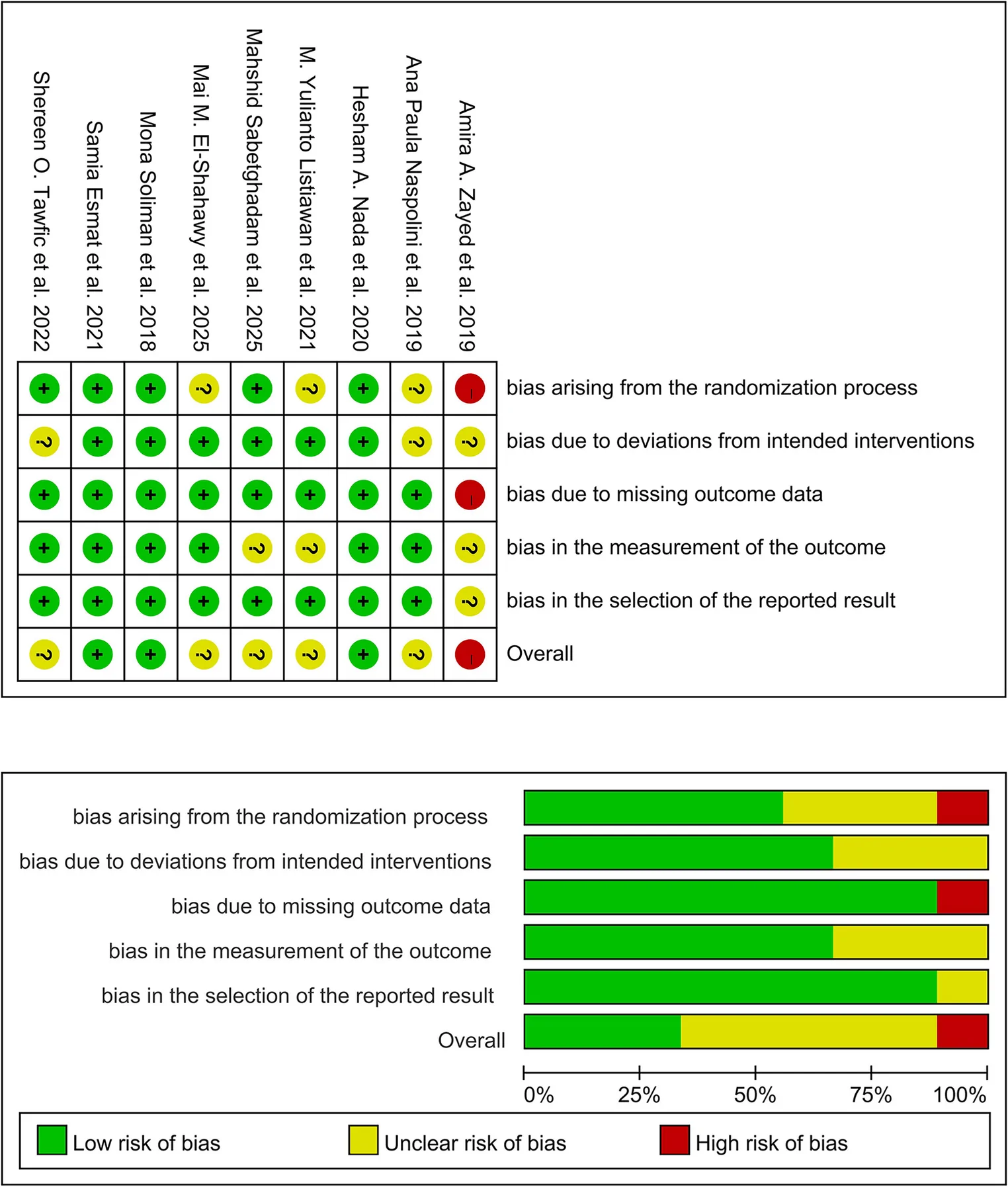
Fig. 2 Risk of bias assessment of the included studies using the Cochrane Risk of Bias tool (RoB 2)纳入研究的 Cochrane 偏倚风险(RoB 2)评估。
A meta-analysis was conducted to compare the efficacy of fractional laser versus microneedling for scar treatment. The primary outcomes analyzed included scar score, collagen fibers, elastic fibers, epidermal thickness, and adverse reactions.
Scar Score: Eight studies compared post-treatment scar scores between the two groups. The heterogeneity test indicated significant statistical heterogeneity ( P < 0.00001, I² = 91%); therefore, a random-effects model was employed. The pooled results showed significant difference in scar score between the fractional laser group and the microneedling group (SMD = -0.99, 95% CI [-1.83, -0.15], P = 0.02) (Fig. 3 A).
Collagen Fibers: Five studies reported data on collagen fiber changes. Analysis revealed high heterogeneity ( P < 0.00001, I² = 94%). A random-effects model was applied, and the pooled analysis indicated that the fractional laser group was significantly superior to the microneedling group in promoting collagen fiber regeneration (SMD = -2.14, 95% CI [-3.57, -0.72], P = 0.03) (Fig. 3 B).
Elastic Fibers: Four studies assessed improvement in elastic fibers. High heterogeneity was observed ( P < 0.0001, I² = 87%); therefore, a random-effects model was used for the meta-analysis. The meta-analysis results showed no statistically significant difference between the fractional laser and microneedling groups regarding elastic fiber improvement (SMD = 0.35, 95% CI [-0.55, 1.24], P = 0.45) (Fig. 3 C).
Epidermal Thickness: Three studies provided data on epidermal thickness. The heterogeneity test yielded P = 0.03 and I² = 72%, prompting the use of a random-effects model. The results indicated no statistically significant difference between the two groups in improving epidermal thickness (SMD = -0.23, 95% CI [-0.90, 0.43], P = 0.49) (Fig. 3 D).
Adverse Reactions: Five studies reported treatment-related adverse events. The heterogeneity test showed moderate heterogeneity ( P = 0.10, I² = 57%), so a random-effects model was used. The analysis revealed no significant difference in the incidence of adverse reactions between the two groups (OR = 4.93, 95% CI [0.33, 74.10], P = 0.25), suggesting that the safety profiles of the two treatments are comparable (Fig. 3 E).
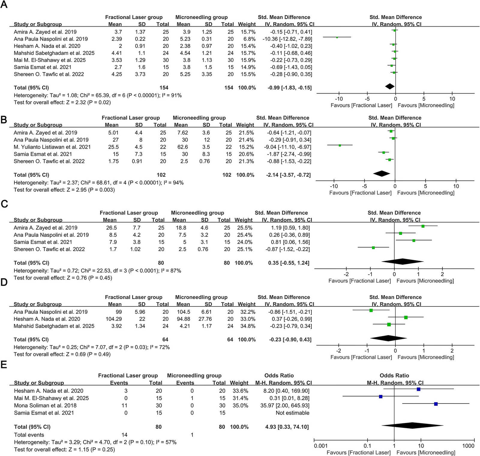
Fig. 3 Forest plots comparing the efficacy of fractional laser versus microneedling for scar treatment. A Comparison of Scar scores. B Comparison of Collagen fibers. C Comparison of Elastic fibers. D Comparison of Epidermal thickness. E Comparison of Adverse reactions点阵激光 vs 微针疗效森林图(A 瘢痕评分、B 胶原纤维、C 弹性纤维、D 表皮厚度、E 不良反应)。
For the CO 2 fractional laser subgroup, the pooled SMD was − 0.31 (95% CI -0.57, -0.04, P = 0.02). The non-CO 2 fractional laser subgroup yielded a pooled SMD of -5.16 (95% CI -15.20, 4.8, P = 0.31), also without statistical significance. The test for subgroup difference between CO 2 and non-CO 2 laser revealed no significant disparity (df = 1, P = 0.34, I 2 = 0%) (Fig. 4 A). When stratified by microneeding type, fractional laser demonstrated significantly lower scar scores compared with mechanical microneedling (SMD = -1.52, 95% CI -2.79, -0.26, P = 0.02). In contrast, no significant difference was detected between fractional laser and radiofrequency microneeding (SMD = -0.34, 95% CI -0.78, 0.11, P = 0.14). The difference between the two microneedling subgroups approached statistical significance (df = 1, P = 0.08, I 2 = 67.0%) (Fig. 4 B).
In the CO 2 fractional laser subgroup, fractional laser therapy significanty outperformed microneedling in facilitating collagen fiber regeneration (SMD= -2.76, 95% CI -4.64, -0.87, P = 0.004). The non-CO 2 fractional laser subgroup showed no statistically significant difference relative to microneedling (SMD= -0.29, 95% CI -0.91, 0.34, P = 0.36). The inter-subgroup comparison demonstrated a statistically significant difference (df = 1, P = 0.01, I 2 = 83.1%) (Fig. 4 C).
Compared with mechanical microneedling. fractional laser achieved superior collagen regeneration outcomes (SMD= -0.87.95% CI -1.68, -0.07, P = 0.03). No significant difference was found between fractional laser and radiofrequency microneedling (SMD = -4.90, 95% CI -12.89, 3.10, P = 0.23). The test for subgroup difference between mechanical and radiofrequency microneedling was non-significant (df = 1, P = 0.33, I 2 = 0%) (Fig. 4 D).
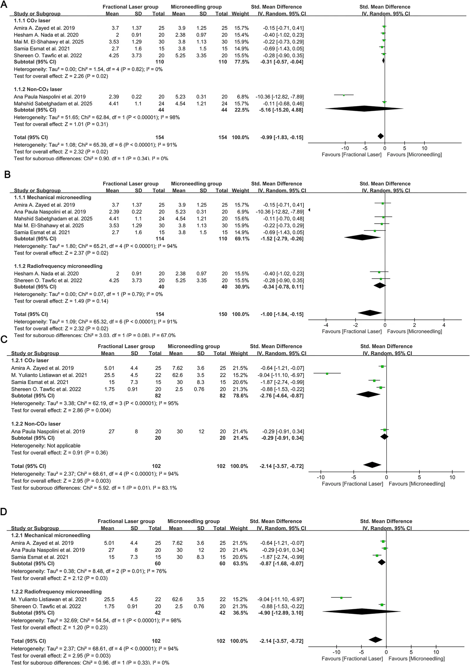
Fig. 4 Subgroup Analysis Forest Plots. A Subgroup analysis of Scar score comparing CO₂ laser versus Non-CO₂ laser. B Subgroup analysis of Scar score comparing Mechanical microneedling versus Radiofrequency microneedling. C Subgroup analysis of Collagen fibers comparing CO₂ laser versus Non-CO₂ laser. D Subgroup analysis of Collagen fibers comparing Mechanical microneedling versus Radiofrequency microneedling亚组分析森林图(A CO₂ vs 非 CO₂ 点阵;B 机械 vs 射频微针;C/D 胶原纤维亚组)。
Meta-regression analyses were performed to explore whether publication year and follow-up duration could account for inter-study heterogeneity of scar score and collagen fiber outcomes (Fig. 5 ). We constructed four regression models: scar score versus publication year (Fig. 5 A, P = 0.323), scar score versus follow-up duration (Fig. 5 B, P = 0.142), collagen fibers versus publication year (Fig. 5 C, P = 0.582), and collagen fibers versus follow-up duration (Fig. 5 D, P = 0.585). The regression plots displayed weak linear trends, yet all covariates failed to reach statistical significance. These results demonstrated that neither publication year nor follow-up duration significantly explained the heterogeneity in treatment effect sizes for scar score or collagen fiber regeneration.
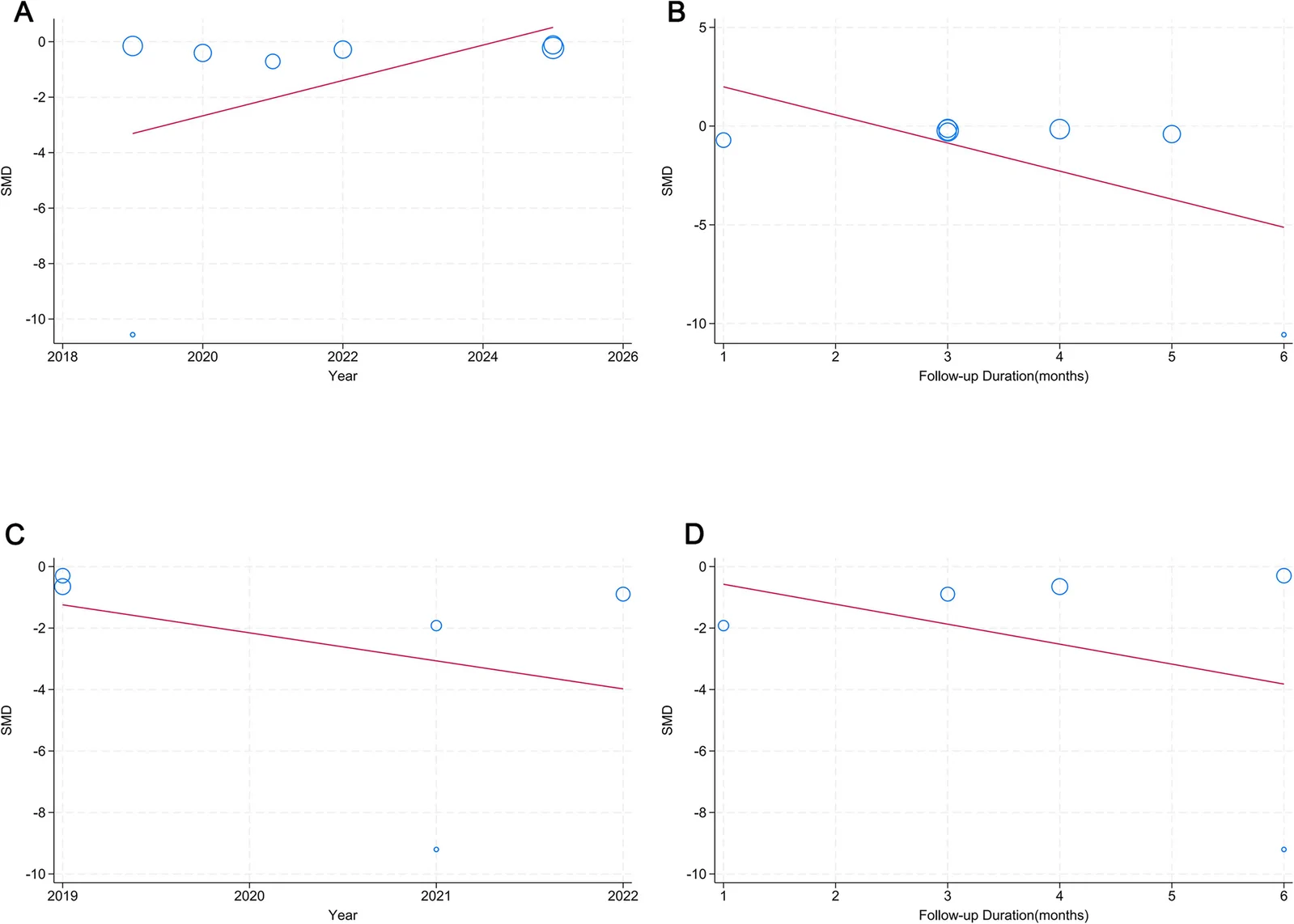
Fig. 5 Meta-regression analyses exploring the effects of publication year and follow-up duration on the standardized mean difference (SMD). A Meta-regression of scar scores against publication year. B Meta-regression of scar scores against follow-up duration (months). C Meta-regression of collagen fiber levels against publication year. D Meta-regression of collagen fiber levels against follow-up duration (months)发表偏倚 meta 回归(按发表年份与随访时长)。
Publication bias for the primary outcomes was visually asessed using funnel plots stratified by laser subtype (Fig. 6 ) exhibited evident asymmetry instead of ideal symmetry. Most studies custered around the apex of the funnel, whereas individual trials displayed substantial deviation from the pooled ffect estimate. In Fig. 6 A (scar score), one non-CO 2 laser study demonstrated prominent outward deviation from the main cluster, corresponding to Ana Paula Naspolini et al. (2019), which largely accounted for the visual asymmetry. Similarly the funnel plot for collagen fibers (Fig. 6 B) also showed an outlying CO 2 laser study distributed far from other observations. These visual patterns raise the possibility of publication bias or small-study effects. Nevertheless, asymmetry may also stem from the limited quantity of included trials, high inter- study heterogeneity, and heterogeneous laser modalities (CO 2 laser versus non-CO 2 laser). Accordingly, the evaluation of publication bias should be interpreted cautiously.
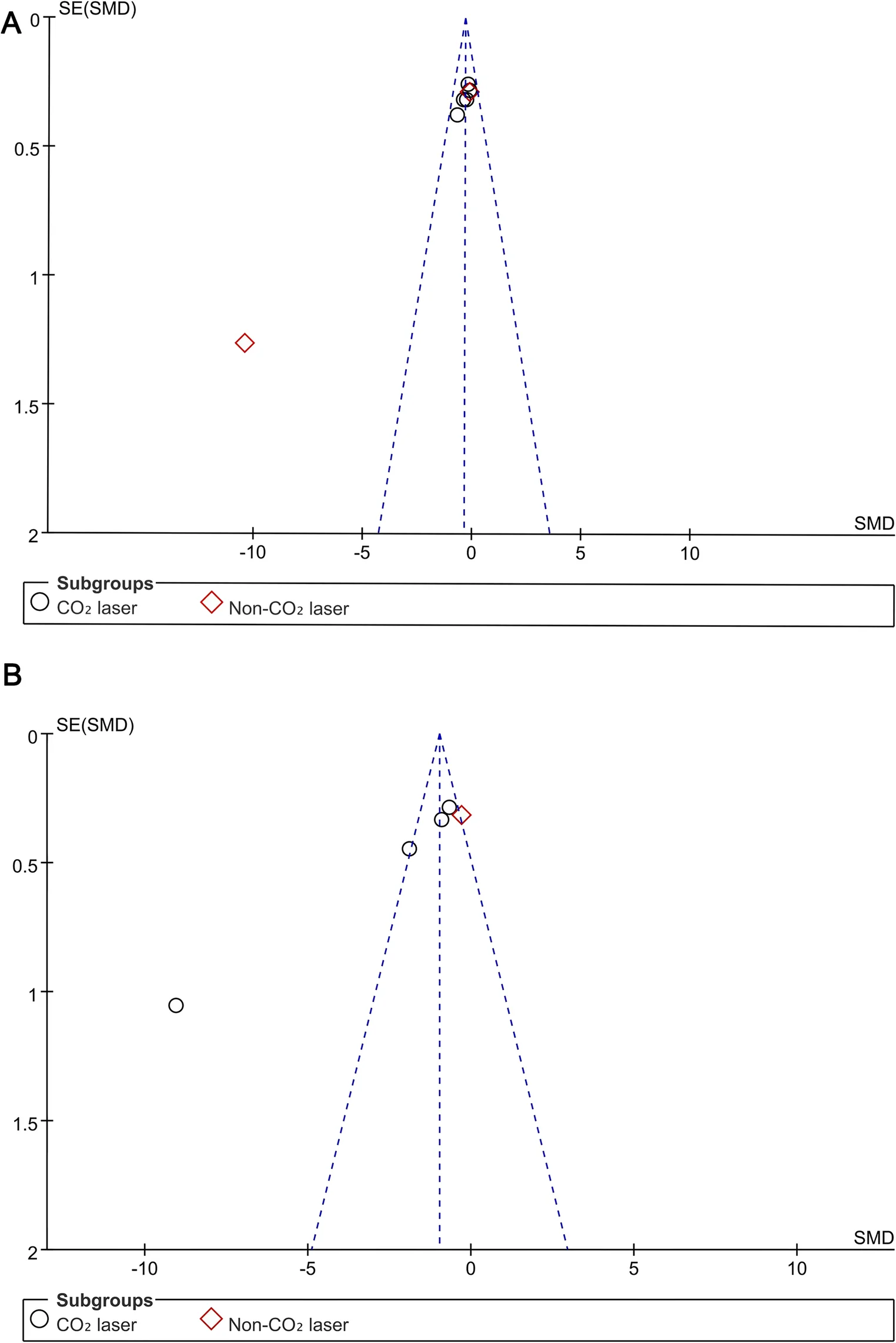
Fig. 6 Funnel plots assessing publication bias. A Comparison of Scar scores. B Comparison of Collagen fibers发表偏倚漏斗图。
Sensitivity analysis adopting the leave-one-out strategy was conducted to assess the robustness of pooled effect estimates for scar score and collagen fibers (Fig. 7 ). Figure 7 A presents sequential exclusion analysis for scar score, and Fig. 7 B corresponds to collagen fiber outcomes. After omitting each individual study iteratively, all recalculated pooled effect sizes remained negative, and their 95% confidence intervals did not cross zero for both endpoints. Notably, removal of the study by Ana Paula Naspolini et al. (2019) did not reverse the direction of the pooled effect for scar score. No single trial was identified to dominate the overall synthetic results. These observations demonstrated that the meta-analytic findings regarding scar score and collagen fiber regeneration were robust and not excessively driven by any individual included study.
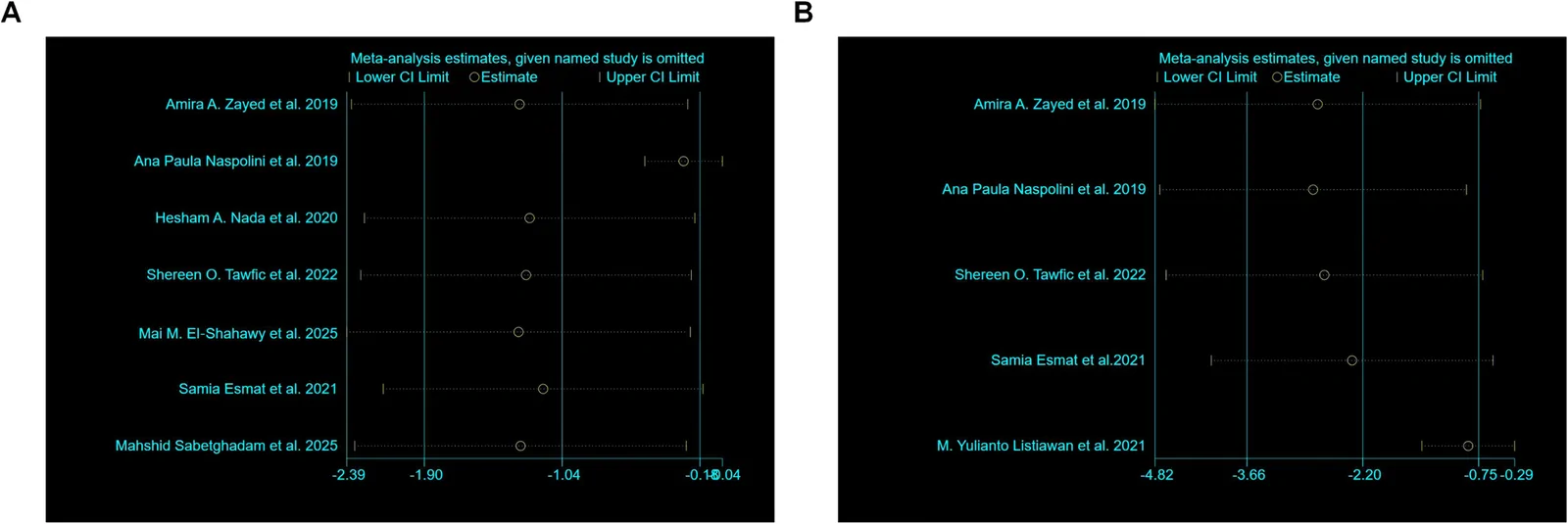
Fig. 7 Sensitivity analysis using the leave-one-out method. A Sensitivity analysis for Scar scores. B Sensitivity analysis for Collagen fibers留一法敏感性分析。
The present meta-analysis indicates that fractional laser therapy and microneedling are both effective modalities for improving non-acne scars and scar-like dermal fibrotic lesions; however, the pooled analysis demonstrated a statistically significant advantage of fractional laser therapy over microneedling in objective scar severity scores. This finding is clinically relevant as it suggests that both modalities may offer comparable benefits for scar management, particularly for patients with hypertrophic or burn scars where collagen remodeling is critical [ 20 ]. Although fractional laser therapy retains a plausible mechanistic advantage, the current quantitative synthesis further supports a measurable overall clinical benefit of fractional laser therapy for global scar improvement. Likewise, although the subgroup analyses did not demonstrate a clear and consistent advantage of specific fractional laser subtypes over microneedling for scar score outcomes, the direction of the pooled overall effect favored fractional laser therapy. In addition, no significant difference in adverse events was observed between groups, indicating that the two treatments appear to have broadly similar safety profiles within the available evidence base. The synthesis of data from randomized controlled trials suggests that the observed benefits of fractional laser therapy may be driven in part by ablative CO₂ laser subtypes, likely because of deeper dermal penetration and more robust neocollagenesis [ 21 ]. However, the clinical significance must be weighed against a higher incidence of transient adverse events, such as prolonged erythema and post-inflammatory hyperpigmentation, associated with fractional laser therapy. These results align with previous literature suggesting that while microneedling offers a favorable safety profile and is effective for scar pliability [ 22 ], fractional laser therapy remains an important treatment option for achieving greater textural improvement in some non-acne scars. The findings directly inform clinical decision-making by guiding practitioners toward personalized treatment protocols based on scar type and patient tolerance for downtime.
The pooled evidence demonstrated a statistically significant superiority of fractional laser therapy over microneedling for overall scar improvement, as reflected by the between-group difference in pooled scar scores. This finding suggests that, although laser-induced photothermolysis and microneedling rely on different biological mechanisms, fractional laser therapy may exert a stronger overall effect on scar remodeling than microneedling. Fractional lasers, particularly ablative types, create microscopic columns of thermal damage that penetrate into the dermis and stimulate wound healing characterized by neocollagenesis and extracellular matrix reorganization [ 23 ]. The high heterogeneity observed suggests that the treatment effect is not uniform and is likely modulated by specific laser parameters, such as wavelength, pulse energy, and density of microthermal zones. This variability highlights that while the laser modality is a critical exposure, its efficacy is highly dependent on technical execution. The significant result for collagen fiber regeneration further supports a mechanistic pathway in which thermal injury directly enhances collagen synthesis, a key biological substrate for scar remodeling [ 24 ]. Nevertheless, because fractional Er: YAG laser and other non-CO₂ platforms were minimally represented in the included studies, no robust comparative conclusion can be drawn for these technologies relative to microneedling. Taken together, the current evidence suggests that fractional laser therapy may confer both a clinical advantage in scar severity reduction and a histologic advantage in collagen remodeling, although these findings should not be generalized across all fractional laser platforms.
In contrast, the analysis revealed no significant differences between fractional laser and microneedling for improving elastic fibers and epidermal thickness. The non-significant result for elastic fibers suggests that neither treatment modality strongly influences neoelastogenesis, a finding that may reflect the inherent biological challenge of regenerating elastic fibers in scar tissue, which are often fragmented and disorganized. This is consistent with the understanding that mature scars are characterized by a paucity of functional elastic tissue, and current resurfacing techniques may have limited capacity to restore this component [ 25 ]. The high heterogeneity in this outcome further indicates that any minor effects are inconsistent and likely dependent on patient-specific factors or measurement techniques. Similarly, the comparable efficacy for epidermal thickness improvement implies that the primary remodeling effects of both fractional laser and microneedling are concentrated in the dermis, with the epidermis undergoing more passive changes. This is biologically plausible, as the dermal matrix provides the structural scaffold for the epidermis, and improvements in dermal collagen and architecture may not directly translate to measurable increases in epidermal thickness. The safety profiles of both interventions were comparable, with no significant difference in adverse reaction incidence. However, this finding should be interpreted cautiously rather than as definitive evidence of safety equivalence. While the wide confidence interval reflects imprecision due to rare events, this finding supports the clinical acceptability of both modalities, suggesting that the choice between them should be guided by efficacy considerations rather than safety concerns.
The exploration of heterogeneity through subgroup and sensitivity analyses provided important methodological insights, but these analyses should be regarded as exploratory. In particular, the revised subgroup analyses did not identify a clear and consistent subgroup effect favoring fractional laser therapy for scar score outcomes. This result weakens the inference that laser type is a definitive effect modifier in this comparison and suggests that previously observed subgroup differences may have been unstable or driven by limited study numbers and between-study variability. This finding is mechanistically grounded in the differential tissue effects of these lasers; traditional ablative lasers create deeper zones of ablation and residual thermal damage, which are known to elicit a more potent neocollagenic response compared to non-traditional or non-ablative lasers [ 26 ]. This provides a clear biological rationale: the strong absorption of CO₂ laser energy by water in the dermis leads to efficient vaporization and coagulation, creating a microenvironment that maximally stimulates fibroblast activity and collagen production [ 27 ]. The robustness of the overall scar score result, confirmed by leave-one-out sensitivity analysis, indicates that the pooled effect is stable and not unduly influenced by any single study. This methodological rigor strengthens the confidence in the observed exposure-outcome relationship. However, the asymmetry noted in the funnel plots for scar scores and collagen fibers raises a cautionary note regarding potential publication bias. This suggests that smaller studies with negative or null results may be unpublished, leading to an overestimation of the true effect size of fractional laser therapy. Accordingly, even where pooled estimates appear to favor fractional laser therapy, the magnitude of benefit in routine clinical practice may be smaller than suggested by the available published literature.
In the pooled analysis, fractional laser-based treatment showed a significant advantage in collagen fiber regeneration, and this effect appeared to be influenced in part by studies of fractional CO₂ laser systems [ 26 ]. However, because the overall evidence base was dominated by CO₂ laser studies and substantial heterogeneity was present, this finding should be interpreted with caution and should not be generalized to all fractional laser platforms. By contrast, the non-significant findings for elastic fiber and epidermal thickness outcomes, coupled with wide confidence intervals, highlight persistent knowledge gaps rather than confirming equivalence. While the wound healing process ultimately aims to restore the extracellular matrix, the specific impact on elastic fiber regeneration following fractional laser treatment is less documented in the histologic literature compared with collagen, which may partly explain the equivocal result.
The superior collagen fiber regeneration associated with fractional laser, particularly driven by CO₂ laser subtypes, is mechanistically supported. Fractional CO₂ lasers create microscopic treatment zones that extend into the dermis, inducing a robust wound healing cascade that promotes neocollagenesis [ 28 ]. Our subgroup result (for CO₂ vs. non-CO₂) strongly validates this mechanism, suggesting that the depth of thermal injury and the specific wound healing response are crucial. The non-significant findings for elastic fiber and epidermal thickness improvements, coupled with wide confidence intervals, highlight a knowledge gap. While the wound healing process ultimately aims to restore the extracellular matrix, the specific impact on elastic fiber regeneration following fractional laser treatment is less documented in the histologic literature compared to collagen, potentially explaining the equivocal result [ 29 ].
Regarding safety, our finding of no significant difference in adverse reactions between treatments is reassuring and consistent with the improved safety profile of fractional laser resurfacing compared to its traditional ablative predecessors. However, the available studies did not show an absence of heterogeneity, and the safety estimate should therefore be interpreted cautiously. The wide confidence interval for the odds ratio underscores the imprecision due to rare events in the included trials. This necessitates comparison with larger safety datasets or registries, as systematic reviews of individual treatments often report low but notable risks like post-inflammatory hyperpigmentation or scarring, particularly with aggressive settings [ 30 ]. Our analysis therefore supports only a provisional conclusion that the overall adverse-event incidence may be broadly comparable between the two interventions; this observation still requires confirmation in larger, prospectively collected safety studies. The methodological robustness of our primary outcome, confirmed by sensitivity analysis, and the noted funnel plot asymmetry underscore the importance of rigorous, transparent meta-analytic methods to minimize bias and produce reliable evidence for clinical practice.
Several limitations of this meta-analysis warrant careful consideration when interpreting the findings. First, substantial and unexplained heterogeneity was observed across multiple primary outcomes, including scar scores, collagen fiber regeneration, elastic fiber improvement, and epidermal thickness. This high heterogeneity, as discussed in methodological literature [ 31 ], indicates considerable variability in study designs, patient populations, laser parameters, and outcome measurement methods across the included trials. Consequently, although some pooled effect estimates favored fractional laser therapy—particularly for collagen fiber outcomes—the true effect magnitude may vary significantly across clinical settings, and these findings should be interpreted cautiously. Second, the analyses for elastic fiber improvement and epidermal thickness were based on a very small number of studies, resulting in wide confidence intervals and non-significant results. This limited statistical power increases the risk of type II error, meaning these non-significant findings should not be interpreted as evidence of equivalence between fractional laser and microneedling. Third, the analysis of adverse reaction incidence demonstrated a wide confidence interval, indicating imprecision due to rare events and small sample size. The non-significant P -value does not rule out clinically meaningful differences in safety profiles. Fourth, funnel plot asymmetry observed for scar scores and collagen fibers suggests potential publication bias, where small studies with negative results may be missing. If present, this could lead to overestimation of the pooled effect sizes, exaggerating the apparent superiority of fractional laser. Finally, the subgroup analyses by laser type and microneedling type should be interpreted as exploratory because of the small number of studies within each subgroup, increasing the risk of unstable estimates. While these exploratory findings are informative, they require validation in prospective trials specifically designed to test these hypotheses. Newer fractional treatment platforms, including picosecond-domain fractional technologies and LIOB-based techniques, were not represented in the pooled analysis because no eligible randomized head-to-head studies were identified. Despite these limitations, this meta-analysis provides a comprehensive synthesis of available evidence, incorporating rigorous statistical methods to account for heterogeneity and exploring potential sources of variation. Future research should focus on large-scale, adequately powered randomized controlled trials with standardized outcome measures and longer follow-up periods to confirm these findings and establish optimal treatment protocols for scar management. Future comparative trials should determine whether these emerging modalities offer distinct advantages over microneedling or established fractional laser systems.
In conclusion, this systematic review and meta-analysis demonstrates that both fractional laser therapy and microneedling are clinically useful and generally well-tolerated interventions for the treatment of non-acne scars and scar-like dermal fibrotic lesions. The current pooled evidence indicates that fractional laser therapy is associated with significantly greater improvement in objective scar severity outcomes and collagen fiber regeneration than microneedling. However, subgroup analyses did not demonstrate a clear and consistent advantage according to laser type, and no significant between-group differences were observed for elastic fiber improvement, epidermal thickness, or adverse events. These findings suggest that fractional laser therapy may provide superior benefit in selected efficacy outcomes, particularly those related to scar appearance and collagen remodeling, while both modalities appear to have broadly comparable safety profiles. These findings provide a crucial evidence-based framework for clinicians, enabling them to tailor scar management strategies to individual patient needs and preferences. Although fractional laser therapy appears to offer an efficacy advantage overall, treatment selection should still take into account scar characteristics, patient tolerance for downtime, device availability, cost considerations, and clinician experience, rather than relying on pooled efficacy estimates alone. Further large-scale, adequately powered randomized controlled trials with standardized outcome measures and longer follow-up are needed to clarify whether specific scar types or device subtypes derive greater benefit from either approach. By identifying key areas of uncertainty and proposing a clear agenda for future research, this work advances the field toward more personalized and effective scar treatment, ultimately aiming to improve both aesthetic and functional outcomes for patients suffering from disfiguring scars.
声明:中文精读 · 仅供学术参考。内容来自公开文献检索,不代表本人观点,不构成诊疗建议。 医疗美容需在正规医疗机构由执业医师实施。
基于相同主题推荐 · 继续深挖