电磁(HIFEM)联合射频身体塑形七项影像学研究的可疑证据、方法学偏倚与利益冲突述评
2026年10月25日31 min read
Annals of Plastic Surgery
结论与实践要点
李哲远博士,本文并非一项阳性疗效研究,而是一篇由独立术者撰写的循证述评,对「电磁(HIFEM)+射频」身体塑形(EmSculpt Neo)的7项影像学研究做了方法学批判。核心结论是:7项研究平均报告的脂肪层减薄8.0 mm、肌肉增厚5.5 mm,差值很可能落在MRI测量误差范围内,且存在轴位层面不一致、照片未标准化、厂商主导拍照等偏倚,临床相关性存疑。
与同类技术比较时须明确:吸脂(作者自引研究)平均减脂45.6%、满意度91.2%、「愿再做」比例93.5%,仍是局部脂肪堆积的金标准;而该联合方案满意度约77.3%、第三方平台「值得做」约74%,且效果可能随时间消退。值得注意的是,所有原始研究均由BTL医学顾问撰写,存在显著财务利益冲突,证据独立性不足。
安全性上,原始研究报告不良事件极少(个别水疱、短暂红斑),但述评提醒能量设备存在反常性脂肪增生、持续性肿胀、烧伤、神经损伤等潜在风险。临床建议:面对以「增肌减脂、免运动」为卖点的设备,应做充分尽职调查,坦诚告知患者疗效证据薄弱与不确定性,优先将局部脂肪堆积患者转诊吸脂,避免以营销话术替代循证沟通。
BackgroundElectromagnetic treatments have recently been combined with radiofrequency to reduce the fat layer and simultaneously increase muscle thickness. Studies report treatment efficacy, using photographs and imaging methods.
MethodsA literature review was conducted. Measurement data were tabulated. The EmSculpt Neo device (BTL Industries, Boston, MA) was used in all studies.
ResultsSeven studies evaluated the fat thickness and muscle thickness using either ultrasound (1 study) or magnetic resonance imaging methods (6 studies). The abdomen was treated in 2 studies. The other studies treated the lateral thighs, flanks, buttocks, inner thighs, and upper arms. The mean reduction in fat thickness was 8.0 mm, and the mean increase in muscle thickness was 5.5 mm. All 7 studies were authored by BTL medical advisors. Measured changes were likely within the margin of error and of questionable clinical relevance. Few adverse events were reported.
DiscussionPhotographs were frequently not standardized, showing changes in treated areas, but unexplained changes in nontreated areas as well. The magnetic resonance imaging scans were affected by differing levels of the axial slice. Measured changes were incremental and likely not indicative of a true treatment effect. The scientific basis for efficacy is tenuous, both for fat reduction and muscle hypertrophy.
ConclusionsA scientific evaluation of the results fails to produce reliable evidence of a clinically meaningful result for this combination energy-based treatment. Financial conflict is a major factor. Liposuction remains the gold standard.
Keywords: body contouring、electromagnetic treatment、radiofrequency、scientific evaluation、HIFEM、EmSculpt、fat thickness、muscle thickness、review
Electromagnetic treatment for body contouring was introduced in 2018. 1 This treatment was labeled HIFEM, an acronym for high-intensity focused electromagnetic. Shortly after its introduction, this method was modified, combining electromagnetic therapy with radiofrequency. 2 The combination is intended to work synergistically to reduce the fat layer and increase muscle. The marketing slogan is “less fat, more muscle.” 3
Over the last 3 years, several investigators have evaluated this combined technology using photographs, measurements, and ultrasound and magnetic resonance imaging (MRI). 2 , 4 – 10 The applicators have been applied to the abdomen, lateral thighs, flanks, buttocks, inner thighs, and upper arms. All publications have documented a significant treatment effect, both in reducing fat and increasing muscle, with almost no adverse effects. This method is recommended as a nonoperative alternative to liposuction with the added benefit of increasing muscle mass without exercise. However, caution is needed. All publications are authored by plastic surgeons and dermatologists who have financial conflicts with the manufacturer. 2 , 4 – 10
The device, which costs about US $135,000, represents a sizable investment. The suggested patient pricing is $3400 for 4 treatments. 11 Part of the sales pitch is that the muscle treatment is temporary, offering a residual income stream from patients who return for maintenance, “like going to the gym, but without all the work.” The fact that any muscle effect is temporary is not mentioned in the publications. 2 , 4 – 10 Before physicians and patients make this investment, both in time and money, due diligence is needed. This review was undertaken to scientifically evaluate the findings and conclusions of these studies.
An electronic literature review was conducted to identify publications that evaluated body contouring using electromagnetic therapy in combination with radiofrequency. The search terms “electromagnetic treatment,” “HIFEM,” and “radiofrequency” were entered into the PubMed search engine. The search included all articles published in English starting in 2018, the year this electromagnetic device was introduced for body contouring, continuing to August 2024. Studies that included measurement data obtained using ultrasound or MRI were evaluated. One study that examined only 3 patients was excluded. 9
Seven studies were identified that evaluated electromagnetic therapy and radiofrequency for body contouring and included imaging of the tissue layers (Table 1 ). 2 , 4 – 8 , 10 All investigators used the same device, EmSculpt Neo (BTL Industries, Boston, MA). Treatment areas included the abdomen, lateral thighs, buttocks, upper arms, inner thighs, and flanks. Six studies used MRIs. 2 , 4 , 6 – 8 , 10 One study, which also included a sham control group, used ultrasound. 5
TABLE 1 Studies Evaluating Electromagnetic Therapy Plus Radiofrequency With Imaging
| # | Study (Year) | No. Patients | Imaging | Area | Follow-up | Difference in Fat Thickness, mm | Difference in Muscle Thickness, mm | Financial Conflict |
|---|---|---|---|---|---|---|---|---|
| 1 | Jacob et al (2021) 2 | 40 | MRI | Abdomen | 1 mo, 3 mo, 6 mo | −8.7*−7.8 | +2.4+2.4 | None reported. CIJ is listed as a speaker and medical advisor for BTL in other studies. |
| 2 | Duncan (2022) 4 | 16 | MRI | Inner thighs | 1 mo, 3 mo | −8.39 | +10.65 | None reported. DID is reported as a BTL clinical investigator in other studies. |
| 3 | Samuels et al (2022) 5 | 40 | Ultrasound | Abdomen | 1 mo, 3 mo, 6 mo | −6.5 | +2.3 | All authors are BTL medical advisors. |
| 4 | Palm et al (2023) 6 | 51 | MRI | Lateral thighs | 1 mo, 3 mo, 6 mo | −18.0 | Not measured | All authors are BTL medical advisors. |
| 5 | DiBernardo et al (2023) 7 | 32 | MRI | Buttocks | 1 mo, 3 mo | −0.2 (NS) | +8.5 | All authors are clinical investigators for BTL. |
| 6 | Jacob and Weiss (2023) 8 | 25 | MRI | Upper arms | 1 mo, 3 mo | −4.9 | +8.9 | None reported. CIJ is listed as a speaker and medical advisor for BTL in other studies. |
| 7 | Cohen et al (2024) 10 | 61 | MRI | Flanks | 1 mo, 3 mo | −6.3†−11.62 | +3.66 | All authors are clinical investigators for BTL. BTL sponsored the study. |
| All studies | 265 | −8.04 | +5.54 | All studies |
Jacob et al 2 reported that 24 patients undergoing abdominal treatment were women; 17 patients were men. Samuels et al 5 did not provide the gender of 40 patients who underwent treatment of the abdomen. In a study evaluating buttock treatment, 29 patients were female, and 3 patients were male. 7 The 16 patients whose inner thighs were treated and all 51 patients having lateral thigh treatment were women. 4 , 6 Most patients undergoing flank treatment were women (50 women, 21 men). 10 The gender of 25 patients undergoing upper arm treatment was not reported, although photographs depict both sexes. 8
Most patients underwent 4 treatments. 4 , 6 – 8 , 10 Two studies used 3 treatments. 2 , 5 All studies reported follow-up times 1 month and 3 months after treatment. 2 , 4 – 8 , 10 Three studies included 6-month follow-up appointments. 2 , 5 , 6 All of the studies measured changes in fat thickness. Six studies measured muscle thickness. 2 , 4 , 5 , 7 , 8 , 10 The mean reported reduction in fat thickness was 8.0 mm, and the mean reported increase in muscle thickness was 5.5 mm. One study reported an increase in buttock muscle thickness, but no significant decrease in buttock fat thickness. 7 Many patients did not keep follow-up MRI appointments because of the COVID-19 pandemic, compromising inclusion rates. 2 , 4 – 6
Few adverse events were reported. One study reported that a patient developed a blister that healed normally and another experienced local erythema. 10 Another study reported mild erythema in several patients that resolved within a few hours. 5 All publications included authors who were listed as medical advisors or clinical investigators for BTL Industries. Six of the 7 studies reported institutional review board approval. 2 , 4 – 6 , 8 , 10
Samuels et al 5 published photographs demonstrating a dramatic 48.9-mm overall reduction in abdominal thickness, greatly exceeding the 6.5-mm mean subcutaneous fat reduction reported in the study. There was also a dramatic, but unexplained, reduction in regions outside the treatment area.
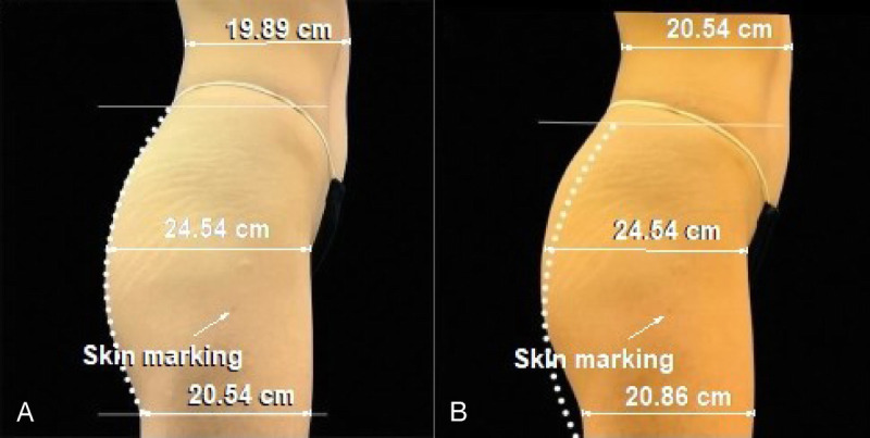
DiBernardo et al 7 reported major changes in buttock muscle thickness. The authors found that combining electromagnetic therapy with radiofrequency produced increases that were significantly greater than electromagnetic therapy alone. The authors provide photographs of a 23-year-old woman treated with electromagnetic therapy alone (Fig. 1 ) and a 21-year-old patient treated with both modalities (Fig. 2 ). Skin markings show that the posttreatment images are moved up vertically compared with the pretreatment images. When the images of the first patient are matched for size and orientation, there is no difference in the anteroposterior measurement of the buttock (Fig. 1 ). The lateral dimension at the level of the untreated abdomen is greater in the posttreatment image.
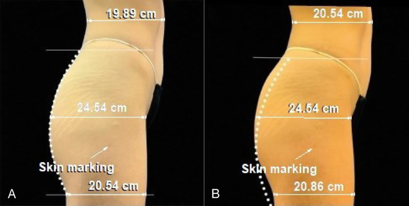
FIGURE 1 A and B, The photographs of a 23-year-old woman before and 3 months after her last of 4 electromagnetic treatments of the buttocks without radiofrequency are compared. The images are matched for size and orientation using the Canfield 7.4.1 Mirror Imaging software (Parsippany, NJ). A skin marking is used for reference. After matching the images, any increase in buttock projection disappears. There is a small increment in the lateral thigh measurement and an increase at the level of the abdomen. Both of these areas are outside the treatment area. The dotted line was superimposed on the image by the authors or manufacturer. The gap at the level of the gluteal fold is unexplained. The dotted line and the horizontal reference line without a measurement were on the original image. This reference line moves up on the pretreatment image as a result of image-matching. A 20-cm abdominal projection is used for standardization. Adapted from DiBernardo et al.723岁女性臀部仅电磁治疗(无射频)前后照片,经软件配准后臀部突出增量消失,治疗区外大腿与腹部反而增大,提示照片变化可能来自摆位而非真实疗效。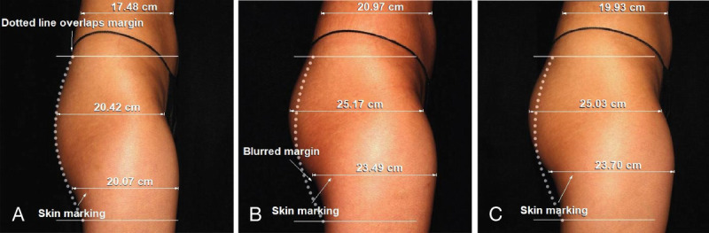
FIGURE 2 A–C, The photographs of a 21-year-old woman before, after the fourth treatment, and at 3 months follow-up are compared. The original images are shown. It was not possible to match the photographs because of nonalignment of reference points. The pretreatment image is lower than the posttreatment images. A skin marking shows a change in vertical level on the 2 posttreatment images. The skin margins are blurry, with an unexplained gap at the gluteal fold. The buttock dimension increases, along with the untreated lateral thigh and abdomen. The 2 horizontal reference lines without measurements were on the original images. A 20-cm abdominal projection is used for standardization. Adapted from DiBernardo et al.721岁女性电磁+射频治疗前后照片,因参考点无法对齐而不可配准,治疗后图像整体上移放大,治疗区外大腿与腹部亦增大,照片标准化存疑。The images in Figure 2 are also not standardized and cannot be matched using imaging software (the reference points cannot be aligned without skewing the orientation). A skin marking at the level of the gluteal fold moves up on the posttreatment images. The posttreatment images appear larger, with increased dimensions not only at the level of the buttock, but also at the level of the untreated abdomen and upper lateral thigh. Although the authors report major increases in thickness of the gluteus maximus, gluteus medius, and gluteus minimus, the MRI scans do not show any differences (Fig. 3 ). The authors state that all images were “reproduced with permission from BTL Industries.” 7
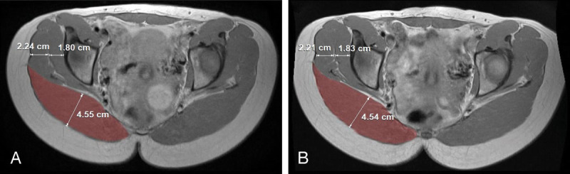
FIGURE 3 A and B, The MRI scans are provided for the 21-year-old woman whose photographs are presented in Figure 2. The muscle thicknesses of the gluteus maximus (red), gluteus medius, and gluteus minimus are unchanged 3 months after treatment. An abdominal width of 34 cm is used for calibration. Adapted from DiBernardo et al.7同一21岁女性的MRI:治疗后3个月臀大肌、臀中肌、臀小肌厚度无变化,与文中「显著增厚」的表述相矛盾。A 2024 publication by Cohen et al 10 includes patient surveys, subjective assessments, photographs, and MRI scans. In addition to measurements of tissue thickness, the authors also measured changes in cross-sectional area on the MRI slices. The authors used an EmSculpt Neo device with 4 “Edge” paddles overlying the anterior and posterior flanks and connected by a circumferential strap. 3 The anterior applicators emit both electromagnetic pulses and radiofrequency; the posterior paddles emit only radiofrequency. 10 The authors report that the heat is distributed evenly to the entire flank area, including the hinge portion of the apparatus. The central abdomen is not treated.
The manufacturer sponsored the protocol and study, which was registered as a clinical trial. 12 The authors recommend this combination treatment as “safe and effective for reducing adipose tissue and strengthening muscle in the area of the lateral abdomen,” 10 although muscle strength was not measured. The study protocol called for measurements of waist circumference, 12 but none are reported.
Cohen et al 10 reported substantial changes in both the subcutaneous fat layer and the flank muscle layer. The mean reduction in the thickness of the fat layer was 28.8%, with a 30.5% reduction in area. The authors reported that the muscle thickness in the flank increased by a similar percentage, 27.2% on average, with a 29.0% increase in area. The authors provide MRI scans of a 52-year-old woman before and 3 months after her last EmSculpt Neo treatment. The authors state that the images were obtained in a prone position to avoid compression of the treated flank areas. However, the images show a supine position. Measurements reveal that the thickness of the posterior flank fat layer decreases by 0.50 cm, or 7.4% (Fig. 4 ). This reduction is much less than the mean 28.8% reduction in fat thickness reported for all patients in the study. The area reduction measures only 2.8%. A 14.3% reduction in area is expected based on a 7.4% reduction in thickness (100 − [92.6% × 92.6%]), suggesting that the change in thickness is not uniform, and possibly one of redistribution. The posterior boundary of the fat layer appears flatter in the posttreatment image, possibly because of compression from the patient lying more on that side. The fat thickness anteriorly increases 6.3%.
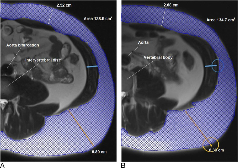
FIGURE 4 A and B, The MRI scans of a 52-year-old woman are compared before and 3 months after treatment. The fat thickness is measured at the posterior flank and over the anterior abdomen. The fat appears white and muscle appears dark on these T2-weighted images. The measurement at the posterior flank decreases by 0.50 cm (7.4%). The fat thickness anteriorly increases 0.16 cm (6.3%). The area decreases by 2.8%. The image taken before treatment is at the level of the intervertebral disc, likely between L3 and L4. The aorta is starting to bifurcate at this level. The image after treatment is at the level of the vertebral body, likely L3. A 34-cm abdominal width is used for standardization. Adapted from Cohen et al.1052岁女性MRI:治疗后后侧flank脂肪厚度减少0.50 cm(7.4%),前腹脂肪反增6.3%,且前后影像轴位层面不同(L3/L4间盘 vs 椎体),存在层面偏倚。Measurements (Fig. 5 ) reveal an increase in flank muscle thickness of 0.30 cm (15.2%). Surprisingly, the thickness of the untreated rectus abdominis decreases 0.19 cm (14.3%). Evaluation of matched photographs of a 40-year-old woman (Fig. 6 ) shows that the flank width decreases 0.54 cm (1.7%).
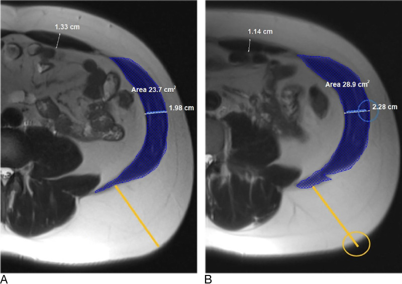
FIGURE 5 A and B, The MRI scans of the same 52-year-old patient depicted in Figure 4 are compared. The muscle thickness at the flank level increases 0.30 cm (15.2%). The muscle thickness of the rectus abdominus decreases 0.19 cm (14.3%). The flank muscle area increases 21.9%. Adapted from Cohen et al.10同一患者MRI:flank肌肉增厚0.30 cm(15.2%),但未经治疗腹直肌反而变薄0.19 cm(14.3%),提示测量可能受摆位或层面影响。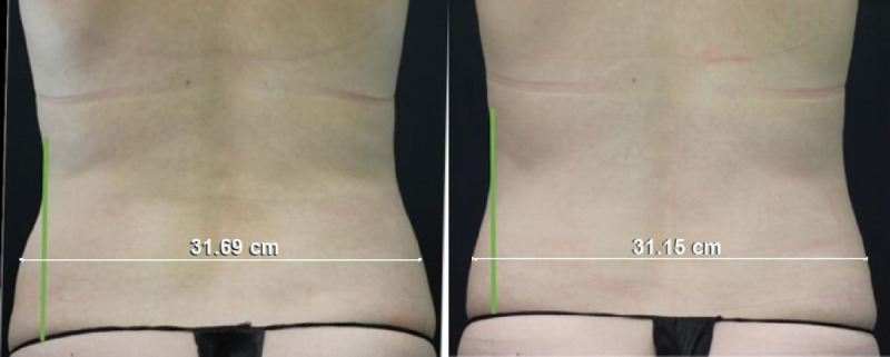
FIGURE 6 A and B, The photographs of a 40-year-old woman before and 3 months after treatment are compared. The photographs have been matched for size and orientation using the Canfield Mirror 7.4.1 imaging software. A 32-cm hip width is used for standardization. The width decreases 0.54 cm (1.7%). Adapted from Cohen et al.1040岁女性照片配准后显示髋宽减少0.54 cm(1.7%),是文中少数看似真实的照片,但绝对变化极小、临床意义有限。
The recent publication by Cohen et al 10 is highlighted on the journal Web site as “Editor's Choice.” The level of evidence rating assigned by the journal is 1. This highest evidence level is usually reserved for a prospective randomized study with a control group for comparison. 13 The study is prospective, but does not include a control group, making it a level 3 study.
It is surprising to see very similar percentage changes in 1-dimensional linear measurements of thickness and 2-dimensional area measurements. Basic geometry dictates that area varies as the square of any change in width. This rule applies to a square, circle, or torus (donut). 14 The abdominal fat layer approximates a donut shape—the inner ring is the peritoneum; the outer ring is the skin. If the width of the layer between the inner and outer rings of the donut is reduced 29%, the reduction in cross-sectional area should be 50% (100 – [71% × 71%]). The same basic geometry issue affects previous studies using energy devices to induce skin contraction. 15 , 16
Previous studies report modest increments, averaging 2.16 mm (range, 1.5–3.0 mm) in muscle thickness after electromagnetic therapy. 17 A change of 2.16 mm represents less than 1/10 of an inch. Measurement variances tend to be high. Changes of this magnitude are likely to fall within the margin of error. 17
It is essential when comparing MRI scans to use the same slice level for comparison. 17 This is especially true when the measurements differ by <5 mm. Otherwise, a fictitious difference caused by varying anatomic levels may exceed any real difference in tissue thickness. 17 Radiological landmarks reveal that the axial slice levels are different (Fig. 4 ). This confounder also affects other studies, published by a co-author, evaluating changes in axial MRI scans after electromagnetic treatments. 2 , 18
Publications often feature photographs showing pronounced changes (Fig. 2 ). 5 , 7 , 19 However, these changes are frequently accompanied by unexplained changes in untreated areas (Figs. 1 and 2 ). 17 Photographic standards, promoted by one of the study authors, 20 are essential when comparing before and after photographs. A blue background is preferred 20 , 21 and is less susceptible to digital editing, 21 such as photoshopping (airbrushing), which can produce indistinct margins. The responsibility for photographs cannot be outsourced to a manufacturer. 21 The manufacturer is not a disinterested party and cannot be expected to be unbiased. The study authors are responsible for photographic integrity. The term “reproduced with permission of the manufacturer” should not appear in captions to photographs in a scientific journal.
A recent review found that the mean reduction in the fat layer among studies using this technology was 5.5 mm and the mean increase in the muscle thickness was 2.16 mm. 17 The change in rectus abdominis separation was not significant. Theoretically, these effects on overall thickness would be expected to offset each other, producing a mean reduction in overall tissue thickness of 3.3 mm (5.5–2.16 mm), or 1/8 of an inch. The muscle and fat tissue layers are easily visible on an MRI scan. However, they are not discernible clinically. The appearance is the same whether the fullness is created by muscle or fat. A treatment that reduces the abdominal fat layer sufficiently to show off the muscle definition (ie, a 6-pack) of the underlying rectus abdominis would be a worthwhile goal for many patients. However, published studies do not show a more chiseled abdominal appearance. 2 , 5 , 17 – 19
The EmSculpt (BTL-899) device received 510(k) clearance for noninvasive lipolysis of the abdomen and reduction in abdominal circumference. 22 This determination was based on clinical trials sponsored by the manufacturer and conducted by a Bulgarian dermatologist. 23 , 24 Evidence of lipolysis was inferred from microscopic signs of fat cell disruption in biopsies of abdominal tissue. Although the histological and clinical studies are listed as completed, no results are available on the clinical trial Web sites, 23 , 24 and no publications are listed on the PubMed Web site. 17
This technology received Food and Drug Administration clearance for muscle toning. 17 Muscle tone is defined as the internal state of muscle-fiber tension within individual muscles and muscle groups or the degree of muscle tension or resistance during rest or in response to stretching. 25 There is no evidence that electromagnetic treatments affect these muscle properties. 17 Although adverse effects are minimal, 17 few are expected if the treatment effects are subtherapeutic.
The criteria for evaluating a new technology are simple. The device must have a sound scientific rationale, and there must be reliable clinical evidence of results. 17 , 26 , 27 Evidence-based medicine does not recognize expert opinion, first principles, bench research 13 (such as histology slides), photographs that defy explanation (Fig. 2 ), 17 or media buzz. 27
The acronym “HIFEM” includes the words “high-intensity.” These words are unnecessary. All energy treatments have a range of intensities, from low to high. In practice, operators try to keep the intensity low because the treatments can be painful. 17 The word “focused” suggests that the energy is directed at a specific site, like a camera lens. However, no specific subcutaneous level is targeted. 17
The proposed mechanism for electromagnetically induced fat reduction is extrapolated from a 2007 Danish exercise physiology study in subjects who were not treated with an energy-based device. 28 Zachary et al 29 studied tissue samples obtained from abdominoplasty specimens and compared electromagnetic treatments with cryolipolysis. The fat cells exposed to electromagnetic treatment remained viable, with no signs of inflammation. By contrast, adipose tissue exposed to cryolipolysis showed a marked inflammatory response and irreversible fat cell injury (apoptosis). The conclusions differed from an earlier study in treated pigs. 30 The lead author of the earlier study was a medical advisor and speaker for BTL Industries. The lack of a dose response (ie, more treatments do not produce a greater effect) 31 is inconsistent with treatment efficacy.
Surface application of a radiofrequency device is limited by the heat needed to cause subcutaneous fat coagulation. The skin would burn (threshold, 42°C) before the fat cells would necrose and coalesce (60°C). This limitation is the reason radiofrequency treatments used for body contouring feature subsurface probes. 32
It is unclear why the 1-month fat layer is reduced (26.2%) almost as much as the 3-month fat thickness (30.5%). 10 If the treatment works by apoptosis, there should be a period of swelling and absorption of the devitalized fat cells. 17 Adipocyte destruction is the common final pathway for any energy-based treatment. The cells cannot be expected to just disappear. Even if the treatments were effective, one must consider the physiological consequences of releasing a large volume of fat cell degradation products (30% of the fat layer) 10 , 33 into a patient's circulation. These fat cells are safely removed from the body during liposuction and can be used for simultaneous fat transfer to the buttocks.
Increasing the muscle cross-sectional area by 10 to 15% requires at least 10 to 14 weeks of dynamic heavy resistance training. 34 , 35 The early increase in muscle thickness after a workout, called the “muscle pump,” is caused by cellular edema, not muscle hypertrophy. 36 DiBernardo et al 7 showed photographs depicting muscle enlargement immediately following the fourth treatment. There is no known mechanism for muscle hypertrophy after just 1 month, or less. 17 Early ultrasound images reveal an echolucent muscle layer, 5 consistent with tissue edema. 17
A 2021 consensus statement published by authors who were all compensated by the manufacturer concluded that electromagnetic therapy reduces fat 30% and increases muscle 25%. 33 These percentages are very similar to the figures reported by Cohen et al. 10 Goldberg et al 33 also claim that this technology reduces muscle separation 19% and reduces waist circumference up to 5.9 cm. These benefits, along with skin tightening and muscle strengthening, 6 , 10 are widely advertised on physician Web sites.
Cohen et al 10 suggest that weak lateral abdominal muscles can lead to poor posture, low “core stability,” lower back pain, increased risk of injuries, and can affect breathing and bowel movements. Presumably, by stimulating the muscles with electromagnetic treatment, these problems may be ameliorated. Another proposed benefit is more comfortable clothing to improve confidence and reduce stress. 10
Realself.com is a highly popular public platform that features reviews of 514 procedures. 37 This site is not influenced by industry sponsorship or conflicted investigators and can provide a “grassroots” impression of patient satisfaction. The “Worth It” score for EmSculpt is 74%, 37 ranking at the 15 th percentile among listed procedures.
Cohen et al 10 report that patients depicted in photographs “feel more toned after the treatment.” Surveys showed that 71.2% of participants “felt improvement in muscles and fat.” Evidently, 28.8% of participants did not feel improvements in muscles and fat. Prospective patients are unlikely to be impressed with the results depicted in the photographs (Fig. 6 ). These photographs do appear authentic, unlike other publications, which often show atypical or unrealistic results. 5 , 7 , 19 The procedure is not painless; 15.1% of the subjects reported pain scores >3 on a 0 to 10 scale. 10
One question not asked in the survey in the survey administered by Cohen et al 10 was, would you redo the procedure? Patients may not find that their result justified their time (4 treatment visits), discomfort, and the expense, which averages US $3000 according to Realself. 37 It is likely that study participants received a financial incentive, although none is reported, 10 which is also a consideration when interpreting survey responses.
Overpromising and underdelivering is not a winning formula, or one that is likely to build a practice. 26 , 27 Trying to convince a patient of a worthy result is not a rewarding experience. Patient disappointment leads to physician disillusionment, which can be a factor in burnout. Many plastic surgeons are familiar with this disappointment, having purchased an expensive device, based on “buzz” in the media, favorable revenue projections, and a fear of being left behind in the marketplace. 26 , 27
The study design used by these investigators 2 , 4 , 6 – 8 , 10 bears similarities to this author's self-funded study using MRIs to evaluate liposuction. 38 The mean reduction in the thickness of the fat layer after liposuction was 45.6%. Two thirds of the swelling had resolved after 1 month, and almost all of the swelling was gone after 3 months. This study was the first publication to rigorously measure changes in the fat layer after liposuction and strongly supports its efficacy. It does not “take up to 3 to 6 months before the results are truly visible.” 10 Liposuction allows confluent areas to be treated, as opposed to spot treatments that, in the case of flank treatment, do not include the central abdomen. Liposuction patients report a 91.2% satisfaction rate, compared with 77.3% for electromagnetic treatment plus radiofrequency, 10 and 93.5% of liposuction survey respondents say they would do it again. 39
Remarkably, all publications evaluating this treatment combination are authored by medical advisors or investigators for BTL Industries. The manufacturer also paid for the MRIs used in the study by Cohen et al. 10 Other studies do not report whether the manufacturer funded these expensive studies, although it would seem likely. This corporate sponsorship creates a huge incentive to report positive findings. It is inconceivable that such a study would be published if the authors concluded that any benefit was nonsignificant or within the margin of error. Corporate funding is not mandatory for imaging studies. 38 Although self-funding involves a financial commitment on the part of the investigators, it ensures that the study findings are credible. The reward for the authors is a worthy contribution to our specialty and the professional recognition that goes with it.
The scientific basis for this treatment combination is questionable. Sudden muscle hypertrophy and reductions in fat thickness without swelling are difficult to reconcile with known physiology. Changes in fat and muscle thickness on MRI scans are small and likely within the margin of error. Incremental changes may not be clinically relevant. Whether this treatment provides sufficient benefit to warrant its cost and time commitment is unclear.
A question for plastic surgeons is whether a scientific journal should serve as a marketing tool for a company that seeks to establish scientific validity for its product. The same issue concerns other energy-based devices. 26 , 27 , 32 The product is marketed before its scientific legitimacy is properly demonstrated. Patients suffer the consequences in the form of adverse effects (eg, paradoxical adipose hyperplasia, 40 prolonged swelling, burns, nerve injuries 32 ) or simply a lack of results. 17
To preserve credibility, investigators should decline direct or indirect reimbursement. To maintain independence and scientific integrity, plastic surgery journals and societies need to reconsider sponsorship from device manufacturers. 41 – 43 Our duty is to protect our patients, and their pocketbooks, from invalid treatments and devices, and inform them of superior options.
Patients interested in treatment of localized adiposity are best referred for liposuction, which remains the gold standard. 38 , 39 Nonoperative alternatives will always have an appeal. However, if the last 2 decades are any indication, one could be forgiven for concluding that energy-based treatments are the way of the future and always will be.
声明:中文精读 · 仅供学术参考。内容来自公开文献检索,不代表本人观点,不构成诊疗建议。 医疗美容需在正规医疗机构由执业医师实施。
基于相同主题推荐 · 继续深挖
本文为一篇针对电磁(HIFEM)联合射频(RF)身体塑形(EmSculpt Neo,BTL公司)的文献述评,系统纳入7项含超声或MRI影像测量的研究,且全部由BTL医学顾问或研究者
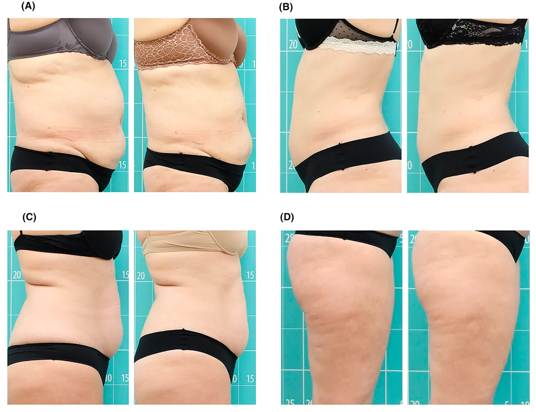
前瞻性单臂研究(西班牙 Asturias 伦理委员会批准),39 例健康女性志愿者(23–68 岁,平均 47 岁,Fitzpatrick II–III),腹部/腰侧 25 例、大
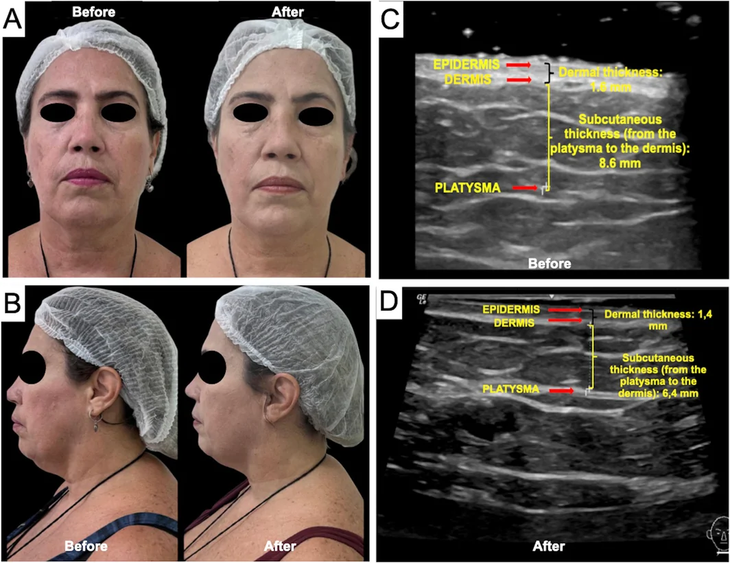
一项巴西前瞻性单中心研究(n=16,PASMFRS 1–4 级颔下脂肪)采用 Megaderme Duo 多层微针射频(40 针镀金微针,深度 2/4/6 mm,15–30 W,1