腋窝下入路臂部提升术(SAB):一项67例女性中度上臂松弛的回顾性队列研究
2026年10月27日24 min read
Plastic and Reconstructive Surgery Global Open
Plastic and Reconstructive Surgery Global Open
结论与实践要点
本研究报道一种名为皮下腋窝入路臂部提升(Subdermal Axillar Brachioplasty, SAB)的微创术式,针对 Iglesias 分级 I–III 的中度上臂皮肤松弛。核心做法是在腋窝皱褶内做单一梭形小切口(平均长度5.4±0.4 cm),先行手臂吸脂(单侧平均抽吸245±62 mL,范围150–360 mL),再以皮下单极射频(每端口至少6–9遍、80%功率、2.5–3.0 L/min 气流、单侧约8 kJ)促进皮肤回缩,最后分层缝合固定组织瓣。其设计目标是替代传统从腋窝延伸至肘部的纵向长瘢痕切口。
在67例均为女性的回顾性队列中(均值年龄50.2岁、BMI 27.8 kg/m²、81.8%为白人),术前73.13%为 III 级、26.86%为 II 级松弛;术后12个月随访显示89.55%改善至 I 级、10.44%为 II 级。并发症共4例:血肿2例(2.98%)、血清肿与感染各1例(均1.49%)。BODY-Q 手臂外观满意度由术前31.9±2.98升至术后94.2±3.45(t=39.2,P<0.001)。
临床适用边界:该术式适用于轻中度(Iglesias I–III 级)松弛、尤其因减重或衰老所致者;Iglesias IV 级、活动性吸烟(术前3个月)、心血管疾病、结缔组织病及严重合并症者被排除,提示重度松弛或高危人群并非适应证。研究局限包括单中心单术者、回顾性设计、无对照、由术者本人(非盲法)测量松弛度,且随访点仅设术前与术后12个月,因此「高满意度、低并发症」应在该证据等级下解读,尚不能直接推断其优于其他术式或具备超过1年的长期稳定性。
Background:Traditional brachioplasty for moderate to severe arm laxity typically requires longitudinal incisions extending from the axilla to the elbow, often resulting in visible scarring and patient dissatisfaction. To address this limitation, we developed subdermal axillar brachioplasty (SAB), a minimally invasive technique combining liposuction, gas-assisted subdermal radiofrequency, and limited axillary skin excision.
Methods:A retrospective observational cohort study was conducted including 67 female patients who underwent SAB between January 2023 and November 2025. Patients with Iglesias grade IV laxity and active smokers were excluded. Arm laxity was objectively measured using a standardized metric protocol preoperatively and at 12-month follow-up according to the Iglesias classification. Outcomes included postoperative laxity grade, surgical parameters, complications, and patient satisfaction assessed using the BODY-Q scale.
Results:Preoperatively, 73.13% of patients presented with grade III laxity and 26.86% with grade II. At 12 months, 89.55% achieved grade I laxity and 10.44%, grade II. The mean lipoaspirate volume was 245 ± 62 mL per arm, reflecting conservative contouring. Mean axillary incision length was 5.4 ± 0.4 cm. Complications included 2 hematomas (2.98%), 1 seroma (1.49%), and 1 infection (1.49%). BODY-Q scores significantly improved from 31.9 ± 2.98 preoperatively to 94.2 ± 3.45 postoperatively ( P < 0.001).
Conclusions:SAB is a safe and effective technique for patients with moderate arm laxity, achieving significant contour improvement and high patient satisfaction with minimal visible scarring. This approach offers a viable alternative to traditional longitudinal brachioplasty in appropriately selected patients.
Takeaways
Question: Is there any surgical procedure to improve arm appearance in patients with notable arm laxity (Iglesias laxity scale grades II–III)?
Findings: Subdermal axillar brachioplasty demonstrated a significant reduction of arm laxity, with most patients exhibiting grade I laxity (81.81%) and grade 0 laxity (18.18%) after surgery, without any complications.
Meaning: Subdermal axillar brachioplasty technique represents a significant advancement in arm contouring surgery, offering a minimally invasive solution for patients with mild to moderate arm laxity. By combining liposuction, subdermal radiofrequency, and layered flap fixation, this method achieves noticeable improvements in arm aesthetics with minimal scarring, avoiding the extensive incisions traditionally required for brachioplasty.
Brachioplasty is a specialized surgical procedure designed to improve the contour of the upper arms and axillary region by removing excess skin and adipose tissue. It is particularly beneficial for patients who have undergone significant weight loss or are of advanced age, as these individuals often present with skin laxity and redundant tissue extending from the shoulder to the elbow and occasionally into the lateral thorax. 1 , 2
With the rise in bariatric surgery and body contouring procedures worldwide, brachioplasty techniques have evolved significantly. Advances in aesthetic goals and surgical innovation have improved outcomes, ensuring better anatomical safety and minimizing complications such as nerve injury. Historically, arm contouring surgery dates back to the 1930s when Thorek introduced an elliptical resection for upper arm skin excess. Subsequent refinements were introduced by Correa-Iturraspe and Fernández in 1954, with Lewis adding Z-plasty in 1970 to address scar contracture, and Pitanguy extending the technique to include lateral thoracic skin. The description given by Lockwood of the superficial fascial system as a structural support further advanced the understanding and approach to upper limb contouring. 3
Brachioplasty remains in high demand, ranking among the most frequently performed cosmetic procedures. According to International Society of Plastic Surgery, the procedure saw a 25.3% increase in 2022 compared with the previous year. 4 This growing interest in reshaping the arms has led to the adoption of various technologies and surgical techniques. For mild laxity, liposuction is often used to contour the arm, 5 or a combination of liposuction and radiofrequency is used to firm the skin. 6 However, moderate to severe laxity often necessitates longitudinal incisions extending from the axilla to the elbow, sometimes reaching the lateral thorax. 7 , 8 These more extensive approaches, though effective, are associated with prominent scarring and higher complication rates, leading to patient dissatisfaction. 9 – 11
To address this clinical gap, we developed the subdermal axillar brachioplasty (SAB) technique, which combines subdermal radiofrequency and a conservative axillary incision. This approach offers favorable aesthetic outcomes in patients with mild to moderate arm laxity (Iglesias laxity scale grades I–III) while minimizing visible scarring. The aim of this study was to describe the SAB technique and evaluate its clinical outcomes.
A retrospective observational cohort study was conducted; data were obtained from the medical records of the patients of Dr. Nora Stanford. The study covered the period from January 2022 to December 2025.
The study population consisted of all patients from the practice of Dr. Nora Stanford in Barranquilla, Colombia, who underwent arm lifting with the SAB technique between January 2023 and November 2025. All patients, aged 30–65 years, who underwent this procedure had a presurgical evaluation that consisted of assessment of the skin laxity and fat distribution with a discussion of the aesthetics goals. All patients were followed up for 1 year. Patients who had arm laxity grade IV (Iglesias scale), were active smokers (3 mo before surgery), had cardiovascular diseases, had a medical history of connective tissue diseases, or had any serious comorbidities such as cancer were excluded.
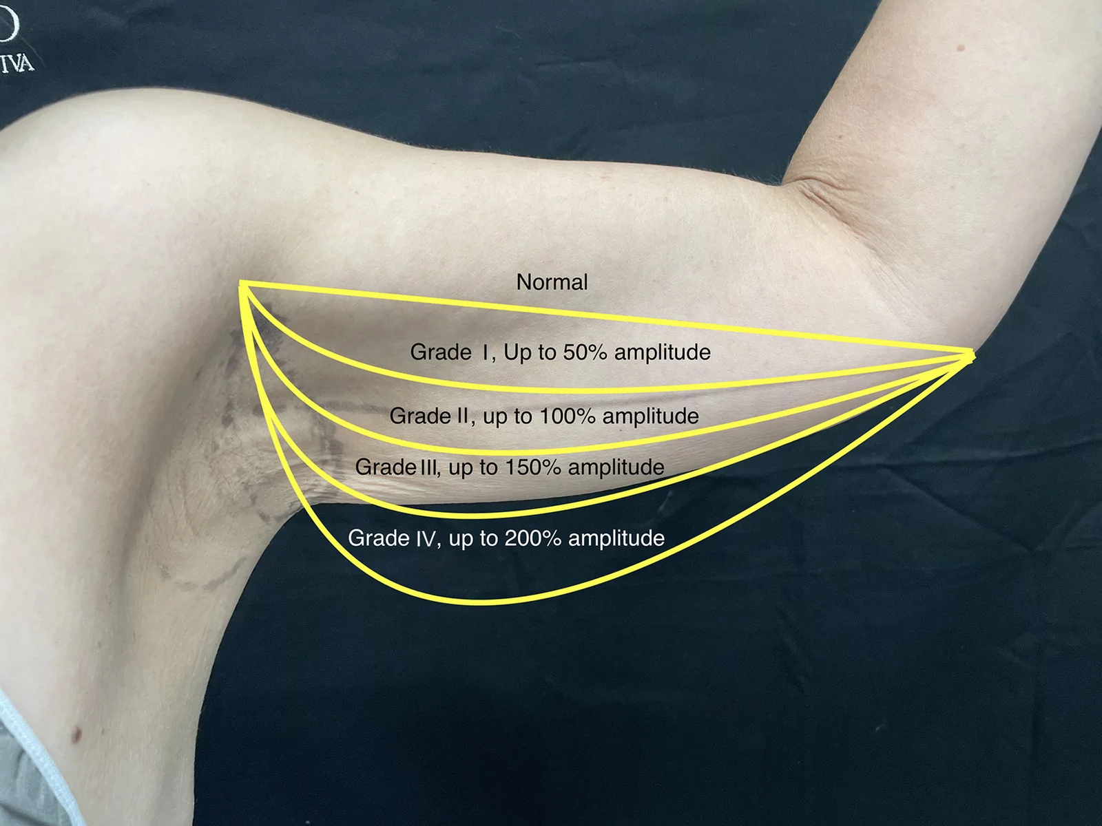
The operational database included sociodemographic information (age, gender) and clinical information (medical history, previous surgical procedures, surgical time, surgical procedure, satisfaction scale results, and surgical complications). During the consultation, Dr. Stanford evaluated the variables of pre- and postsurgical Iglesias arm laxity classification 12 using a meter to determinate objectively the grade of laxity (before surgery and 1 year after the procedure) (Table 1 and Fig. 1 ).
Table 1. Iglesias Arm Laxity Classification
| Grade | Description |
|---|---|
| Grade I | - The skin flap is up to 50% of the arm amplitude |
| Grade II | - The skin flap is greater than 100% of the arm amplitude |
| Grade III | - The skin flap is greater than 150% of the arm amplitude |
| Grade IV | - The skin flap is greater than 200% of the arm amplitude |
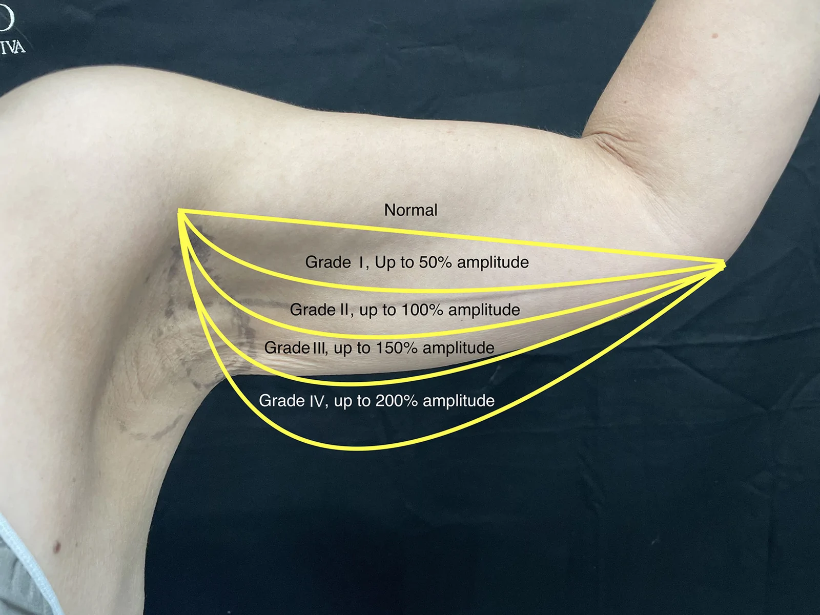
Fig. 1. Iglesias arm laxity classification. Schematic representation of the Iglesias classification system used to grade upper-arm skin laxity. Laxity is quantified as the percentage of redundant skin measured from the muscle border to the most distal portion of the skin flap in 90-degree angle arm abduction.Iglesias 上臂松弛分级示意图,以手臂外展90°时冗余皮肤占臂幅百分比界定松弛程度(I 级≤50%、II 级>100%、III 级>150%、IV 级>200%)。
Eligibility for SAB is determined based on preoperative assessment of arm laxity, selecting patients classified between grades I and III according to the Iglesias scale. ( See Video 1 [online] , which demonstrates the preoperative marking process for SAB.) ( See Video 2 [online] , which shows the surgeon explaining the anatomical references used to determine the anterior and posterior borders of the axillary fossa, the central axis marking, and the location of the fusiform excision.)
Preoperative markings are performed with the patient in standing position. The arm is positioned in alternating adduction and abduction to accurately delineate the anterior and posterior anatomical boundaries of the axillary fossa. This dynamic evaluation ensures that the planned incision remains concealed within the axillary fold when the arm is adducted. The central axis of the axilla is identified and marked, and a line is drawn 0.5 cm distal to this axis to designate the superior margin of the fusiform excision while remaining within the safe anatomical limits of the axillary region. A pinch test is conducted to confirm the extent of the skin flap and to define its anterior and posterior margins within the hollow part of the axilla. Careful manual retraction of the flap is performed to assess tissue mobility, with attention to minimizing tension to reduce the risk of postoperative wound dehiscence and hypertrophic scarring.
The patient is then positioned in lateral decubitus, and liposuction of the arm is carried out. Depending on the patient’s needs, this may involve limited aspiration of the lower third of the arm or circumferential 360-degree wet liposuction performed with power-assisted liposuction. Following aspiration, subdermal monopolar radiofrequency is applied through both proximal and distal access points, with a minimum of 6–9 passes performed per port, at 80% of power with a gas flow of 2.5–3.0 L/min per arm (8 kJ per arm). The treatment extends along the arm toward the elbow.
After repositioning the patient in the supine position, a fusiform incision is made along the axillary marking, extending down to the level of the axillary fascia. Meticulous hemostasis is achieved throughout the procedure to minimize bleeding and optimize wound healing. Layered closure is performed, beginning with 3 initial interrupted sutures using 2-0 Vicryl to secure the underlying tissues, followed by a single-pass subdermal anchoring suture with 3-0 Vicryl. The skin is then approximated using 3-0 Monocryl for a precise and aesthetically favorable closure.
To evaluate the success of the intervention, results were compared using the Iglesias arm laxity scale both before and after surgery. Patient satisfaction was assessed by analyzing changes in BODY-Q scale scores presurgery versus postsurgery, providing a measure of satisfaction with the outcomes of the procedure.
Arm laxity was objectively assessed using a flexible metric measuring tape during the preoperative consultation. With the patient standing and the arm positioned at 90-degree abduction and 90-degree elbow flexion, the midpoint of the arm was identified by palpating the superior and inferior borders of the muscle mass. Arm amplitude was defined as the linear distance between the upper muscular border and the lower muscular border. The redundant skin flap was then measured from the inferior muscular border to the most distal portion of the hanging skin in a relaxed, nonstretched position. No external traction was applied during measurement to avoid artificial elongation. Measurements were taken in centimeters and subsequently categorized according to the percentage relative to the total arm amplitude, in accordance with the Iglesias classification.
Informed consent (for surgical procedure, use of images, and use of data for research) was signed by each patient before surgery. Any patient could refuse to participate in the study or could withdraw consent to participate at any time. This research was conducted according to the Declaration of Helsinki.
Descriptive statistics were used to show the patient information results. To demonstrate differences between preoperative versus postoperative arm laxity, Student t tests were used. The level of significance was defined as a P value of less than 0.05. Jamovi statistical software (The Jamovi Project, Australia; version 2.5; retrieved from https://www.jamovi.org) was used for both univariate and multivariate analyses.
补充视频 / 附件(原文未随文提供原图,可于出版方页面查看)gox-14-e8160-s001.mp4Video 1. This video demonstrates the preoperative marking process for Subdermal Axillar Brachioplasty (SAB).补充视频 / 附件(原文未随文提供原图,可于出版方页面查看)gox-14-e8160-s002.mp4Video 2. The surgeon explains the anatomical references used to determine the anterior and posterior borders of the axillary fossa, the central axis marking, and the location of the fusiform excision
A sample of 67 patients was obtained, with a mean age of 50.2 years (SD, 7.1 y). Regarding gender, all patients were women, and most of them were White (81.8%), with a mean body mass index of 27.8 kg/m 2 . Of the 67 patients, 16 (23.88%) reported having a medical history. The most common responses related to medical history were no medical history (76.11%), hypertension (10.44%), and hypothyroidism (7.46%) (Table 2 ).
Table 2. Patient Characteristics
| Variable | Measure of Central Tendency or Percentage | Dispersion Measurement, SD |
|---|---|---|
| Age, y | Mean: 50.2 | 7.1 |
| Sex, n (%) | Total: 67 | NA |
| Female | 67 (100) | |
| Male | 00 (0) | |
| Height, m | Mean: 1.69 | 0.06 |
| Weight, kg | Mean: 79.4 | 4.3 |
| Body mass index, kg/m 2 | Mean: 27.8 | 2.1 |
| Race, n (%) | Total: 67 | |
| White | 54 (80.59) | |
| Latino | 13 (19.40) | |
| Medical diagnoses, n (%) | Total: 67 | |
| - None | 51 (76.11) | |
| - Hypertension | 07 (10.44) | |
| - Hypothyroidism | 05 (07.46) | |
| - Diabetes | 01 (01.49) | |
| - Asthma | 01 (01.49) | |
| - Facial paralysis | 01 (01.49) | |
| - Gastroesophageal reflux disease | 01 (01.49) |
Regarding surgical time, the average duration was 108 minutes (SD, 12.9 min). All patients underwent general anesthesia. Additionally, most patients requested this procedure due to significant weight loss (62.68%), whereas the remaining patients cited skin laxity due to age. Of the patients, 73.13% had grade III arm laxity as per the Iglesias scale, and the remaining 26.86% had grade II laxity. After the intervention, most patients exhibited grade I laxity (89.55%), with the rest achieving grade II laxity (10.44%) (Figs. 2 – 4 ; notice the changes before and after surgery). The average length of the surgical wound in the axillary region was 5.4 cm; the mean lipoaspirate volume was 245 mL per arm (SD, 62 mL; range, 150–360 mL), reflecting conservative contouring rather than extensive volume reduction (Table 3 ).
Table 3. Surgical and Clinical Information
| Variable | Measure of Central Tendency or Percentage | Dispersion Measurement, SD |
|---|---|---|
| Surgical time, min | Mean: 108 | 12.9 |
| Anesthesia, n (%) | Total: 67 | NA |
| General | 67 (100) | |
| Local | 00 (00) | |
| Presurgical arm laxity, n (%) | Total: 67 | NA |
| Grade 0 | 00 (0) | |
| Grade I | 00 (0) | |
| Grade II | 18 (26.86) | |
| Grade III | 49 (73.13) | |
| Grade IV | 00 (0) | |
| Postsurgical arm laxity, n (%) | Total: 67 | NA |
| Grade 0 | 00 (0) | |
| Grade I | 60 (89.55) | |
| Grade II | 07 (10.44) | |
| Grade III | 00 (0) | |
| Grade IV | 00 (0) | |
| Surgical wound length, cm | Mean: 5.4 | 0.4 |
| Lipoaspirate volume per arm, mL | Mean: 245 | 62 |
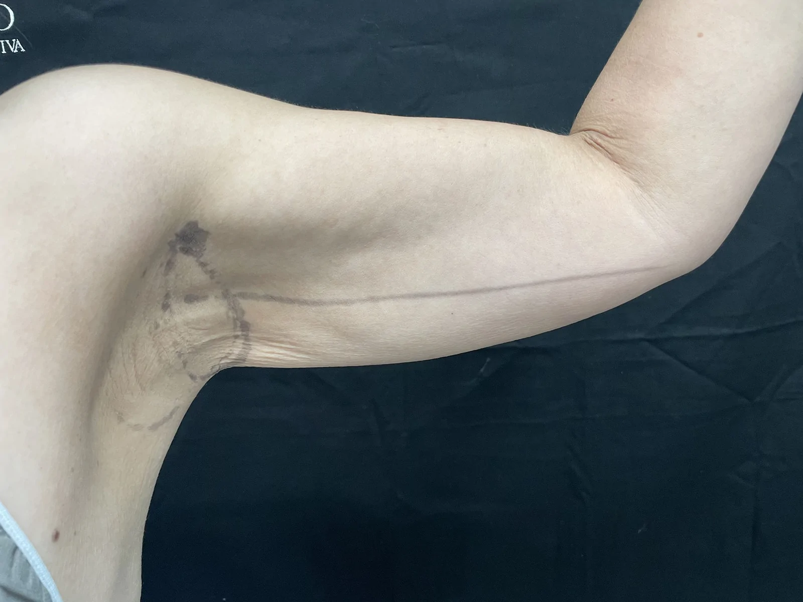
Fig. 2. Preoperative markings for SAB. Clinical photograph showing preoperative markings that define the anterior and posterior limits of the axillary fusiform excision while the arm is positioned in the adduction–abduction position. The patient is a 45-year-old woman with a starting laxity grade of III.SAB 术前标记:于手臂内收—外展动态下界定腋窝梭形切除的前、后边界,使切口在手臂下垂时隐藏于腋窝皱褶内。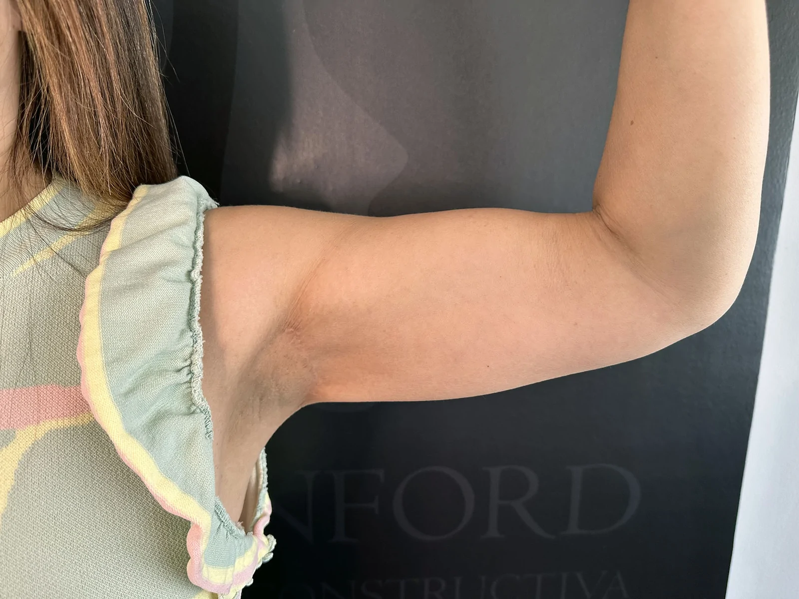
Fig. 3. Postoperative result demonstrating concealed axillary scar. Standardized postoperative photograph demonstrating correction of arm laxity with the incision fully hidden inside the axillary fossa. The patient is a 48-year-old woman, shown 6 months postoperatively with a laxity grade of I.术后示意:切口完全藏于腋窝,展示松弛矫正后的外观(48岁女性,术后6个月,松弛 I 级)。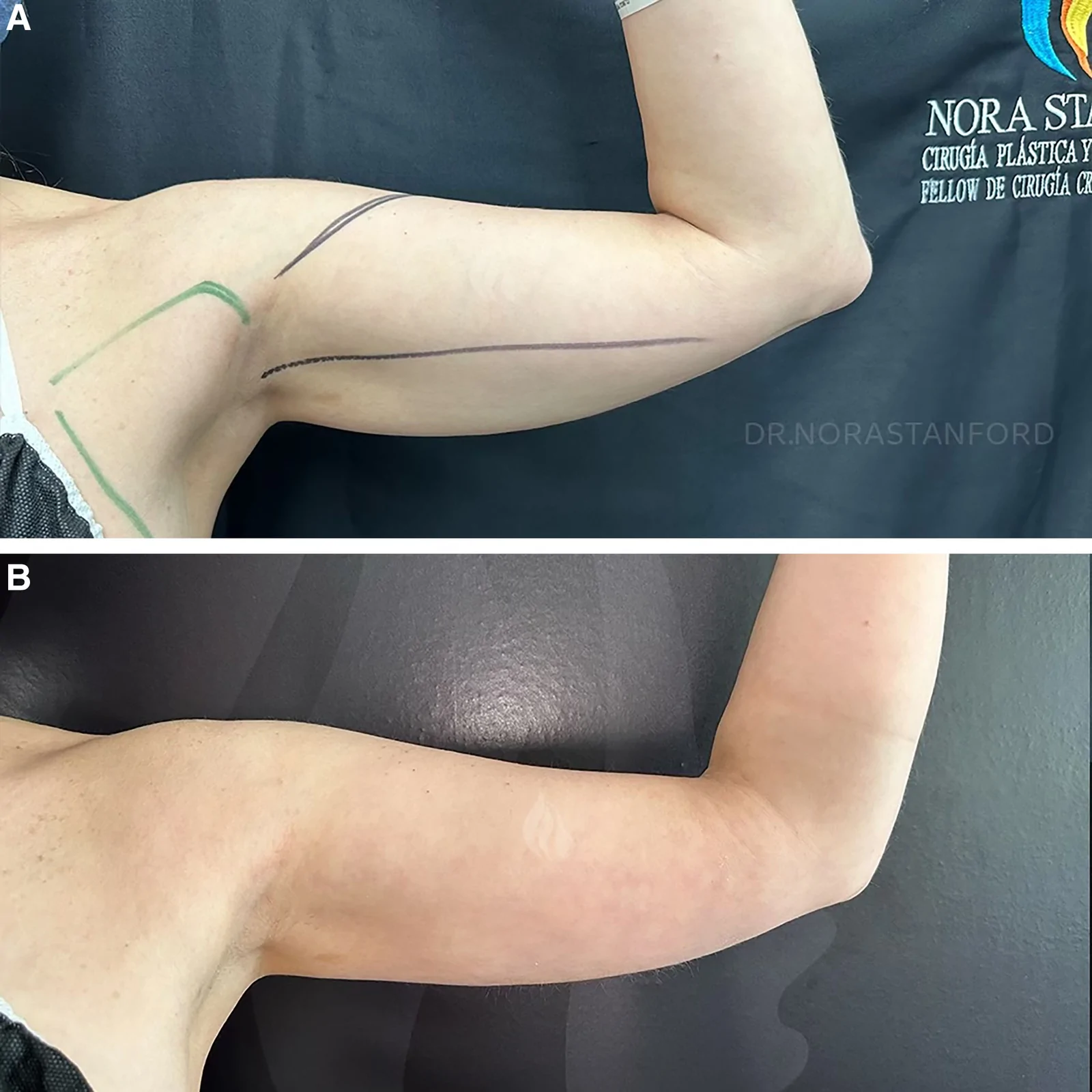
Fig. 4. Pre- and postoperative comparison in a 51-year-old woman who underwent SAB. A, Preoperative photograph showing arm laxity grade II and marking of incision trajectory. B, Twelve-month postoperative image demonstrating improved contour and nonvisible axillary scar.51岁女性术前(II 级松弛及切口轨迹标记)与术后12个月对比,可见轮廓改善且腋窝瘢痕不可见。Some complications occurred during and after the 1 year of follow-up: 2 cases of hematoma, 1 case of seroma, and 1 case of infection. Patient satisfaction with the procedure was high, based on the BODY-Q scale. Patients’ satisfaction with the appearance of their arms increased from low (31.9 out of 100) to high (94.2 out of 100). This difference is statistically significant (Student t test, 39.2; P < 0.001) (Table 4 ). Also, we achieved aesthetic results with no common complications because the location of the incision site (axillar fossa) has a higher risk of infection, dehiscence, and scar deformities (Fig. 5 ).
Table 4. Satisfaction Score
| Variable | Measure of Central Tendency | Dispersion Measurement | Test |
|---|---|---|---|
| BODY-Q score | Mean | SD | Student t test = 39.2 |
| Before SAB | 31.9 | 2.98 | P < 0.001* |
| After SAB | 94.2 | 3.45 |
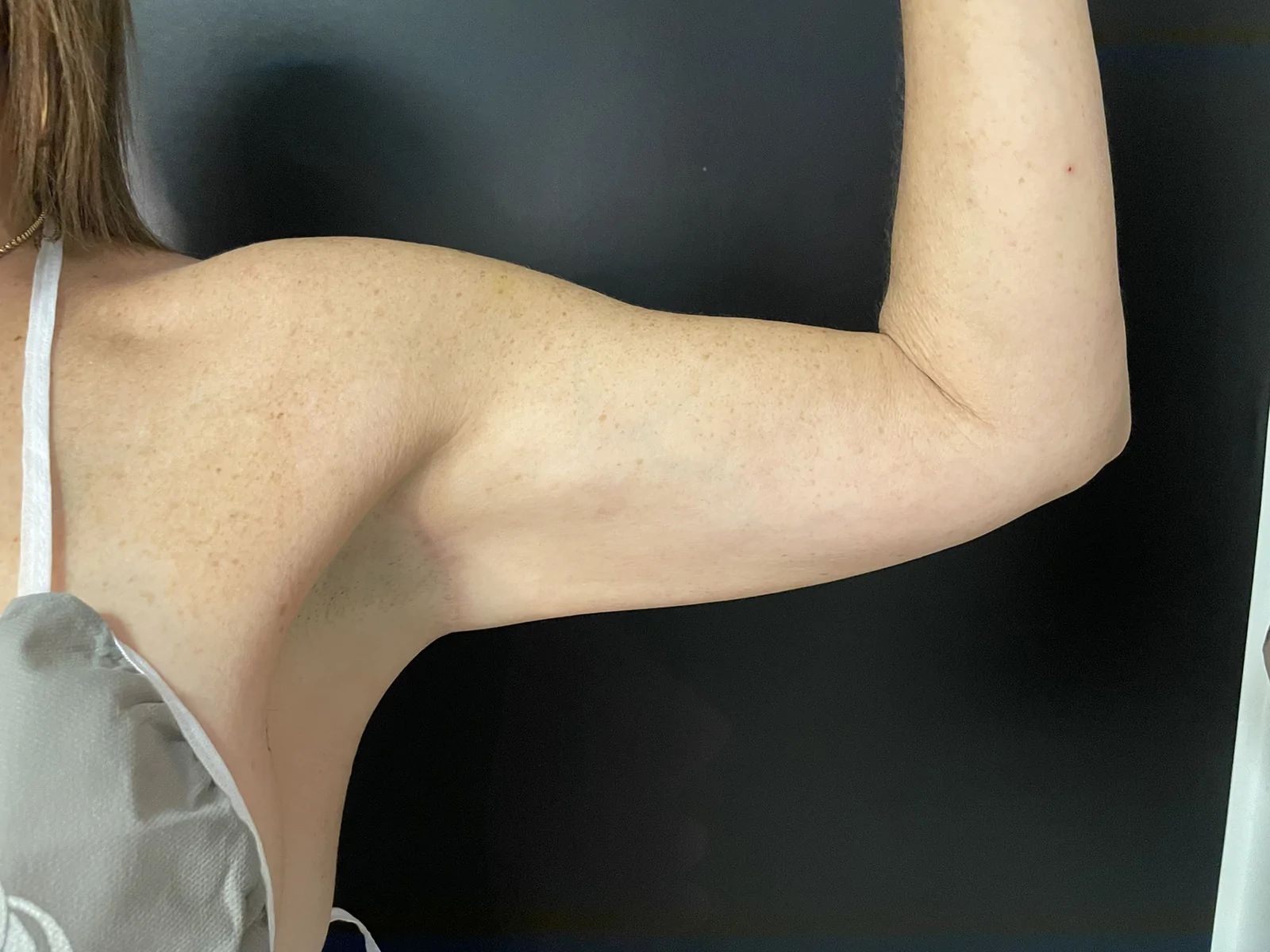
Fig. 5. Twelve-month postoperative arm contour in a 47-year-old woman, showing grade I laxity. Lateral view with arm flexed at 90 degrees, demonstrating smooth arm contour and scar concealed within the axilla.47岁女性术后12个月侧位,手臂屈曲90°,轮廓平滑、瘢痕隐于腋窝内。
This study introduces SAB as a novel arm lifting technique that combines liposuction, subdermal radiofrequency application, and layered flap fixation—all achieved through a single, small incision in the axillary region. This method addresses the problems of excess skin (Iglesias scale grades I–III) and skin laxity in patients who are either older or have undergone significant weight loss. The innovative approach discussed offers highly noticeable, effective results in arm contouring while minimizing scarring and avoiding the traditional long incision extending down to the elbow (Figs. 6 , 7 ).
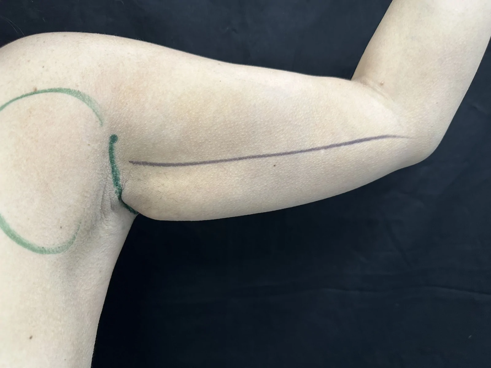
Fig. 6. Preoperative marking before liposuction and radiofrequency in a 42-year-old woman with a starting laxity grade of II. Photograph demonstrating liposuction access markings and axillary incision design before SAB.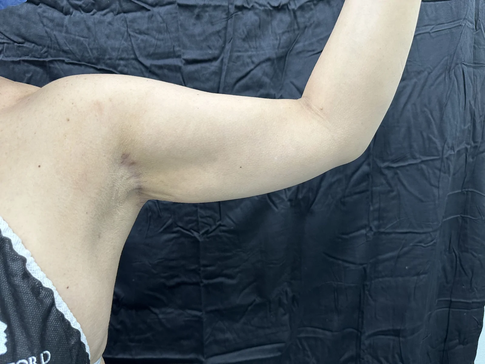
Fig. 7. Three-month postoperative result showing axillary scar integration in a 42-year-old woman. Postoperative image showing natural arm contour with a fine, inconspicuous scar inside the axillary fold and a postoperative laxity grade of I.As individuals age or lose a significant amount of weight, various structural and functional changes occur in their arms. Muscle mass and strength decline, leading to reduced muscle size, along with the infiltration of fat and fibrous tissue, which negatively affects muscle quality. 13 These changes are often accompanied by a reduction in bone mineral density, weakening the structural integrity of the arm. 14 Substantial weight loss can also lead to severe skin laxity, with excess skin particularly prominent in the upper arms, contributing to contour deformities. 2 In addition, aging affects the vascular system, causing the brachial artery walls to thicken and arterial elasticity to decrease, further contributing to functional decline. 15 Together, these changes impact both the appearance and strength of the arms, often prompting patients to seek aesthetic interventions.
To address the issue of “bat wings,” or excess skin and fat in the upper arms, several brachioplasty techniques have been developed. Liposuction-assisted brachioplasty is recommended for patients with mild skin laxity (arm laxity grade I). Although this technique offers good results with minimal surgical scarring, it is limited to those with only mild arm laxity 16 ; for patients with moderate to severe laxity (Iglesias arm laxity classification grades II–IV), longitudinal incision brachioplasty or L-brachioplasty are more commonly performed. These procedures, often required after significant weight loss or because of aging, remove excess fat and skin to improve contour of the arm. However, patients often express dissatisfaction with the long, visible scars that extend down the arm. 3 , 7 The SAB technique, as demonstrated in this study, achieved excellent results in patients with grade II–III arm laxity, with only minimal scarring in the axillary region, thus avoiding the discomfort of a long, visible scar for these patients (Fig. 8 ).
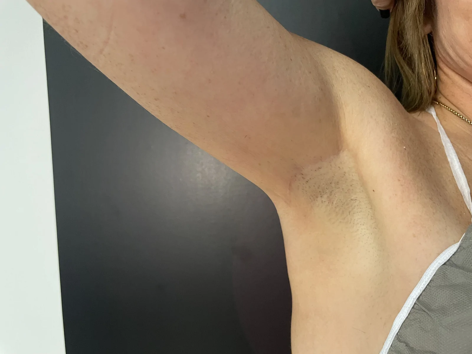
Fig. 8. Close-up view of healed axillary incision. Magnified postoperative image highlighting the scar hidden within the axillary crease, demonstrating optimal maturation and minimal visibility.愈合后腋窝切口特写,瘢痕位于腋窝皱褶、成熟度好、可见度低。The SAB technique has 2 key advantages that make it effective for patients with moderate to severe laxity. First, the layered fixation technique provides stability to the tissue, preventing dehiscence. Second, subdermal radiofrequency application promotes skin contraction by subdermal fibroseptal coagulation with reduction in skin laxity, 17 , 18 thus providing a youthful appearance and toned arms.
Complications from traditional arm lift procedures can vary. Hypertrophic scarring is the most frequently reported issue, affecting up to 24% of patients, whereas aberrant scarring occurs in about 9.9% of cases. 10 , 11 Infections are reported in 3.64%–14.6% of cases, with wound dehiscence affecting around 6.81% of patients. 11 Seroma formation is seen in 5.91% of cases, especially when multiple procedures are performed. 19 Hematomas occur in roughly 2.06% of patients, 20 and nerve-related complications and lymphedema affect 2.47% and 2.46% of patients, respectively. 11 , 21 , 22 Fortunately, the design of the SAB technique reduces the likelihood of many of these complications. In this study, 2.98% of the cases presented hematoma, and 1.49% presented seroma and infection. The minimal incision significantly lowers the risk of hypertrophic and aberrant scarring that has been reported in previous studies. 23 Layered sutures reduce the chances of wound dehiscence, and the minimally invasive nature of the procedure decreases the risk of infections, hematomas, and seromas compared with more extensive open surgery. In this study, no complications related to the SAB technique were observed in the patient cohort.
Overall, patient satisfaction with the SAB procedure was high, thanks to the significant aesthetic improvements achieved with minimal scarring in the axillary region. The natural-looking arm contour and the absence of a long, visible scar gave patients a more youthful appearance, enhancing their confidence and satisfaction with the procedure. Also, subdermal radiofrequency treatment has shown good results for skin traction.
This study has some limitations that must be acknowledged. First, its retrospective observational design limits the ability to establish causal inferences, and the absence of a control group prevents direct comparison between SAB and other brachioplasty techniques or energy-based procedures performed without surgical resection. Second, all procedures were performed by a single surgeon in a private practice setting, which may limit the generalizability of the results to other surgeons or institutions with different patient populations. Third, arm laxity measurements were performed by the operating surgeon, who was not blinded to the intervention, potentially introducing measurement bias. Although a standardized protocol was used, the absence of blinded independent evaluators represents a methodological limitation. Fourth, the arm laxity measurements were only taken at the preoperatory consultation and at 12 months follow-up.
The SAB technique offers a minimally invasive solution for patients with mild to moderate arm laxity by combining liposuction, subdermal radiofrequency, and layered flap fixation. This method achieved improvements in arm aesthetics with minimal scarring, avoiding the extensive incisions traditionally required for brachioplasty. The results of this study demonstrate high patient satisfaction, a reduction in postoperative complications, and effective correction of arm laxity. As body contouring techniques continue to evolve, the SAB approach stands out as a safe, effective, and patient-friendly option for addressing excess skin and fat in the upper arms.
The authors have no financial interest to declare in relation to the content of this article.
声明:中文精读 · 仅供学术参考。内容来自公开文献检索,不代表本人观点,不构成诊疗建议。 医疗美容需在正规医疗机构由执业医师实施。
基于相同主题推荐 · 继续深挖
针对 Iglesias I–III 级中度上臂松弛,SAB 以腋窝单一小切口联合吸脂、皮下单极射频与有限皮肤切除,避开传统从腋窝延伸至肘部的长瘢痕。
本文为一篇针对电磁(HIFEM)联合射频(RF)身体塑形(EmSculpt Neo,BTL公司)的文献述评,系统纳入7项含超声或MRI影像测量的研究,且全部由BTL医学顾问或研究者
本文为遵循 PRISMA-ScR 的范围综述,系统检索 PubMed、Scopus、Embase、LILACS,共纳入 1998–2025 年发表的 17 项研究(6 项回顾性、2